T6 Cytoskeleton BIO 107
1/45
There's no tags or description
Looks like no tags are added yet.
Name | Mastery | Learn | Test | Matching | Spaced |
|---|
No study sessions yet.
46 Terms
Cytoskeleton function in prokaryotes
Internal network of divers
maintenance of cell shale in many species
Cell separation during cell division
Movement of some cell contents
Composition of cytoskeletal strands
chains of globular protein
Bundles of fibrillar protein
different elements have different kinds of protein providing different functions
Green Fluorescent Protein (GFP)
Gree fluorescent protein from green fluorescent jellyfish
allows to label things in cell (cytoskeleton)
Can image LIVE CELLS
Tubulin superfamily (FtsZ)
Globular proteins, FtsZ is protein of tubulin superfamily
Forms a band around midpoint of cell (Z ring)
Creates strangulation in cell during cell division that leads to septatuon, eventually resulting in two daughter cells
segments cell

Inhibition of FtsZ
results in filamentous morphology and lysis
New avenue for strains of pathogenic bacteria with multiple resistance to current treatments
antiobiotics
Actin superfamily
Globular proteins
MreB
ParM
MreB
Globular protein, protein of actin superfamily
gives rod-shaped bacteria their rod shape
Probably used building scaffold in the deposition of new cell wall during growth
ParM
Protein of actin superfamily
moves plasmids to opposite ends of cell prior to division
Only for low count plasmids
1 copy not high count (100 of copies)
Crescentin
A coiled-coil filamentous protein (CCRP), family bending bacilli into curved shape
without crescentin is rod shaped, needs to bend bacilli into curved rod
Cell movement
Mobility: the cell can move but it is not propelling itself .
Ex. Red blood cells (cells are mobile)
Motility: cell can move by propelling itself with its own means of locomotion
ex. E.coli bacteria
Power own movement
Prokaryotic flagellum
Unlike in eukaryotes, flagellum is not part of cytoskeleton in prokaryotes
hollow tube filament made of self organizing flagellum proteins
Not related to cytoskeletal proteins
Grows by addition at the tip
Basal apparatus (motor) anchored in the cell wall, protruding into cytoplasm
The flagella filament is attached to motor by the hook
own support mechanisms in membrane
Different from cytoskeleton proteins

Flagellar arrangements
Flagella not part of cytoskeleton, but shape of cell is controlled by the cytoskeleton and that influence movement
e.coli is petrichous it means that flagella are all around perimeter
Locomotion dependent on shape of cell which is controlled by cytoskeleton
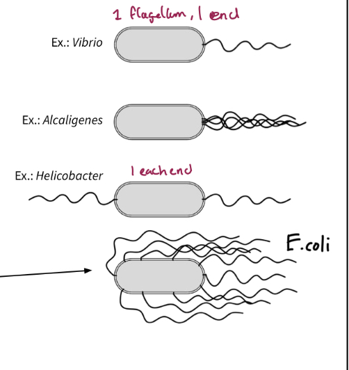
Flagella form bundles
Because geometry is the sane when turning direction is the same, flagella bundle into propelling unit
multiple flagella bundle together to form one large flagellum
Direction of flagella movement
Run: Counterclockwise motor rotation causes flagellar bundling and directional movement ( forward )
Tumble: clockwise rotation causes unbundling and spreading of flagellation and the cell rotates (in place)
opposite direction if twist unbundled→ separate whip around independently
Flagella move independently to rotate bacteria

Chemotaxis
Guidance system; movement directed by the concentration gradient of chemicals
concentration gradient is external to cell
chemicals must be present to direct movement
cycles of runs and tumbles
Higher concentration of chemicals → less bacteria tumbles
Longer runs, less tumbles to go towards chemical attractant
Type IV pili
Twitchy motion,
Rectractile protein filaments that move the cell closer to anchor point
like using grappling hook
Lengths of protein extend from cell and retract into cell
Archaellum
Archael flagellum
shorter and simpler in structure
Archael flagellum similar to type IV pili of bacteria
Grows by addition at base of archaellum, not tip
Rotational motion
Powered by ArP, not proton gradient
Lacking detailed knowledge about subject
Periplasmic flagellum
known as axial filaments
found in spirochaetes
External structures: internal to the outer membrane, but eternal to the plasma membrane
outside cell between plasma membrane and outer membrane (periplasmic space)
make entire cell rotate in corkscrew motion
Is flagella or axial filament more suitable for mucuous environment?
Flagella: have to push big bacteria through thick medium not efficient
Axial filaments: thin bacterium corkscrew entire body
How e.coli bacteria move?
Runs- swims in straight line, counterclockwise, flagella moves as singular bundle
Tumble - rotation and spreading of flagella, clockwise, flagellar bundle falls apart
Flagellum vs archaellum
Size
archaellum is smaller and simpler
Filament growth
Flagellum grows from tip
Archaellum grows from base
Energy source
flagellum is powered by proton gradient
Archaellum is powered by ATP
Hook
flagellum has hook, archaellum does not
Comparison of eukaryotic and prokaryotic cytoskeleton function
Maintaining shape: both
Move things during replication: both
Move things during regular physiological processes: eukaryotes
Propel the cell or its surroundings: eukaryotes
Cytoskeleton
Composed of different classes of fibers, it forms vast network throughout the cell
dynamic scaffolding and locomotion of Eukaryotic cell
Cytoskeletal fibers:Microfilaments (cortex underneath membrane),micro tubules ( in every cell), intermediate filaments
DNA in nucleus
Broad functions of cytoskeleton
Maintain and change cell shape
Spatially organize cell contents
Connect cells and environment
3 classes of cytoskeletal fibres
Microtubules (tubulin family)
polar (directional) (not charged)
Globular monomers
Microfilaments (actin family)
polar (directional in different way)
Globular monomers
Intermediate filaments
apolar
Filamentous monomers
Microtubules
hollow tubes made of tubulin heterodimers
Stiff structural tubes
Strong compression
They can help maintain the cell shape under pressure
can push not pull
Function as tracks to move cell contents and cell
microtubulin track for cell contents to move on
tubulin assembles into hollow Microtubules made of dimers of alpha and beta tubulin
polar : 1 end alpha. 1 end beta
Microtubules as tracks for vesicle transport
Motor proteins attach to both Microtubules and cargo (usually vesicle) to transport it along Microtubules to specific destination

Motor proteins on Microtubules are polar
Kinesin moves toward plus end ( anterograde transport)
Dye in moves toward the minus end (retrograde transport)
Both walk on tube, hand over hand fashion
Kinesins and dyenins are ATP powered and move in opposite directions

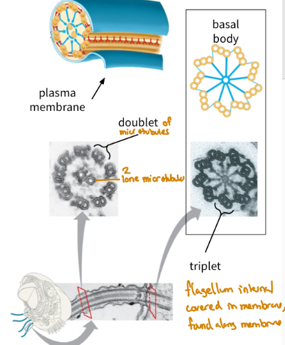
Eukaryotic cilia and flagella composition and structure
Eukaryotic cilia and flagella are both internal structures (within cell membrane) made of Microtubules
both are ATP powered with same configuration
Basal body: 9 Microtubules triplets it’s no central Microtubules (9+0)
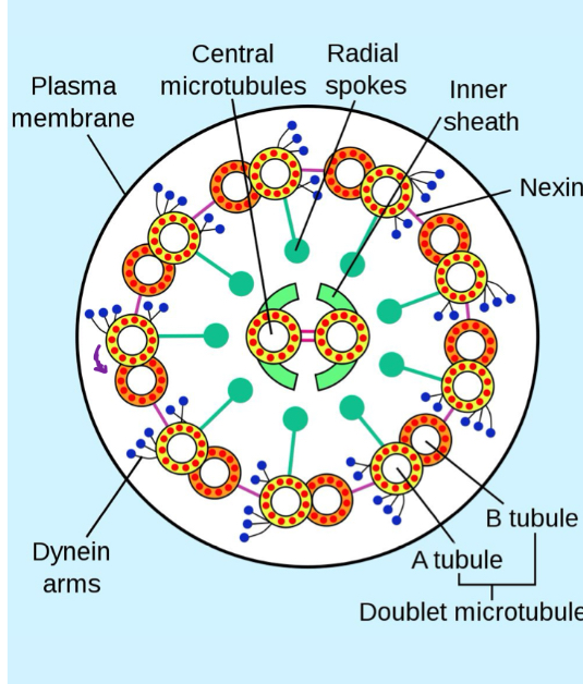
Motile portion: 9 Microtubules doublets with a central Microtubules pair (9+2)
Dyenins move flagellum
Dyenins attached to neighbouring doublet “walk” in surface pfmicrotubule outlet facing it
dyenin moves toward minus end
Dynein arms attach:The dynein motor domain on the A-tubule binds to the B-tubule of the adjacent doublet.
ATP binds dynein → releases microtubule.
ATP causes dynein to detach from the B-tubule.
ATP hydrolysis → conformational change (“power stroke”).
Dynein undergoes a large structural shift that slides the A-tubule relative to the B-tubule.
ADP + Pi released → dynein reattaches at a new position.
The cycle repeats, producing continuous sliding motion between adjacent microtubules.
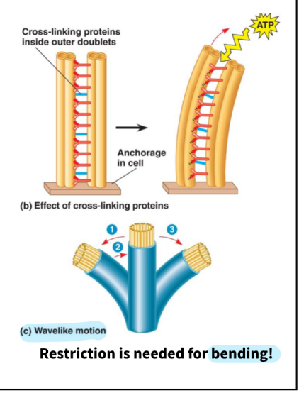
How flagella bend
Movement is restricted to cause bending, dyenin prevented from walking past each other
Bending motion required to bend outlet, anchorage (basal body) , elastic component
If microtubule doublets could slide freely, the flagellum would just extend and contract (like a telescope).
But nexin links and radial spokes restrict that sliding.
So, when dynein on one side of the flagellum is active, that side shortens, and the other side stays extended.
This asymmetric activity causes the flagellum to bend.
Compare eukaryotic cilia and flagella structure
Eukaryotic cilia and flagella have same structure and same evolutionary origin
They differ in size and type of novement:
flagellum: undulates symmetrically side to side
Cilium: beats asymmetrically with a different stroke on each side
depending on which side bearing towards
Power stroke, extended recovery strokes
extend straight then push
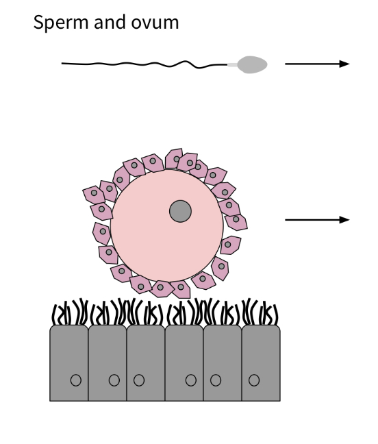
Motility vs mobility: sperm and ova
Sperm is motile : soerm cells propel themselves with flagellum
Ova. (Eggs) are mobile: beating cilia on epithelial cells of Fallopian tubes propel themselves ovum
do not propel themselves
Propelled by cilia n fallopian tube
Microtubules radiate from MTOCs
Centrosome is an MTOC; Centrosome organize Microtubules
Centrosome functions as Microtubules organizing center (MTOC)
Other structures serve the sane purpose in a scenes of centrioles (such as I plants and fungi)
Microtubules role in mitosis
Microtubules guide chromosomes during mitosis
Microtubules are part of spindle apparatus (cell division lectures)
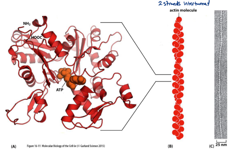
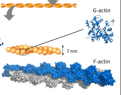
Microfilaments (actin filaments/ thin filaments)
G actin = protein monomer if Microfilaments (g for globular)
G-actin is polymerized into F-actin to form filaments (f for filamentous)
Microfilaments provide tensile strength to various parts of cell
good under tension, cannot resist compressive forces
Not strong under compression
Help shape cell
Provide movement to the cell and cell membrane
Formation of F-actin requires ATP
polymerization of G-actin
Microfilaments form cell cortex (sub membrane shape cortex)
Microfilaments give structure to some cells
microvilli (outfolds) on intestinal epithelial cells are internally supported by actin (Microfilaments)
Actin myosin system used for cell motion and cell contraction
How muscle cells are able to contract individually and collectively to produce muscle contraction; how muscle cells generate motion
ATP powered
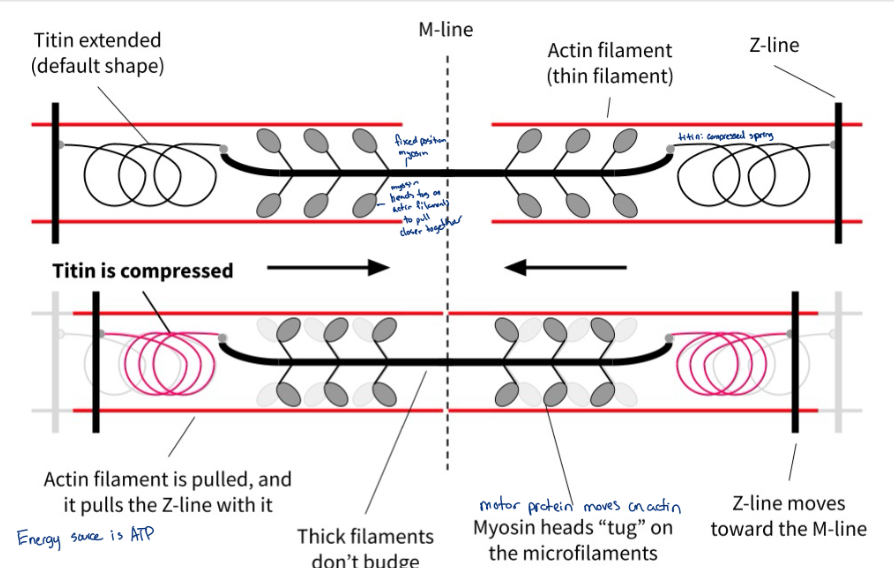
Actin myosin system for muscle cell contraction steps
The Cross-Bridge Cycle (Actin–Myosin Interaction)
ATP binds myosin head →
Myosin detaches from actin.ATP hydrolyzed (ATP → ADP + Pi) →
Myosin head “cocks” into a high-energy position.Myosin binds to actin (if Ca²⁺ present) →
Cross-bridge forms between actin and myosin.Power stroke:
Pi released → myosin head pivots, pulling actin toward the M-line → sarcomere shortens.Aline is pulled closer together towards M line in center to make cell contact
ADP released, new ATP binds → cycle repeats.

Amoeboid movement
Most common types of crawling movement in Eukaryotes driven by actin filaments and myosin motor proteins pulling on them, forming pseudopodia
shape membrane by changing shape of actin myofilaments
Projections called pseudopodia
Extension of a pseudopodium
At the leading edge of the cell, actin filaments polymerize (assemble) beneath the plasma membrane.
This pushes the membrane forward, forming a bulge — the pseudopodium.
Attachment to the surface
The pseudopodium adheres to the substrate using adhesion proteins (like integrins).
This anchors the front end of the cell.
Cytoplasmic streaming
The internal fluid (cytoplasm) flows forward into the pseudopodium — called endoplasmic streaming or sol–gel transformation.
The actin cortex near the front remains more gel-like (dense), while the back becomes more fluid.
Contraction at the rear
Myosin II interacts with actin filaments at the rear (posterior) of the cell, pulling the cell body forward.
This contraction drags the rest of the cytoplasm toward the pseudopodium.
Detachment of the rear
The old adhesion points at the rear detach, completing one “step.”
Then the process repeats: extend → attach → pull → detach.
Cellular streaming in plants
An actin myosin system
actin myosin used to move organelles in plants
Chloroplast move by actin and myosin heads
Bacteria that manipulate actin based cytoskeleton
Echlira chaffeensis (gram negative) is an obligate intracellularvpaparsite that hijacks cytoskeleton of neutrophils to find other neutrophils for infection
parasitic bacteria hijack cytoskeleton of neutrophils → membrane reshaped to find other host cells
Intermediate filaments
Large family of 50+ proteins
Twist to form cable like structures of very high tensile strength
Toughen parts of cell and morph cell into specific shape
ANIMAL ONLY STRUCTURE
diameter/ thickness is between Microfilaments and Microtubules
Intermediate filaments bundle formation
Ifs come in bundles of coiled coils
monomers intertwine to form coiled coil dimers with strong disqualified bridges (and other interactions)
Dimers pair up staggered to form tetramers
Filamentous proteins , threads not globular , pair of intertwined threads stagger to form ropes of intermediate filaments
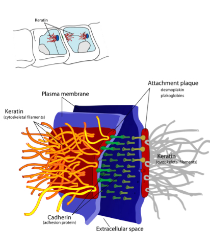
Desmosome fibers composition
Desmosome fibers are made of keratin (an IF protein)
Desmosome needs high tensile strength to keep cells together
Keratin IFs project from the Desmosome plate to inside both cells
Lamin
Lamin: protein that forms nuclear lamina, type of intermediate filaments
net structure lines nuclear envelope where chromosome attached
Extracellular Keratin
Extracellular keratin is a type of extracellular intermediate filament
extracellular keratin forms tough structures like claws, horns, beaks, feathers, hair, nails, etc.