Chapter 13- Occipital Lobe Neuropsych
1/63
There's no tags or description
Looks like no tags are added yet.
Name | Mastery | Learn | Test | Matching | Spaced |
|---|
No study sessions yet.
64 Terms
When we stain V1 what is revealed?
Blobs(cytochrome oxidase rich): color perception
Interblobs: Form and motion perception
Where is the occipital lobe located in terms of anatomy and list its function?
It is located in the posterior part of the brain and it is responsible for vision.
parietal-occipital sulcus
The occipital love is distinguished from the parietal lobe by this sulcus.
What are the three landmarks within the visual cortex?
Calcarine sulcus: divides the upper and lower halves of the visual word
Lingual gyrus: part of the visual cortical regions V2 and VP
Fusiform gyrus: V4
Why do human's have a larger visual cortex compared to Old World Monkeys?
This is a result of the development of language in Humans. Occipital ->Parietal -> Temporal
Visual areas extend well beyond the occipital lobe
What is the striate cortex?
Cortical layer IV features four distinct layers and appears a s a thick stripe.
What is cytochrome oxidase?
an enzyme crucial in making energy available to cells.
When we stain area V2 what is revealed by cytochrome oxidase?
Thick stripes and pale stripes: Form and motion perception
Thin stripes: Color perception
How does color perception affect our ability to detect motion and form?
enhances our ability to detect motion and form.
Where does visual processing start?
starts in V1 and then advances to other regions of visual processing.
What is V2's responsibility?
The secondary processing location and it projexts onward to other visual areas.
What is the dorsal stream?
Visual stream responsible for visual guidance of movement: Where pathway
What is the ventral stream?
Stream responsible for objection perception and motion perception: what pathway.
What the STS stream?
Stream also known for its object perception and motion perception. Lateral projections.
What did the Macaque Monkey study reveal?
a dorsal action pathway and a ventral perceptual pathway were proposed for the visual system.
-Ventral pathway contains two parts: one that is on the lateral surface and one that projects on the ventral surface of the temporal lobe.
-Visual streams project to many higher visual areas throughout the temporal, parietal, and frontal lobes, and these areas have specific functions, but they interact.
Define Vision for Action
-Visual processing is necessary to direct specific movements, such as reaching for a mug or a cell phone, and to shape the hand to pick up the object.
-This can be done with or without attention and awareness.
-Processing visual information about a moving target in order to catch it requires the interaction of multiple visual areas, largely in the parietal lobe and part of the dorsal stream.
Define Action for Vision
-When you look at an object, you visual system focuses on only part of that object
-Looking at a face the observer focuses on the eyes and the mouth of the left visual field. This bias is specific to facial recognition
-When a subject performs a mental rotation task, the task is often accompanied by eye movements.
Define Visual Space
-Objects can be described in terms of their relation to the observer(egocentric space) or to each other (allocentric space).
- the visual system likely encodes both perspectives to guide movements appropriately and to remember a visual scene.
We are able to recognize a wide variety of objects, but we likely do not have brain areas dedicated to recognizing every category of object. Some biologically important objects, such as faces or hands, do appear to have brain regions dedicated to them.
Visual scenes have an enormous amount of detail, and attention is a limited resource. Both top-down and bottom-up factors influence what we attend to in the visual system.
Define Visual Recognition
- We enjoy the ability to recognize and respond to visual info.
- We do not have specialized regions for each object, but we do have specific areas for biologically significant information, hands and faces, but we also have ones for objects and places.
Define Visual Attention
-We select specific aspects of visual input and attend to them selectively.
-Neurons respond selectively to stimuli in particular places, times, or when a movement needs to be executed.
-Independent mechanisms of attention are probably required both for guiding movements(parietal) and for recognizing objects(temporal)
What is visions primary function?
guide movement, not to recognize objects.
Simple animals move toward or away from light without perceiving light versus dark.
dorsal stream damage
patients can see objects, but cannot accurately reach for them.
What does the dorsal stream provide?
real-time visual control of action.
How were the Imaging Studies of Dorsal and Ventral streams constructed and what were the results?
Subjects are asked to compare two faces
They showed activity in the temporal lobe when asked to say which faces were the same
They showed activity in the parietal lobe when asked to consider where on the face a dot or box was superimposed
When asked to detect motion, area V5 showed increased activity
When asked to detect color, area V4 showed activity
Results from functional imaging studies match results expected from studies of nonhuman primates and from lesion studies.
Where is information from both eyes combined?
Information from both eyes about a particular location in space is combined in V1
If a visual disturbance affects both eyes, the disturbance must be in V1
What is macular sparing?
Damage to the visual cortex typically spares information from the central part of the visual field
The reason is uncertain, but could be because the region of the visual cortex corresponding to vision receives blood from multiple cerebral arteries, making a stroke in that area less likely
What are scotomas?
are small blind spots that are often unnoticed because the eyes are constantly moving and the brain fills in the blind spots with information from the previous position of the eyes
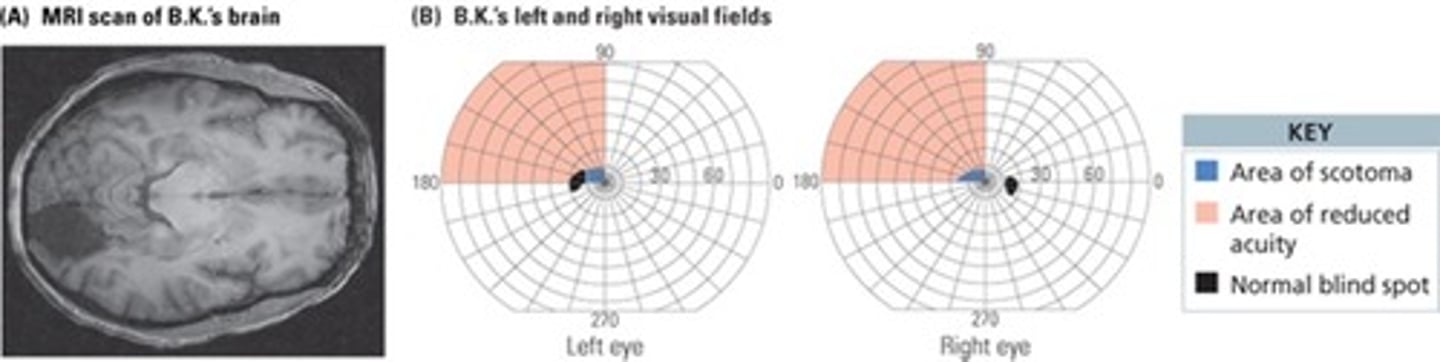
V1 Damage and Scotoma
Patient awoke to discover loss of vision in left visual field
Vision returned to the lower left visual field, but not the upper left field
MRI scan revealed evidence of a stroke in the right occipital lobe
Perimetry maps regions of blindness by asking patient to detect a small light moving against a dark background
Shortly after the stroke, was unable to detect the light in the area of the stroke, but could report that it had been there after researchers moved the light to a different part of the visual field
Over time, some vision returned, but the scotoma remained, and form vision was still poor in the upper left quadrant
Knowing something about the visual stimulus, even though the patient says they cannot see it, is called blindsight. This indicates that while there is damage in V1, higher visual areas remain intact.
V1 Damage and Blindsight
Patient had surgery to remove abnormal blood vessels, which removed part of the right calcarine fissure
Patient typically reports they see nothing in the left visual field, but sometimes has a "feeling" that something is there; and they guess correctly about the shape of the object
Accurately indicates the location of stimuli and the orientation of lines they did not see
Cortical blindness occurs when a patient has no conscious awareness of visual stimuli but can accurately indicate locations, directions, forms, or colors of the stimuli
In other patients with blindsight, fMRI shows that they have activity in V5 when there is a stimulus moving in their blind spot, as well as activity in the prefrontal cortex.
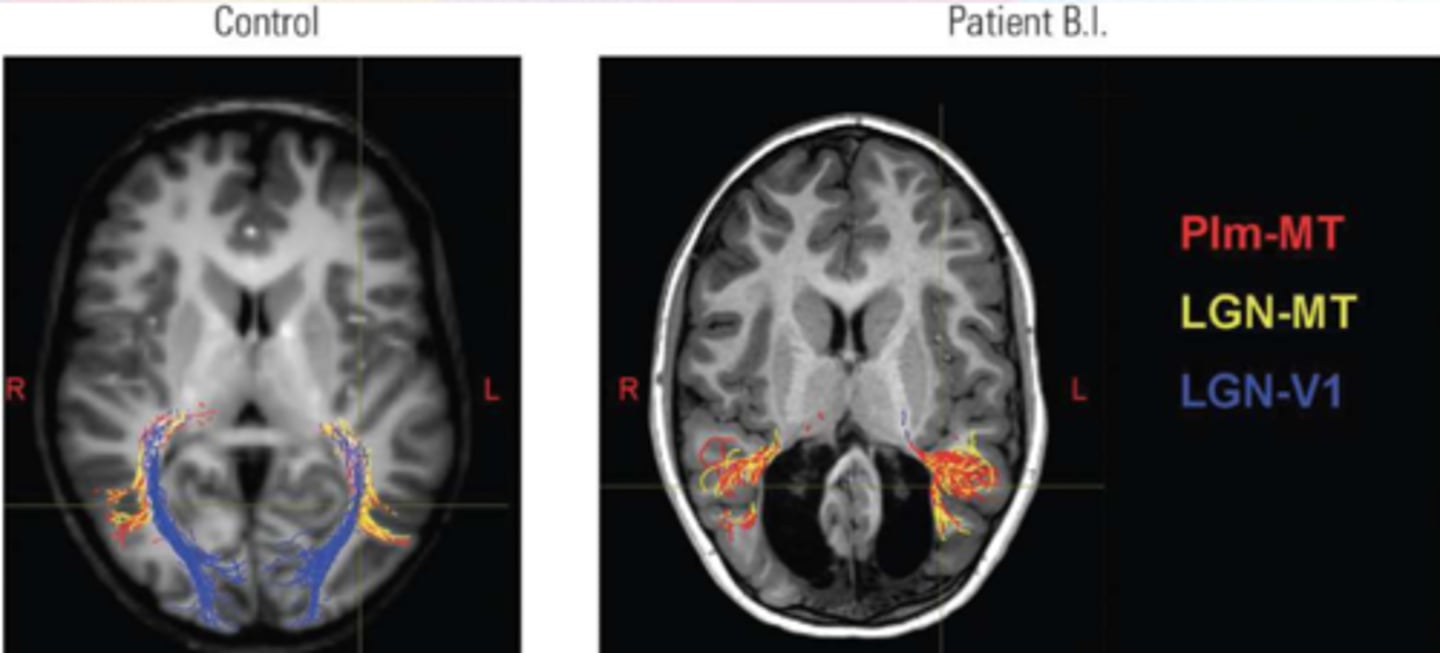
More than blindsight
Child sustained damage to the occipital lobe bilaterally in the first 2 weeks of life due to a genetic disorder
At age 6, he is not blind
He can recognize faces and colors, and navigate his environment
Significant plasticity to rearrange his visual system and strengthen pathways from subcortical areas to V5
V4 Damage and Loss of Color Vision
Artist lost color vision following a car accident
Visual acuity improved, but the world appeared as shades of gray
He lost the ability to remember or imagine colors
He even dreamed in black and gray
Conscious Color Perception
Man was electrocuted and had to be resuscitated, resulting in significant damage to the posterior cortex
He could detect the presence of light, but was otherwise blind
Unlike J.I., he retained the ability to imagine colors
V5(MT) Damage and Movement
Following damage to area V5, patient was unable to detect motion
Pouring fluids was difficult because she could not see the level rise in the cup
Interacting with people was disturbing because she could not see them move
Vision was otherwise normal
Case suggests that the brain processes the movement of a form separate from the form itself
Symptoms can be mimicked by applying TMS to V5/MT
A patient with a condition similar to L.M. described the terror of trying to cross a street, when they could not see the traffic moving and know when it was safe to go.
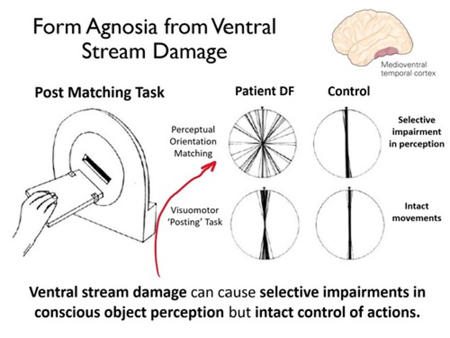
Occipital Damage and Visual Agnosia
Visual agnosia is the inability to recognize objects or pictures of objects—or the inability to draw a copy of the objects
Patients with carbon monoxide poisoning leading to damage to area LO have visual form agnosia and cannot recognize line drawings of objects
Patients cannot draw pictures of objects when looking at them and cannot reproduce line drawings
No difficulty reaching for and grasping objects, even though they cannot recognize them
Unable to correctly judge lines as horizontal or vertical, but can orient movements to match horizontal or vertical lines
Results suggest that the dorsal stream is intact (guiding movements to a target), but the ventral stream is not (recognizing objects).
Optic ataxia
a deficit in visually guided movements, such as reaching
Associated with damage to posterior parietal lobe
Patient had bilateral strokes in the occipitoparietal regions
Normal form and color vision, but could not reach for objects or form her hand into the correct shape to pick up objects
D and T
Higher-Level Visual Processes
D has a lesion to the right occipitotemporal region and has some difficulties reading, but is unable to recognize faces
Prosopagnosia is the inability to recognize familiar faces
T has a lesion to the left occipitotemporal region and has lost the ability to read, but has no problem recognizing faces
Alexia is difficulty reading
Define Visual Agnosia
the inability to recognize objects or pictures of objects or the inability to draw a copy of the objects.
What is prosopagnosia?
patients cannot recognize familiar faces, including their own
Rely on recognizing a person based on the sound of their voice, their hair, or the way they walk
Visual perception is normal, and patients can differentiate between human and nonhuman faces
Studies find bilateral damage at the occipitotemporal junction
What is Alexia?
is the inability to read
Associated with damage to the left fusiform gyrus and lingual area
Left hemisphere is specialized to combine letters to form words
considered a visual agnosia where the patient is unable to combine parts (letters) into a whole (words)
V3 specialty
process dynamic form, or the shapes of objects in motion
V4 specialty
predominantly processes color information
V5 specialty
involved in motion processing; also known as MT
What are the ventral visual regions beyond the Occipital lobe and what are their functions?
-(LO: Lateral Occipital: Object analysis)
-FFA, Fusiform Face Area, Face Analysis
-EBA, Extrastriate Body Area, Body Analysis
-FBA, Fusiform Body Area, Body Analysis
-STS, Superior Temporal Sulcus, Analysis of Biological Motion
-STSp, Superior temporal sulcus(posterior), moving-body analysis
-PPA, Parahippocampal place area, analysis of landmarks.
What are the dorsal stream visual regions beyond the Occipital Lobe and their functions?
-LIP, Lateral Intraparietal Sulcus, voluntary eye movement
-AIP, Anterior Intraparietal Sulcus, Object-directed grasping
-VIP, Ventral Intraparietal Sulcus, Visuomotor guidance,
-PRR, Parietal Reach Region, visually guided reach
-cIPS, Intraparietal Sulcus, Object-directed action.
What do Dorsal-stream neurons provide?
A connection between the visual world that is perceived and the intent to take action in that world.
What kind of information does Dorsal-stream neurons send?
Send information about shape, movement, and location is sent to the parietal lobe over different pathways.
How are parietal-cortex lesions that impact visual areas characterized?
Characterized as visuospatial or visuomotor.
When does dorsal stream neuronal information processing occur?
It occurs below conscious awareness.
Explain Visual Processing Hierarchy
the ventral stream takes part in object recognition to allow us to identify objects such as mugs and pens.
The dorsal stream takes part in visual action to guide our movements, such as the hand postures for grasping a mug or pen. The dorsal and ventral streams exhange info through polysnsory neurons in the STS stream.
Where is information of the visual world mapped?
Information from the visual world is mapped onto a specific location in the retina, and this mapping is retained up to the visual cortex
What is monocular blindness?
it is when one of the visual fields is completely blind. occurs when there is a destruction of the retina or optic nerve of one eye
What is bitemporal hemianopia and what is it caused by?
A lesion of the medial region of the optic chiasm severs the crossing fibers, loss of of both temporal fields, half of your vision on opposite sides, split moon being reflected across a y axis.
What is a nasal hemianopia?
a lesion of the later chiasm results in a loss of vision of one nasal field.
The side in which the visual field closest to your nose is lost, is damaged. For example right nasal hemianopia the lesion is located in the right hemisphere before the crossing.
What is homonymous hemianopia?
complete cuts of the optic tract, lateral geniculate body, or area V1 result in the loss of half of the field of view on the same side in both eyes.
What is quadrantanopia?
quarter of the visual field in both eyes is lost due to a partial lesion of the visual cortex.
What is optic ataxia?
a deficit in visually guided movements, such as reaching.
What did the Grasp pattern study reveal?
D.F. does not recognize the object but perceives enough information about shape to control her grasp as she picks it up.
-V.K. recognizes objects cannot control her movements in relationship to them. Unstable grasp points that do not pass through the center of an object.
Define Apperceptive Agnosia.
an object agnosia in which the patient fails to recognize a basic feautre of the object, color or motion.
Define simultagnosia
the patient is able to perceive an object, but is unable to perceive more than one object at a time.
What is associative agnosia?
A pateient can perceive the object, but they cannot recognize the object, associated with damage to the ventral stream.
What are apperceptive and simultagnosia, a result of?
bilateral damage to the lateral aspects of the occipital lobe.
How do patients demonstrate intact imagery?
Patients who are unable to recognzie an object or draw an object that is in front of them, but can draw it from memory.
What does visual imagery research suggest?
Imagery results from top-down activation of visual areas and may be initiated by prefrontal areas.
Which hemisphere of dorsal stream areas does mental rotation use?
The right hemisphere dorsal stream.