(25.3.4) Physiology of Kidney & Step 1 Glomerular Filtration

0.0(0)
studied byStudied by 0 people
0.0(0)
full-widthCall Kai
learnLearn
examPractice Test
spaced repetitionSpaced Repetition
heart puzzleMatch
flashcardsFlashcards
GameKnowt Play
Card Sorting

1/24

encourage image

There's no tags or description

Looks like no tags are added yet.

Study Analytics
Name
Mastery
Learn
Test
Matching
Spaced

No study sessions yet.

25 Terms

1
New cards

How much is fluid is processed daily by the kidneys?

  • Approximately 180 L of fluid processed daily BUT → only 1.5 L of urine is formed

  • Kidney’s filter body’s entire plasma volume 60 times each day

  • Consume 20-25% of oxygen used by body at rest

2
New cards

Define Filtrate

  • Produced by glomerular filtrationbasically blood plasma - proteins

  • Urine is produced from filtrate

3
New cards

Define Urine

  • <1% of original filtrate

  • Contains metabolic wastes and unneeded substances

4
New cards

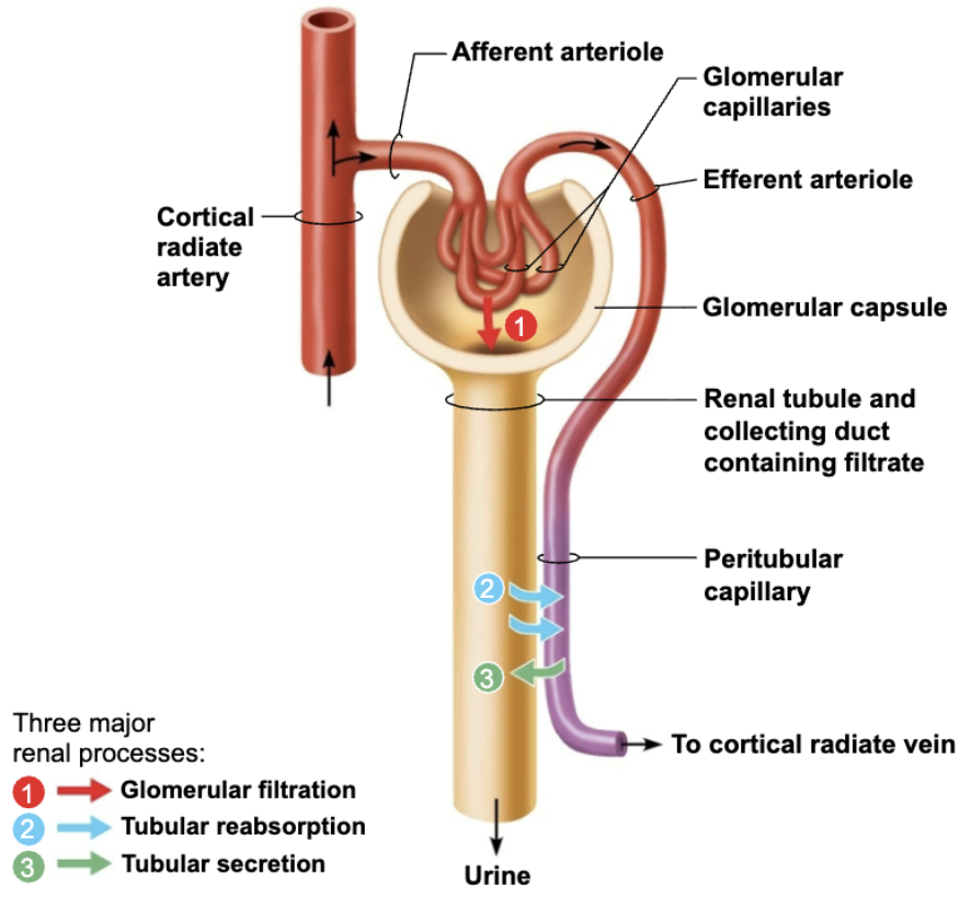
List and Define the 3 Major Renal Processes

Three processes are involved in urine formation and adjustment of blood composition:

  1. Glomerular filtration

    • Produces cell-free and protein-free filtrate

  2. Tubular reabsorption

    • Selectively returns 99% of substances from filtrate to blood in renal tubules and collecting ducts

  3. Tubular secretion

    • Selectively moves substances from blood to filtrate in renal tubules and collecting dicts

<p><strong><em>Three processes are involved in </em></strong><span style="color: red;"><strong><em>urine formation</em></strong></span><strong><em> and </em></strong><span style="color: red;"><strong><em>adjustment of blood composition</em></strong></span><strong><em>: </em></strong></p><ol><li><p><strong>Glomerular filtration </strong></p><ul><li><p>Produces <em>cell-free and protein-free filtrate</em></p></li></ul></li><li><p><strong>Tubular reabsorption </strong></p><ul><li><p><em>Selectively returns</em> 99% of substances from filtrate to blood in renal tubules and collecting ducts</p></li></ul></li><li><p><strong>Tubular secretion </strong></p><ul><li><p><em>Selectively moves</em> substances from blood to filtrate in renal tubules and collecting dicts </p></li></ul></li></ol><p></p>
5
New cards

Explain Glomerular Filtration

  • A PASSIVE PROCESS (no metabolic energy required)

  • Hydrostatic pressure forces fluids and solutes through filtration membrane glomerular capsule

  • NO reabsorption into capillaries of glomerulus occurs

6
New cards

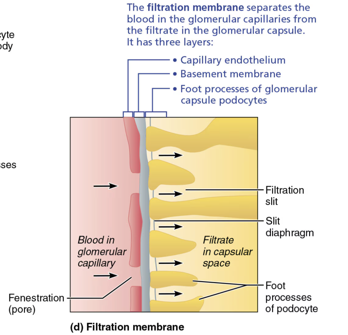
Role of the Filtration Membrane

STRUCTURE

  • Porous membrane between blood and interior of glomerular capsule 

FUNCTION

  • Allow water and solutes smaller than plasma proteins to pass → NORMALLY NO CELLS CAN PASS 

<p><span style="color: blue;"><strong>STRUCTURE</strong></span><span style="color: red;"><strong> </strong></span></p><ul><li><p>Porous membrane between blood and interior of glomerular capsule&nbsp;</p></li></ul><p><span style="color: red;"><strong>FUNCTION</strong></span></p><ul><li><p>Allow water and solutes smaller than plasma proteins to pass → NORMALLY NO CELLS CAN PASS&nbsp;</p></li></ul><p><span style="color: red;"><strong> </strong></span></p>
7
New cards

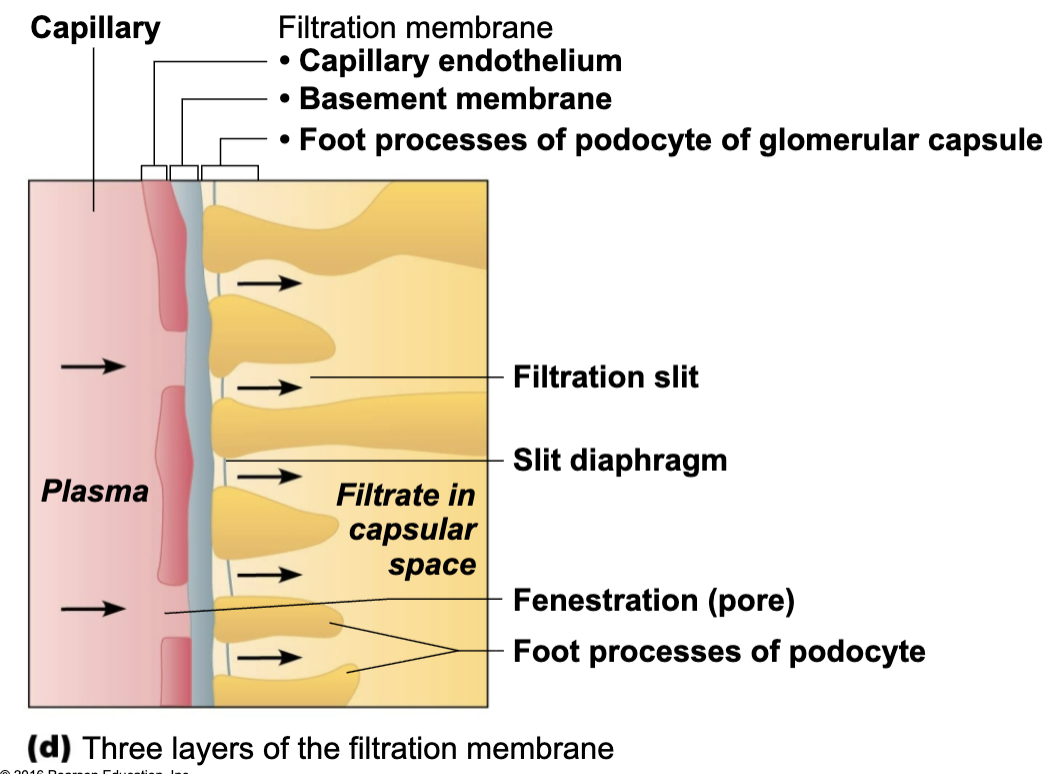
Name and Describe of the Layers of the Filtration Membrane

  1. Fenestrated endothelium 

    • of glomerular capillaries

  2. Basement membrane 

    • Fused basal laminae of two other layers

  3. Foot processes of podocytes (Visceral layer of glomerular capsule)

    • with filtration slits → slit diaphragms repel macromolecules

<ol><li><p><strong>Fenestrated endothelium&nbsp;</strong></p><ul><li><p>of glomerular capillaries</p></li></ul></li><li><p><strong>Basement membrane&nbsp;</strong></p><ul><li><p>Fused basal laminae of two other layers</p></li></ul></li><li><p><strong>Foot processes of podocytes&nbsp;</strong>(Visceral layer of glomerular capsule) </p><ul><li><p>with filtration slits → slit diaphragms repel macromolecules</p></li></ul></li></ol><p></p>
8
New cards

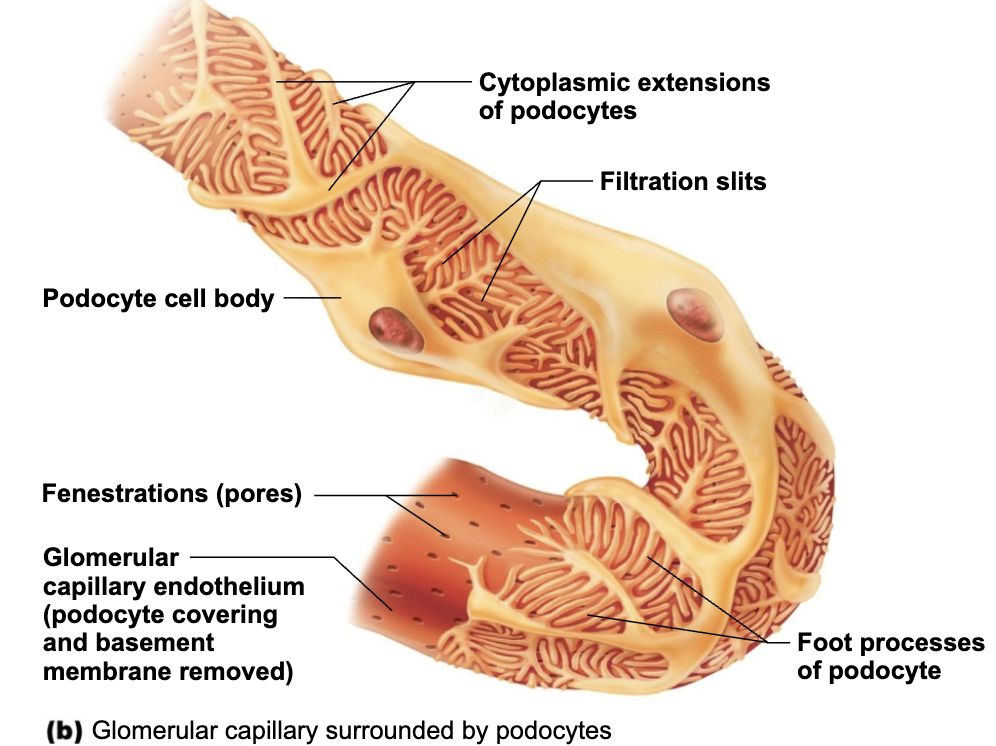
Explain how the filtration membrane" “decides” to let substances into renal tube

  1. Macromolecules “stuck” in filtration membrane are engulfed by glomerular mesangial cells

  2. Allows molecules smaller than 3nm to pass

    • EX: Water, Glucose, Amino acids, Nitrogenous wastes

  3. Plasma proteins remain in blood to maintain colloid osmotic pressure

<ol><li><p>Macromolecules “stuck” in <span style="color: purple;"><strong><span>filtration membrane</span></strong></span> are engulfed by <span style="color: blue;"><strong><span>glomerular mesangial cells</span></strong></span></p></li><li><p>Allows molecules <span style="color: blue;"><strong>smaller than 3nm</strong></span> to pass</p><ul><li><p><span style="color: green;"><strong><span>EX: Water, Glucose, Amino acids, Nitrogenous wastes</span></strong></span></p></li></ul></li><li><p><span style="color: blue;"><strong>Plasma proteins</strong></span> remain in blood to maintain colloid osmotic pressure</p></li></ol><p></p>
9
New cards

Purpose of Plasma proteins in Blood

Plasma proteins remain in blood to maintain colloid osmotic pressure

  1. Prevents loss of all water to capsular space

  2. Proteins in filtrate indicate membrane problem

10
New cards

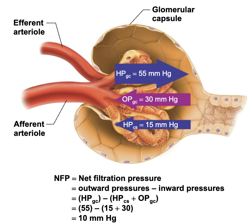
Explain how Fluid moves out of Glomerulus and into the Renal Tubule

Pressures that Affect Filtration

  1. Outward Pressure → Forces that promote filtrate formation

    • Hydrostatic pressure in glomerular capillaries (HPgc) is essentially glomerular BP

      • Chief force pushing water, solutes out of blood

      • 55 mmHg

      • Reason why HIGH is that efferent arteriole is a high-resistance vessel with a diameter smaller than afferent arteriole

  2. Inward Pressure → Forces inhibiting filtrate formation

    • Hydrostatic pressure in capsular space (HPcs)

      • Filtrate pressure in capsule

      • 15 mmHg

    • Colloid osmotic pressure in capillaries (OPgc)

      • “Pull” of proteins in blood

      • 30 mmHg

  3. Net Filtration Pressure (NFP) → sum of forces

    • 55 mmHg forcing out minus 45 mmHg opposing = vet outward force of 10 mmHg

    • PRESSURE RESPONSIBLE FOR FILTRATE FORMATION

<p><strong><em>Pressures that Affect Filtration</em></strong></p><ol><li><p><strong>Outward Pressure → <mark data-color="green" style="background-color: green; color: inherit;">Forces that promote filtrate formation</mark></strong></p><ul><li><p><span style="color: blue;"><strong><span>Hydrostatic pressure in glomerular capillaries (HP</span><sub><span>gc</span></sub><span>) </span></strong></span>is essentially glomerular BP</p><ul><li><p>Chief force pushing water, solutes <strong>out of blood</strong></p></li><li><p><span style="color: green;"><strong><span>55 mmHg</span></strong></span></p></li><li><p>Reason why HIGH is that efferent arteriole is a high-resistance vessel with a diameter smaller than afferent arteriole</p></li></ul></li></ul></li><li><p><strong>Inward Pressure → <mark data-color="red" style="background-color: red; color: inherit;">Forces inhibiting filtrate formation</mark></strong></p><ul><li><p><span style="color: red;"><strong><span>Hydrostatic pressure in capsular space (HP</span><sub><span>cs</span></sub><span>)</span></strong></span></p><ul><li><p>Filtrate pressure in capsule</p></li><li><p><span style="color: green;"><strong><span>15 mmHg</span></strong></span></p></li></ul></li><li><p><span style="color: red;"><strong><span>Colloid osmotic pressure in capillaries (OPgc)</span></strong></span></p><ul><li><p>“Pull” of proteins <strong>in blood</strong></p></li><li><p><span style="color: green;"><strong><span>30 mmHg</span></strong></span></p></li></ul></li></ul></li><li><p><strong>Net Filtration Pressure (NFP) → <mark data-color="purple" style="background-color: purple; color: inherit;">sum of forces</mark></strong></p><ul><li><p><span style="color: green;"><strong><span>55 mmHg forcing out minus 45 mmHg opposing = vet outward force of 10 mmHg</span></strong></span></p></li><li><p>PRESSURE RESPONSIBLE FOR FILTRATE FORMATION</p></li></ul></li></ol><p></p>
11
New cards

If the osmotic pressure in the glomerular capillaries increased from 28 mmHg to 35 mmHg due to dehydration, would net filtration increase or decrease?

→ Net filtration would decrease.

12
New cards

Explain Effect and Cause of Anuria

  • EFFECT

    • Abnormally low urinary output (less than 50ml/day)

  • CAUSE

    • May indicate that glomerular BP is too low to cause filtration

    • Renal failure and anuria can also result from situations in whicj nephrons stop functioning

      • EX: Acute nephritis, Transfusion reactions, and crash injuries

13
New cards

Define Glomerular Filtration Rate (GFR)

  • GFR = Volume of filtrate formed/minute by both kidneys

  • Normal = 120-125 ml/min

14
New cards

Name and Describe the Factors GFR is directly proportional to

  1. Net Filtration Pressure (NFP)

    • Primary pressure is glomerular hydrostatic pressure

  2. Total surface area available for filtration

    • Glomerular mesangial cells control by contracting

  3. Filtration membrane permeability

    • Much more permeable than other capillaries

15
New cards

Role of GRS

Constant GRS is important as it allows kidneys to make filtrate and maintain extracellular homeostasis

16
New cards

The rate of kidney filtrate formation would normally be dependent upon all of the following factors except__________.

A.) systemic blood pressure

B.) filtration membrane integrity

C.) blood calcium level

D.) renal artery/arteriole diameters

→ C.) blood calcium level

  • Systemic blood pressure

  • Filtration membrane integrity

  • Renal artery/arteriole diameters

17
New cards

Name Two Types of Controls of GFR

  1. Intrinsic controlsmaintain GFR in kidney

    • Constant GFR is important as it allows kidneys to make filtrate and maintain extracellular homeostasis

    • Renal auto-regulation

  2. Extrinsic controls → maintain systemic BP

    • GFR affects systemic blood pressure

    • Nervous system and Endocrine mechanisms

18
New cards

T/F: Increased GFR causes increased urine output, which lowers blood pressure, and vice versa

→ TRUE

19
New cards

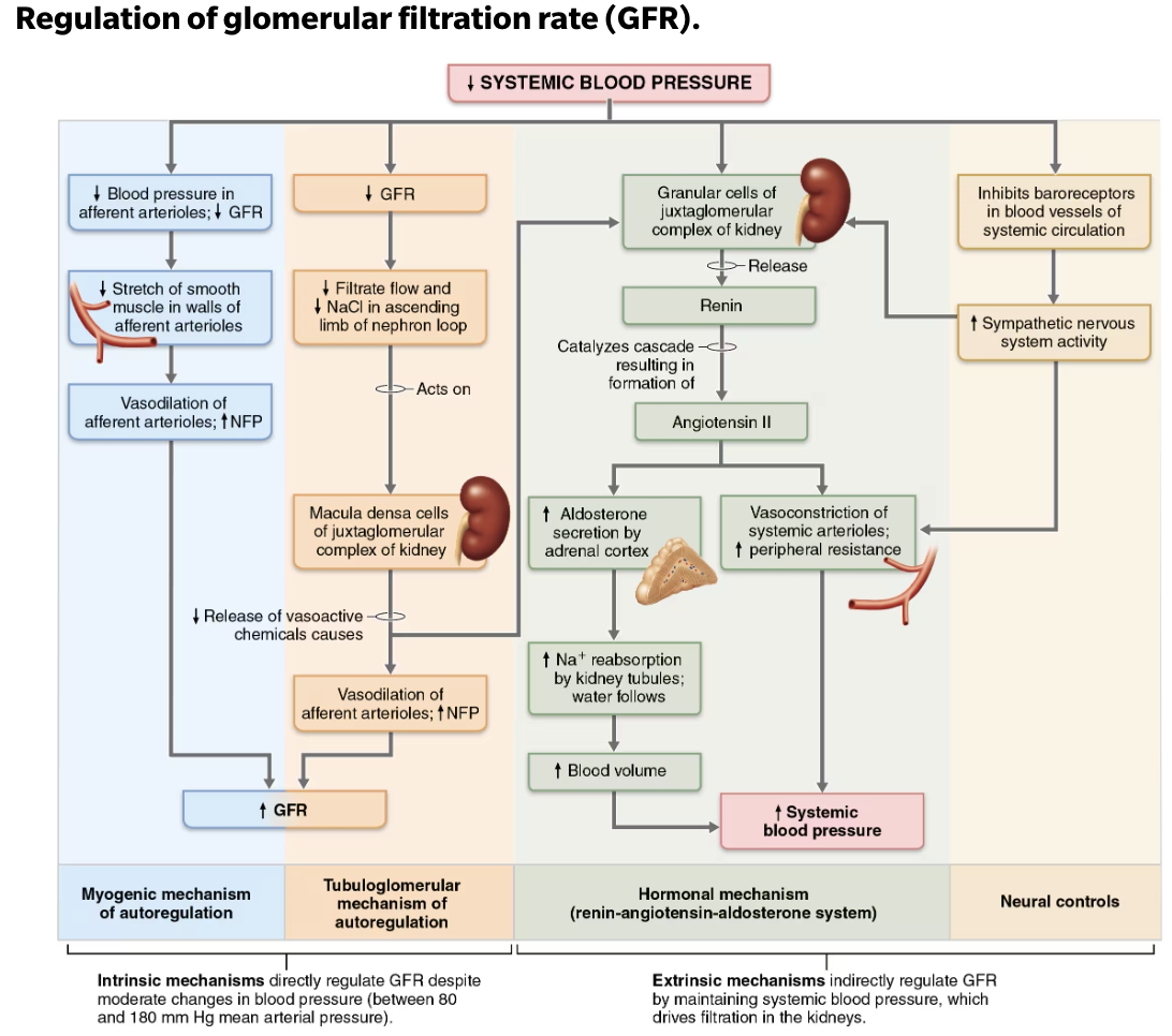
Define Intrinsic Controls

  • Maintains nearly constant GFR when MAP is in range of 80-180 mmHg

    • Auto-regulation ceases if out of that range

  • Two types of renal auto-regulation

    1. Myogenic mechanism

    2. Tubuloglomerular feedback mechanism

20
New cards

Explain how Myogenic Mechanism works to regulate glomerular filtration

Intrinsic Control

Local smooth muscle contracts when stretched:

  1. INCREASED BP causes muscle to stretch, leading to constriction of afferent arterioles

    • Restricts blood flow into glomerulus

    • Protects glomeruli from damaging high BP

  2. DECREASED BP causes dilation of afferent arterioles

Both help maintain normal GFR despite normal fluctuations in BP

<p><code>Intrinsic Control</code></p><p><strong>Local </strong><span style="color: purple;"><strong><mark data-color="purple" style="background-color: purple; color: inherit;"><span>smooth muscle </span></mark></strong></span><strong>contracts when stretched:</strong></p><ol><li><p><span style="color: green;"><strong><mark data-color="green" style="background-color: green; color: inherit;"><span>INCREASED BP</span></mark></strong><mark data-color="green" style="background-color: green; color: inherit;"><span> causes muscle to </span></mark><strong><mark data-color="green" style="background-color: green; color: inherit;"><span>stretch</span></mark></strong><mark data-color="green" style="background-color: green; color: inherit;"><span>, leading to constriction of </span></mark><strong><mark data-color="green" style="background-color: green; color: inherit;"><span>afferent arterioles</span></mark></strong></span></p><ul><li><p>Restricts blood flow into glomerulus</p></li><li><p>Protects glomeruli from damaging high BP</p></li></ul></li><li><p><span style="color: red;"><strong><mark data-color="red" style="background-color: red; color: inherit;"><span>DECREASED BP </span></mark></strong><mark data-color="red" style="background-color: red; color: inherit;"><span>causes </span></mark><strong><mark data-color="red" style="background-color: red; color: inherit;"><span>dilation</span></mark></strong><mark data-color="red" style="background-color: red; color: inherit;"><span> of </span></mark><strong><mark data-color="red" style="background-color: red; color: inherit;"><span>afferent arterioles</span></mark></strong></span></p></li></ol><p></p><p><strong><em>Both help maintain normal GFR despite normal fluctuations in BP</em></strong></p><p></p>
21
New cards

Explain how Tubuloglomerular Feedback Mechanism works to regulate glomerular filtration

Intrinsic Control

Flow-dependent mechanism directed by macula densa cells → Responds to filtrate NaCl concentration

  1. If GFR INCREASES → filtrate flow rate INCREASES

    • Leads to DECREASED reabsorption time → causing HIGH NaCl levels in filtrate

    • Feedback mechanism causes constriction of afferent arteriole → which LOWERS NFP and GFR, allowing more time for NaCl reabsorption

  2. Opposite mechanism for DECREASED GFR

<p><code>Intrinsic Control</code></p><p><strong>Flow-dependent mechanism directed by </strong><span style="color: purple;"><strong><mark data-color="purple" style="background-color: purple; color: inherit;"><span>macula densa cells</span></mark></strong></span><strong> → Responds to filtrate NaCl concentration</strong></p><ol><li><p><span style="color: green;"><strong><mark data-color="green" style="background-color: green; color: inherit;"><span>If GFR INCREASES → filtrate flow rate INCREASES</span></mark></strong></span></p><ul><li><p>Leads to DECREASED reabsorption time → causing HIGH NaCl levels in filtrate</p></li><li><p>Feedback mechanism causes constriction of afferent arteriole → which LOWERS NFP and GFR, allowing more time for NaCl reabsorption</p></li></ul></li><li><p><span style="color: red;"><strong><mark data-color="red" style="background-color: red; color: inherit;"><span>Opposite mechanism for DECREASED GFR</span></mark></strong></span></p></li></ol><p></p>
22
New cards

When the macula densa detects an increase in NaCl concentration in the renal filtrate, what happens to the glomerular filtration rate (GFR)?

GFR decreases

23
New cards

Define Extrinsic Controls

  • Purpose is to regulate GFR to maintain systemic BP

    • Will OVERRIDE renal intrinsic controls if blood volume needs to be increased

  • Two types of renal auto-regulation

    1. Sympathetic Nervous System

    2. Renin-Angiotensin-Aldosterone Mechanism

24
New cards

Explain how Sympathetic Nervous System works to regulate glomerular filtration

Extrinsic Control

  • Under normal conditions @ rest

    • Renal blood vessels dilated

    • Renal auto-regulation mechanism prevail

  • Under abnormal conditions, such as extremely low ECF volume (low BP)

    • Norepinephrine is released by sympathetic nervous system and epinephrine is released by adrenal medulla causing

      1. Systemic vasoconstriction → increases BP

      2. Constriction of afferent arterioles → decrease GFR

      3. Blood volume and pressure increases

      4. Sympathetic Nervous System

<p><code>Extrinsic Control</code></p><ul><li><p><span style="color: purple;"><strong><mark data-color="purple" style="background-color: purple; color: inherit;"><span>Under normal conditions @ rest</span></mark></strong></span></p><ul><li><p>Renal blood vessels dilated</p></li><li><p>Renal auto-regulation mechanism prevail</p></li></ul></li><li><p><span style="color: purple;"><strong><mark data-color="purple" style="background-color: purple; color: inherit;"><span>Under abnormal conditions</span></mark></strong></span>, such as extremely low ECF volume (low BP)</p><ul><li><p><span style="color: blue;"><strong><span>Norepinephrine</span></strong></span> is released by sympathetic nervous system and epinephrine is released by adrenal medulla causing</p><ol><li><p><span style="color: green;"><mark data-color="green" style="background-color: green; color: inherit;"><span>Systemic vasoconstriction </span></mark><strong><mark data-color="green" style="background-color: green; color: inherit;"><span>→ increases BP</span></mark></strong></span></p></li><li><p><span style="color: red;"><mark data-color="red" style="background-color: red; color: inherit;"><span>Constriction of afferent arterioles</span></mark><strong><mark data-color="red" style="background-color: red; color: inherit;"><span> → decrease GFR</span></mark></strong></span></p></li><li><p><span style="color: green;"><strong><mark data-color="green" style="background-color: green; color: inherit;"><span>Blood volume and pressure increases</span></mark></strong></span></p></li><li><p>Sympathetic Nervous System</p></li></ol></li></ul></li></ul><p></p>
25
New cards

Explain how Renin-Angiotensin-Aldosterone Mechanism works to regulate glomerular filtration

Extrinsic Control

Main mechanism for increasing BP

  • Three pathways to renin release by granular cells

    1. Direct stimulation of granular cells by sympathetic nervous system

    2. Stimulation by activated macula densa cells when filtrate NaCl concentration is low

    3. Reduced stretch of granular cells

<p><code>Extrinsic Control</code></p><p><strong>Main mechanism for increasing BP</strong></p><ul><li><p><strong>Three pathways to </strong><span style="color: red;"><strong><span>renin</span></strong></span><strong> release by </strong><span style="color: purple;"><strong><mark data-color="purple" style="background-color: purple; color: inherit;"><span>granular cells</span></mark></strong></span></p><ol><li><p><span style="color: blue;"><strong><mark data-color="blue" style="background-color: blue; color: inherit;">Direct stimulation of granular cells by</mark></strong><mark data-color="blue" style="background-color: blue; color: inherit;"> sympathetic nervous system</mark></span></p></li><li><p><span style="color: blue;"><strong><mark data-color="blue" style="background-color: blue; color: inherit;">Stimulation by activated macula densa cells </mark></strong><mark data-color="blue" style="background-color: blue; color: inherit;">when filtrate NaCl concentration is low</mark></span></p></li><li><p><span style="color: red;"><strong><mark data-color="red" style="background-color: red; color: inherit;">Reduced stretch of granular cells</mark></strong></span></p></li></ol></li></ul><p></p>