Cell Bio chp. 17 - Cytoskeleton
1/48
There's no tags or description
Looks like no tags are added yet.
Name | Mastery | Learn | Test | Matching | Spaced | Call with Kai |
|---|
No analytics yet
Send a link to your students to track their progress
49 Terms
Cytoskeleton general functions
cell support
transport of organelles and vesicles
movement
cell division
response to environment
where do intermediate filaments expand from and where are they anchored
Expand from center of cell
Anchored at cell membrane
desmosomes
cell junction at plasma membrane that link intermediate filaments of neighboring cells
can intermediate filaments be found in the nucleus
Yes
What is the secondary structure of an intermediate filament monomer
a-helix
dimer structure for intermediate filaments
coiled-coil
tetramer structure of intermediate filaments
staggered antiparallel coiled coils
What can eight intermediate filament tetramers make
helical array that makes up rope like structure and can be added to growing filament
4 major classes of intermediate filaments and location
keratin filaments (cytoplasm)
Vimentin and vimentin-related filaments (cytoplasm)
neurofilaments (cytoplasm)
nuclear lamins (nucleus)
Most diverse intermediate filament
Keratin filaments
What cells contain keratin filaments
epithelial cells
Cells containing vimentin and vimentin related filaments
connective tissue cells
muscle cells
glial cells
cells containing neurofilaments
nerve cells
cells containing nuclear lamins
all animals cells
(all animal cells have a nucleus so they have nuclear lamins)
Epidermolysis bullosa simplex
Keratin filament mutation that interferes with keratin filament formation at epidermis.
Skin cells become easily ruptured and blisters result
Plectin
Makes intermediate filaments stable and strong
Plectin mutations
muscular dystrophy (muscle weakness)
neurodegeneration (death of neurons)
What is nuclear envelope supported by
meshwork of intermediate filaments (nuclear lamins)
nulcear lamina functions
structural support to nuclear envelope
attachment site for chromosomes
nuclear lamina phosphorylation and dephosphorylation significance
When phosphorylated nuclear lamina dissemble during mitosis
When dephosphorylated nuclear lamina come back together during telophase
What do defects in nuclear lamina cause
Progeria - impaired cell division and increased cell death
specialized functions of microtubules
build mitotic spindle for cell div.
Bundled to form cilia
Where do microtubules grow out of
centrosomes
basal bodies
What are tubulin subunits made of
aB heterodimer
Protofilament structure
single strand of aB subunits stacked head to tail
plus end with B
minus end with a
microtubule structure
tubulin dimers pack together with the same orientation in all 13 protofilaments to give definite polarity to microtubule
centrosome structure
spherical matrix
pair of centrioles oriented at right angle made of short microtubules
Y-tubulin ring
How do microtubules grow
a minus end is embedded in centrosome
microtubule grows out of Y-tubulin ring complexes from plus end
B plus end extends into cytoplasm
Instability of growing microtubules
There is a rapid between some microtubules growing out of centrosome while others simultaneously shrink
What contributes to microtubule instability
B-dimer GTPase activity
Growing microtubule process
polymerization is faster than GTPase activity of B-dimer
GTP-cap is formed at plus end
Shrinking microtubule process
Polymerization of microtubule is slow
GTPase activity of B-dimer is faster than polymerization
B-dimers are associated to GDP which has weak association
GDP bound B-dimers are released to cytoplasm as microtubule shrinks
GTP vs GDP binding
GTP associated dimers bind much more strongly to neighbors
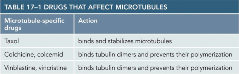
Drugs that affect microtubules
What stabilizes microtubules
Microtubule capping proteins on cell cortex
Do microtubules organize cell interior
Yes
Orientation of microtubule in axon and importance
All are the same with plus end facing the axon terminal
This allows for outward transport of materials in nerve cell body and inward transport of materials at nerve terminals
How do motor proteins move along microtubules
Through motor proteins globular heads
motor protein structure
globular head at one end that interact with microtubule
tail at one end that interacts with cargo
Two major motor proteins direction across microtubule
dynein (moves toward minus end)
kinesin (moves toward plus end)
Process of motor proteins moving across microtubule
leading globular head is associated to ADP and lagging head is associated to ATP
hydrolysis of ATP releases attachment of lagging head to microtubule
The release of ADP and binding of ATP causes conformational change to leading head
conformational change allows for ADP associated lagging head to be moved ahead of ATP associated leading strand
original orientation is retained
ADP positioning difference for dynein vs kinesin
dynein: ADP is closer to minus end than ATP
kinesin: ADP is closer to plus end than ATP
Different types of kinesin transport
Can either:
directly bind cargo to tail
bind cargo to adaptor protein associated to tail
Dynein transport
Always uses adaptor proteins to bind cargo and deliver to minus end
Importance of microtubules and motor proteins with organelles
They determine position of organelles in cytoplasm
Microtubules in cilium or flagellum structure
2 central microtubules
9 outer doublet microtubules
Two rows of dynein arms on outer microtubule surface that contact adjacent microtubule to generate beating force
nexin that link 9 microtubule doublets together
radial spoke
Cilia movement
Power stroke: fully extended cilium quickly moves fluid over cells surface
recovery stroke: cilium curls back into position and minimally interacts with outside fluid
Cilium cycles between these two phases
Kartangers syndrome
mutation in ciliary dynein