Ch. 16 - Special Senses
1/78
There's no tags or description
Looks like no tags are added yet.
Name | Mastery | Learn | Test | Matching | Spaced |
|---|
No study sessions yet.
79 Terms
Hearing
a response to vibrating air molecules
sense resides in inner ear
Equilibrium
the sense of motion, balance, and body orientation in 3D space
sense resides in inner ear
Sound
any audible vibration of molecules
A vibrating object pushes on air molecules
These air molecules push on other air molecules
the air molecules hitting the eardrum cause it to vibrate
Pitch
our sense of whether a sound is “high” or “low”
Determined by vibration frequency: hertz or cycles/second
in hearing loss, we can hear the outer ranges of sound, but struggle with the middle range
Most hearing loss with age is in the range of 250 to 2,050 Hz
Loudness
the perception of sound energy, intensity, or amplitude of the vibration
Expressed in decibels (dB)
Prolonged exposure to sounds > 90 dB can cause damage
Outer, Middle, and Inner Ear
Outer - what we can see
Middle - starts at ear drum and goes to auditory tube
Inner - cochlea and vestibule
What happens in the outer, middle, and inner ear?
the outer and middle ear only deal with the transmission of sound to the inner ear
The inner ear deals with vibrations and converts them to nerve signals
Otitis Media
Middle-ear infection
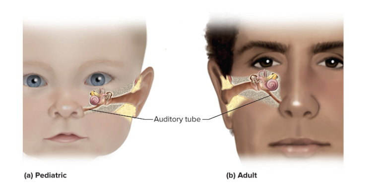
is common in children
Auditory tube is short and horizontal
infections easily spread from throat to tympanic cavity and mastoid air cells
Symptoms
Fluid accumulates in tympanic cavity producing pressure, pain, and impaired hearing
Can spread, leading to meningitis
Can cause fusion of ear ossicles and hearing loss
makes patient have a hard time hearing bc fluid build up doesn't allow eardrum to work properly
Components of Inner Ear
Bony Labyrinth
Membranous Labyrinth
Labyrinth
Cochlea
Bony Labyrinth
passageways in temporal bone
Membranous Labyrinth
fleshy tubes lining bony labyrinth
Filled with endolymph: similar to intracellular fluid
Floating in perilymph: similar to cerebrospinal fluid
Labyrinth
Vestibule and three semicircular ducts
Cochlea
organ of hearing
Winds coils around a screw-like axis of spongy bone called the modiolus
Threads of the screw form a spiral platform that supports the tube of the cochlea
Cochlea has three fluid-filled chambers separated by membranes
Scala vestibuli
Scala tympani
Scala media
Scala vestibuli
superior/top chamber
Filled with perilymph
Begins at oval window and spirals to apex
Scala tympani
inferior/bottom chamber
Filled with perilymph
Begins at apex and ends at round window
Secondary tympanic membrane: covers round window
Scala media (cochlear duct)
middle chamber
Filled with endolymph
is lined with spiral organs
Spiral Organ
acoustic organ that converts vibrations into nerve impulses
has epithelium composed of 4 rows of hair cells and supporting cells
Hair cells have stereocilia
Stereocilia
long, stiff microvilli on apical surface
has a tectorial membrane

Tectorial Membrane
Gelatinous membrane that rests on top of stereocilia
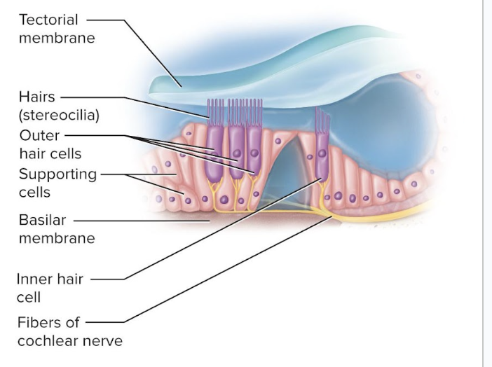
Inner Hair Cells
One row of about 3,500 cells
Provides for hearing
cannot tell difference between sounds like outer hair cells
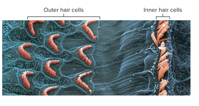
Outer Hair Cells
three rows of about 20,000 cells
Adjusts response of cochlea to different frequencies
Increases precision
help us tell where sounds are coming from
tips of stereocilia of outer hair cells are imbedded in the tectorial membrane

Tympanic Membrane
eardrum
Ossicles (bones of the middle ear) concentrate the energy of the vibrating tympanic membrane to a smaller area
Ossicles create a greater force per unit area at the oval window and overcome the inertia of the perilymph
Ossicles and their muscles have a protective function
Lessen the transfer of energy to the inner ear
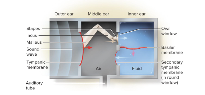
Vibration of ossicles causes…
vibration of the basilar membrane under hair cells
hair cells move with the basilar membrane
Endolymph
a high K+ fluid that bathes the stereocilia of outer hair cells
the high concentration of K+ creates a gradient and makes the outside of the cell positive and the inside negative
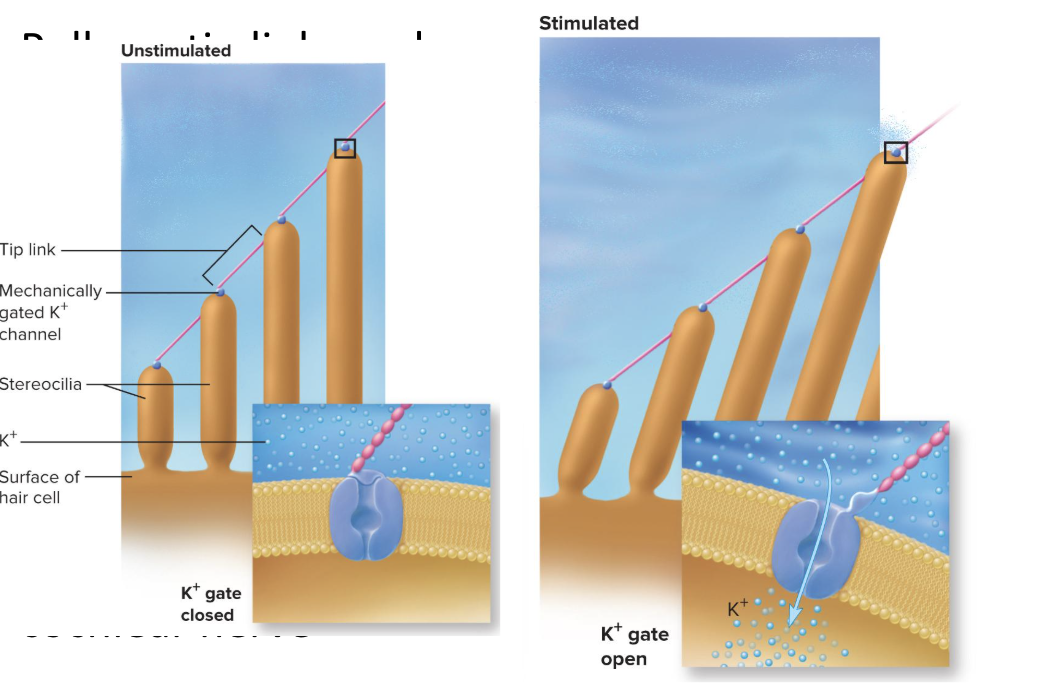
Stimulation of Stereociliia of Inner Hair Cells:
Stretchy protein filament (called a tip link) connects the ion channel of one stereocilium to the sidewall of the next
Tallest stereocilium is bent when the basilar membrane rises up toward the tectorial membrane
the bending pulls on tip links and opens ion channels
K+ flows in —depolarization causes release of a neurotransmitter
Stimulates sensory dendrites and generates action potential in the cochlear nerve
Variations in loudness (amplitude) cause…
variations in the intensity of cochlear vibrations
Soft sound produces a slight up-and-down motion of the basilar membrane
Louder sounds make the basilar membrane vibrate more aggressively
Triggers higher frequency of action potentials
Brain interprets this as louder sound
Pitch depends on …
which part of the basilar membrane is vibrating
low pitch noises go more towards the distal end, farther down the cochlea
high pitch noises go towards the proximal end, the beginning area of the cochlea
Conductive Deafness
conditions interfere with transmission of vibrations to inner ear
caused by damaged tympanic membrane, otitis media, blockage of auditory canal, and otosclerosis
Otosclerosis
fusion of auditory ossicles that prevents their free vibration
Sensorineural (nerve) Deafness
death of hair cells or any nervous system elements concerned with hearing
Common in factory workers, musicians, construction workers
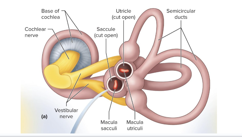
Vestibular Apparatus
makes up the receptors for equilibrium
Three semicircular ducts
Detect only angular acceleration (dynamic equilibrium)
Two chambers
Anterior saccule and posterior utricle
Responsible for static equilibrium and linear acceleration
Static Equilibrium
the perception of the orientation of the head when the body is stationary
our idea of where we are in space (upright, where is up and down, etc)
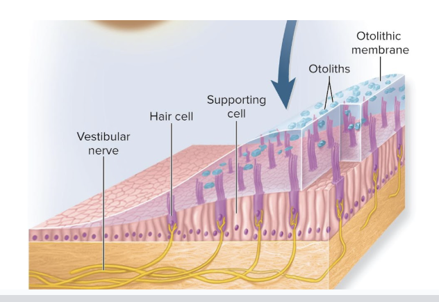
when head is tilted, the heavy otolithic membrane sags, bending the stereocilia and stimulating the hair cells
Dynamic Equilibrium
the perception of motion or acceleration
the equilibrium of movement
in car, linear acceleration detected as otoliths lag behind, bending the stereocilia and stimulating the hair cells
Linear acceleration—change in velocity in a straight line
Angular acceleration—change in rate of rotation
Macula
a 2 x 3 mm patch of hair cells and supporting cells in the saccule and utricle
Macula sacculi: lies vertically on wall of saccule
Macula utriculi: lies horizontally on floor of utricle
used for static equilibrium
Each hair cell has 40 to 70 stereocilia and one true cilium called a kinocilium
embedded in a gelatinous otolithic membrane

Are the macula sacculi and macula utriculi parallel or perpendicular?
they are perpendicular to each other so they can bend in their directions
Because macula sacculi is vertical, it responds to vertical acceleration and deceleration
Otoliths
calcium carbonate–protein granules (little tiny bits of bones) imbedded in a gel like substance, so when they bend, they pull of the gel like substance, thereby pulling the hairs and opening mechanically gated channels
add to the weight and inertia and enhance the sense of gravity and motion
Semicircular Ducts
there are 3 of them
detect rotary movements
are held by the bony semicircular canals of the temporal bone
Each duct is filled with endolymph and opens up as a dilated sac (ampulla) next to the utricle
the 3 ducts are perpendicular to each other
allows them to detect movement in 3d space
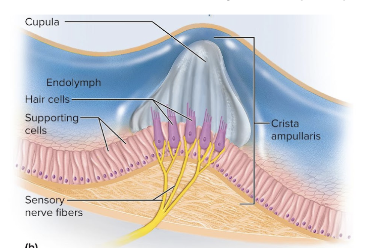
Each ampulla contains crista ampullaris
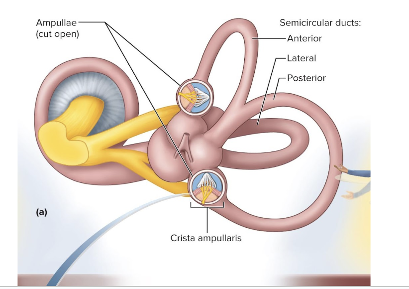
Crista Ampullaris
mound of hair cells and supporting cells
Consists of hair cells with stereocilia and a kinocilium buried in a mound of a gelatinous membrane called the cupula
one in each duct
Vision
perception of objects in the environment using the light they emit or reflect
components include the retina and the optic nerve
Light
visible electromagnetic radiation
wavelengths of light range from 400 to 700 nm in humans
Light must cause a photochemical reaction to produce a nerve signal
Ultraviolet Radiation
< 400 nm
has too much energy and destroys macromolecules
Infrared Radiation
> 700 nm
too little energy to cause photochemical reaction, but does warm the tissues
Retina
attached to eye only at the optic disc (back exit of the optic nerve) and ora serrata (front edge of retina)
is pressed against the rear of the eyeball by the vitreous humor
Detached retina causes blurry areas of vision and can lead to blindness
includes the macula lutea and fovea centralis
retina has blood vessels

Vitreous humor
a jelly-like substance that keeps retina in place
Macula lutea
patch of cells on the visual axis of the eye
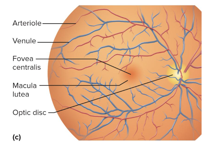
Fovea centralis
pit in center of macula lutea

Optic Disc
blind spot
where the optic nerve exits retina
there are no receptors there
Visual Filling
brain fills in the “picture” across the blind spot area
Brain ignores unavailable information until fast eye movements redirect gaze
Formation of an Image
Light passes through the lens to form tiny inverted image on retina
the iris diameter is controlled by two sets of contractile elements
pupillary constrictor
pupillary dilator
when we see something, it is first upside down, but our brain turns it back to right-side-up
Pupillary Constrictor
smooth muscle encircling the pupil
Parasympathetic stimulation narrows the pupil

Pupillary Dilator
spoke-like myoepithelial cells
Sympathetic stimulation widens pupil
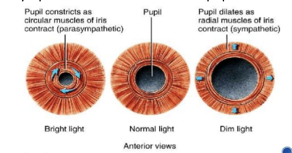
Pupillary constriction and dilation occurs when…
light intensity changes
when gaze shifts between distant and nearby objects
Photopupillary Reflex
the constriction of the pupil in response to light
Mediated by autonomic reflex arc
Brighter light is signaled to the pretectal region of the midbrain
Excites parasympathetic fibers in oculomotor nerve that travels to ciliary ganglion in orbit
Postganglionic parasympathetic fibers stimulate pupillary constrictor
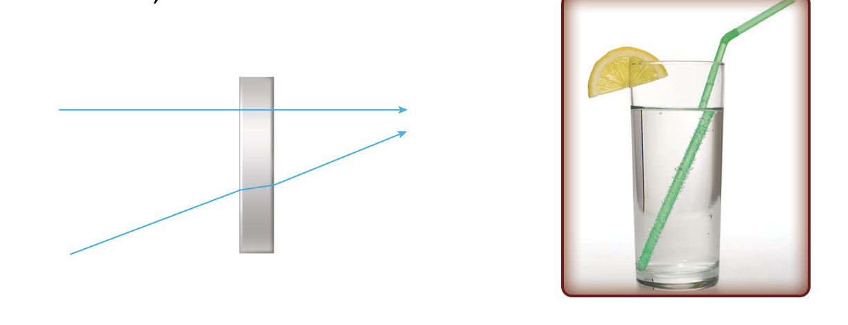
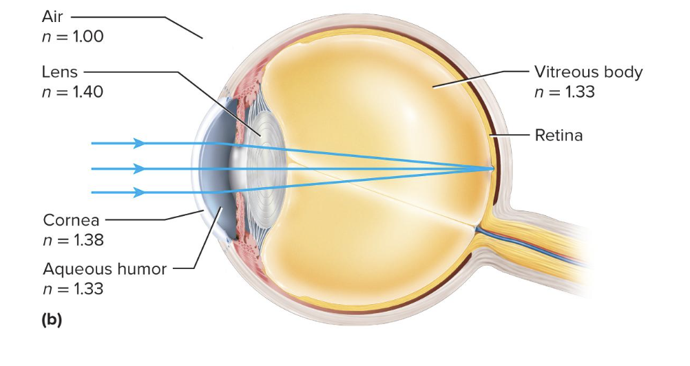
Refraction
the bending of light rays
change in speed of light causes change in direction of light
Refractive Index
A measure of how much it reduces light rays relative to air
Angle of incidence at 90° light slows but does not change course
Any other angle, light rays change direction (are refracted)
The greater the refractive index and the greater the angle of incidence, the more refraction
Refraction in the Eye
the light passing through the center of the cornea is not bent
any light striking off-center is bent toward the center
the aqueous humor and the lens do not greatly alter the path of light
Cornea refracts light more than lens does
Lens merely fine-tunes image
Lens becomes rounder to increase refraction for near vision
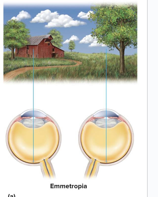
Emmetropia
state in which the eye is relaxed and focused on an object more than 6 m (20 ft) away
Light rays coming from that object are parallel, so we can dilate without damage
Rays focused on retina without effort
Light rays coming from a closer object are too divergent to be focused without effort
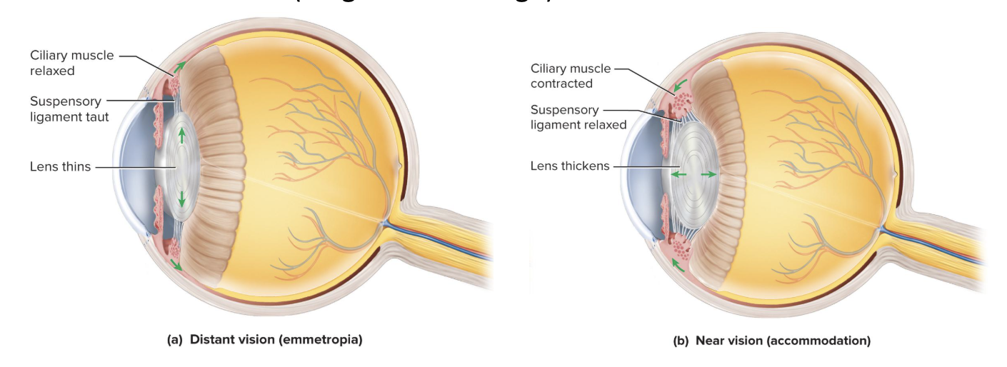
Adjustment to close-range vision requires 3 things:
Convergence of eyes
we have to angle our eyes to the thing were looking at
Constriction of pupil
Blocks peripheral light rays and reduces spherical aberration (blurry edges)
Accommodation of lens
change in the curvature of the lens lets you focus on nearby objects
Accommodation of Lens:
Ciliary muscle contracts, suspensory ligaments slacken, and lens takes a thicker shape
causes light to be refracted more strongly and focused onto the retina
Near Point of Vision
closest an object can be and still come into focus
lengthens with age because the lens of our eyes become stiffer as we age
Retina converts light energy into…
action potentials
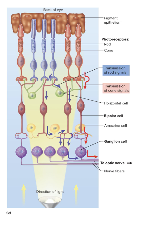
Structure of the Retina
Pigment epithelium
Most posterior part of retina
Absorbs stray light so visual image is not degraded
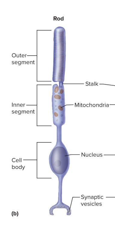
Rod Cells
Cone Cells
Rod Cells
light-absorbing cell best for night vision or monochromatic vision
Uses visual pigment rhodopsin
a photoreceptor
anterior segments are what pick up light
more sensitive to light
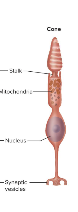
Cone Cells
light-absorbing cells best for color, photopic, or day vision
good for higher resolution vision
not as sensitive to light as rods are
contain photopsin (iodopsin)
a photoreceptor
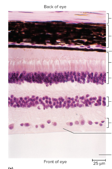
Histology of the Retina
Pigment epithelium
Rod and cone cells
Bipolar cells
Rods and cones synapse on bipolar cells
Bipolar cells synapse on ganglion cells
Ganglion cells
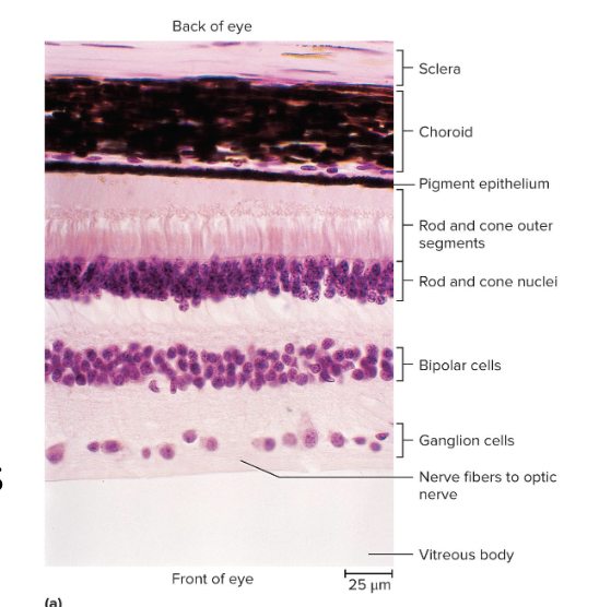
Ganglion Cells in the Retina
Single layer of large neurons near vitreous (front of the eye)
Axons form the optic nerve
Some absorb light with the pigment melanopsin and transmit signals to the brainstem
Detect light intensity for pupil control and circadian rhythms
do not contribute to visual image
Ganglion cells are the only retinal cells that produce action potentials
How light changes rhodopsin:
pigment receives light, high energy electrons of the photon cause pigment to change shape
In the dark, retinal is bent (cis-retinal) and retinal and opsin are together
In the light, the retinal molecule straightens (trans-retinal), and retinal separates from opsin
called bleaching of rhodopsin
To reset, it takes five minutes to regenerate 50% of bleached rhodopsin
How light changes photopsin
function similarly to rhodopsin
But are faster in regenerating their photopsin
90 seconds for 50% of bleached photopsin
What neurotransmitter do rods release when it is dark and how does that change when it is light?
rods constantly release glutamate
glutamate goes from axons of rods to the bipolar cells
acts as an inhibitor to the bipolar cells
bipolar cells will be hyperpolarized
makes it so that bipolar cells do not release neurotransmitters to our ganglion cells
When light hits, rods stop releasing glutamate, which allows neurotransmitters to be sent to ganglion cells
bipolar cells are excited by increasing light intensity
when rods are not activated they are releasing neurotransmitters
when rods are activated, they stop releasing neurotransmitters
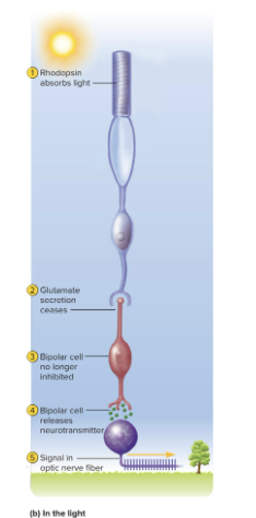
Generating the Optic Nerve Signal
When bipolar cells detect fluctuations in light intensity, they stimulate ganglion cells directly or indirectly
Ganglion cells respond to the bipolar cells with rising and falling firing frequencies
ganglion cells are only retinal cells that produce action potentials
Using the optic nerve, these changes provide visual signals to the brain
Duplicity Theory of Vision
explains why we have both rods and cones
A single type of receptor cannot produce both high sensitivity and high resolution
It takes one type of cell and neural circuit for sensitive night vision
It takes a different cell type and neuronal circuit for high-resolution daytime vision
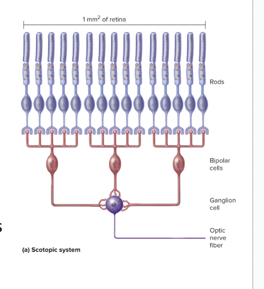
Characteristics of Rods
are very sensitive and react even in dim light
have extensive neural convergence
lots of rod cells converge onto one bipolar cell
many bipolar cells converge onto a single ganglion cell
results in a high degree of spatial summation
multiple signals from different locations are being combined to produce a stronger overall effect
rods are on the outer sides of the retina
made for low resolution
cannot focus finely detailed images
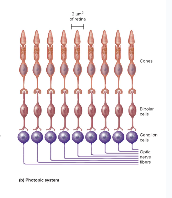
Fovea Centralis
has only cones, no rods
No neuronal convergence
Each foveal cone cell has a “private line to the brain”
concentration of cones gives us high-resolution color vision
little spatial summation so less sensitivity to dim light
3 Types of Cones
named for absorption peaks of their photopsins
Short-wavelength (S) cones
peak sensitivity at 420 nm
Medium-wavelength (M) cones
peak at 531 nm
Long-wavelength (L)cones
peak at 558 nm
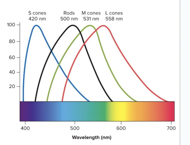
Color perception is based on..
the mixture of nerve signals representing cones of different absorption peaks

Stereoscopic Vision
depth perception
ability to judge distance to objects
Requires 2 eyes with overlapping visual fields which allows each eye to look at the same object from different angles
Fixation Point
the point in space that the eyes are focused on
Looking at an object within 100 feet, each eye views it from a slightly different angle
Provides brain with information used to judge the position of objects relative to the fixation point
Senescence of Vision
Loss of flexibility of lenses (presbyopia)
Cataracts (cloudiness of lenses) becomes common
Night vision is impaired due to fewer receptors, vitreous body less transparent, pupil dilators atrophy, and enzymatic reactions become slower
half-lives increase as we age, making the enzymatic reactions slower
Glaucoma risks increase
Senescence of Hearing
Tympanic membrane and ossicle joints stiffen
Hair cells and auditory nerve fibers die
Death of vestibular neurons results in dizziness
Taste and smell are blunted as receptors decline
we can regenerate our taste and smell receptors