Lesson 67 Female Reproductive system
1/63
There's no tags or description
Looks like no tags are added yet.
Name | Mastery | Learn | Test | Matching | Spaced | Call with Kai |
|---|
No analytics yet
Send a link to your students to track their progress
64 Terms
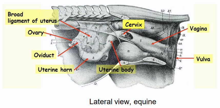
Equine and Bovine
Species where vaginal fornix form circumferential ring like recess that can impede advancement of artificial insemination catheter
Major component female repro system
bovine
Species where the uterine horn is curved inward
Canine ovaries topography
ventral to lumbar vertebra and caudal to kidneys. Right more cranial than left.
Canine right ovary
dorsal to descending duodenum and ascending colon
Canine Left ovary
between descending colon and abdominal wall
Canine inactive Ovaries morphology
elliposidal/oval/round in shape
Canine Active Ovaries Moprhology
irregular shaped because of copus luteum or follicles
Polytocous
produces many offspring
Monotocous
Produce one offspring
Ovarian bursa image
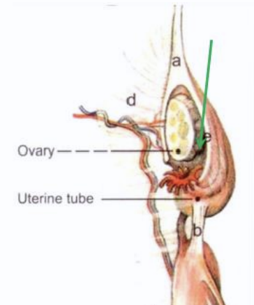
Ovarian Bursa
Space formed by fusion of distal mesovarium and mesalpinx. Basically pouch that holds ovaries.
Canine Ovarian Bursa opening
small slit like opening
Feline ovarian bursa opening
fairly large opening
Large species ovarian bursa opening
Mesovarium and mesosalpinx form cape or jacket covering over the ovaries
Fallopian tube/ uterine tube function
transport ovum to uterus(site of fertilization). Supported by mesosalpinx
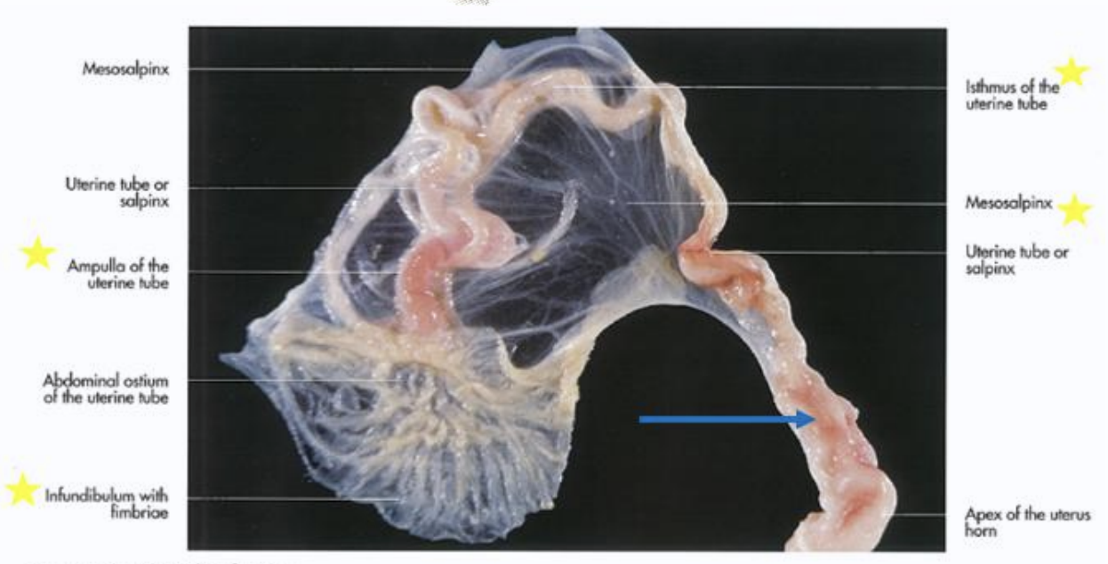
Uterine tube components
infundibulum, ampulla, isthmus(blue arrow)
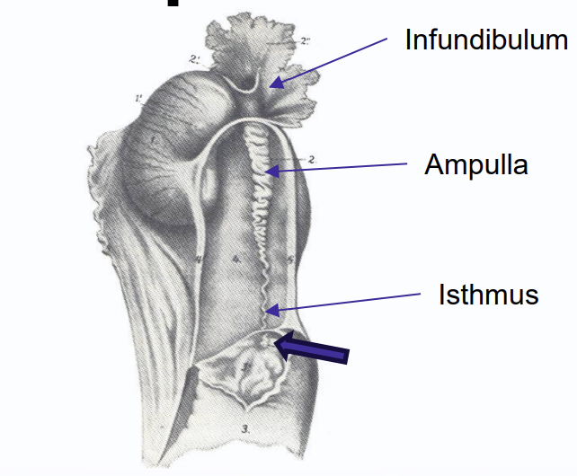
Infundibulum
Funnel shaped ovarian end of the tube. Lined by fibria/fingerlike projections on its free border that traps released ovum
Amupulla
site of fertilization. contues the infundibulum. widest part of uterine horn
Isthmus
continues the ampulla and narrows in diameter and straightens out. Joins uterine horn at spinctered papilla called uterotubal junction
Uterine horn in ruminants
widens in diameter as it connects to uterine horn
Canine Uterus topography
dorsolateral to loops of intestines and ventral to digestive tract in pelvic cavity
Canine Uterine horns
Y shaped. Site of implantation. suspended by broad ligament in dorsolateral region in abdominal cavity
Canine Uterine body
located between horns and cervix. Located dorsal to bladder and proximal urethra. Short in dogs. longer in cats
Canine uterus image
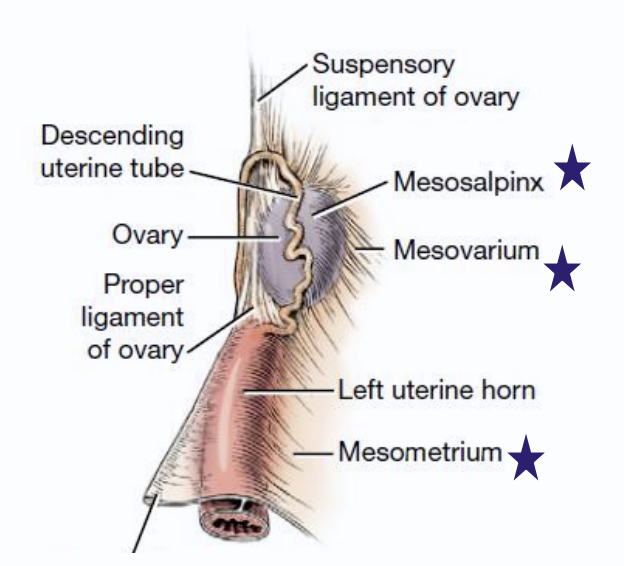
Broad ligament
bilateral sheets suspends ovaries, uterine tube, uterus and vagina to abdominal roof and pelvic walls. Composed mesometrium, mesovarium, mesosalpinx
mesometrium
largest part of the broad ligament in the dog/cat uterus. It attaches the ,uterine horn, uterus and cranial vagina to the dorsolateral body wall. Houses uterine vessels
mesovarium
cranial part of the broad ligament and ovarian pedicle that connects ovaries of the dogs/cats to the dorsolateral abdominal wall
mesosalpinx
fold arrising from mesovarium that attaches to the two uterine tubes onto the mesovarium, not directly attached to the body wall
ovarian pedicle
arterial and venous structures within the mesovarium which supply and return blood to and from the ovary. This is ligated and trisected in spays.
Proper ligament
caudal continuation of free edge of mesovarium. Connect ovary to uterine horn
Round Ligament
Caudal continuation of proper ligament, passes through vaginal ring/inguinal canal. Forms vaginal process in bitches. Supports pregnant uterus.
suspensory ligament
cranial end of mesovarium. forms proper ligament of ovary, Attaches ovary to last few ribs and transversalis fascia. Normally ripped to free ovaries during spay.
Cervix Canine
sphincter of the uterus. found in pelvic cavity. Lumen has longitudinal folds
Carnine cervix-Portio vaginalis
caudal projection of cervix into vaginal lumen
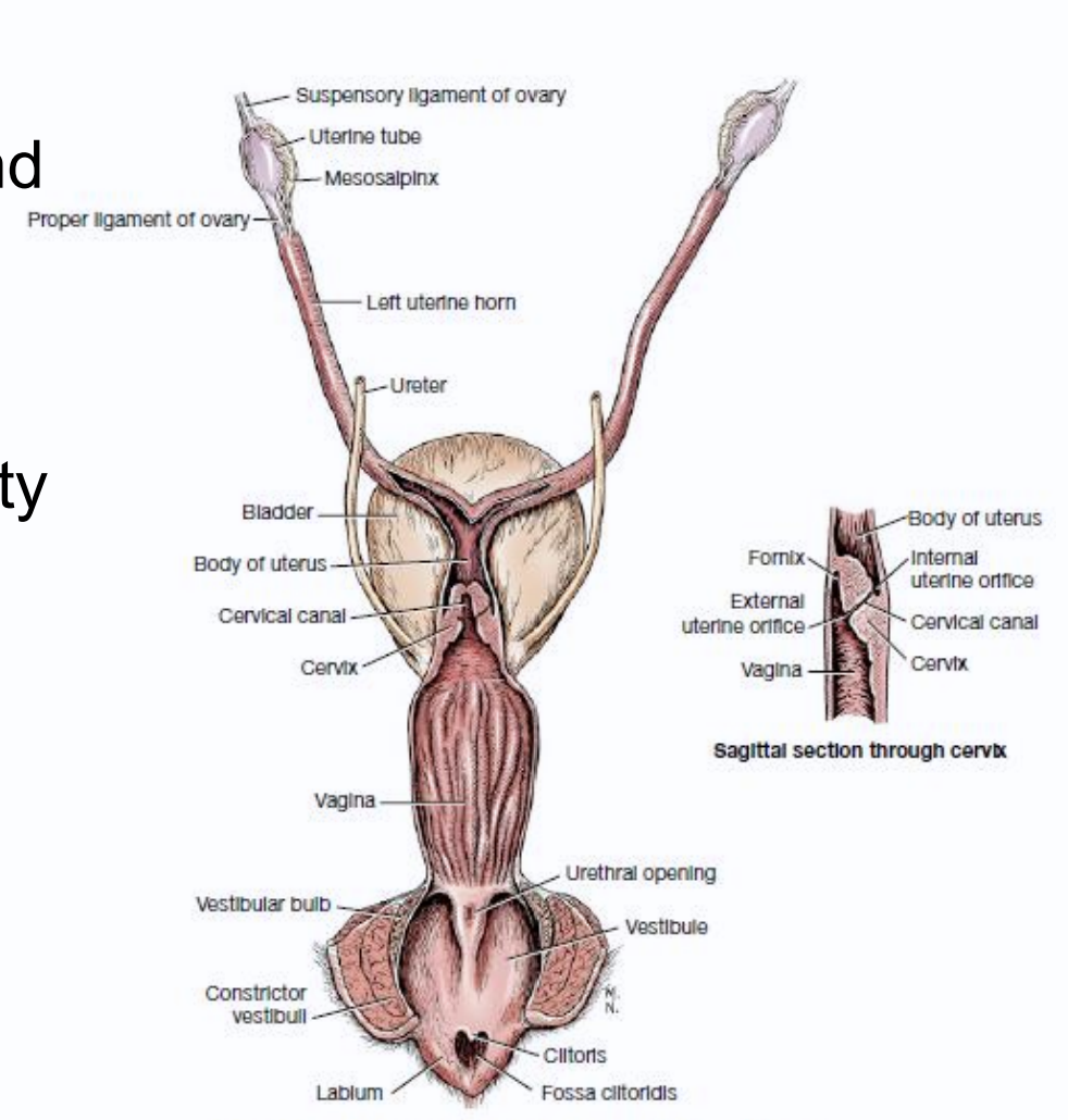
Fornix vagina
Recess around vaginal portion of the cervix, site of obstruction when passing AI catheter
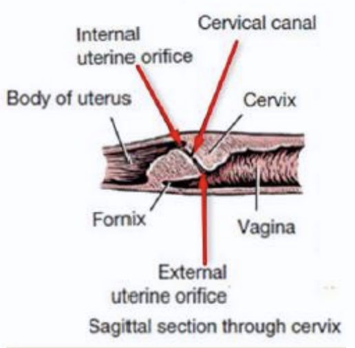
Interal ostium/os
opening towards the body of the uterus
external ostium/os
opening towards vagina and forms the ends of cervical canal
Vagina borders-Canine
proximally-vaginal fornix
caudally-external urethral opening
transverse-mucosal ridge, urethra tubercle (dog) and groove (cats). Transverse hymen(horse)
Vestibule-canine
Female copulatory organ. together with vagina forms the birth canal. Also part of urinary tract. Hold accessory sex glands
Vestibule accessory sex glands
dog-minor vestibular gland
cats- minor and major(Bartholin) vestibular gland
Vestibular bulbs(dog/horse)
dark patch on mucosa wall of vestibule, concentrated erectile tissue with venous sinus. Helps with “tie” when mating
Fossa clitoridis and clitoris
Invagination that houses clitoris/ homologue to penis
Vulva-canine
lips fused dorsally and ventrally to form vulva comissure
Most Species
Dorsal commisure is rounded which ventral commissure is pointed in ______
Arrangment of reproductive tract from external to internal
vulva, vesibule, vagina, cervix, uterine body and horns, uterine tube, ovaries
Ligaments of female reproductive system image
Pudendal nerve
What innervates muscles of the vulva/vestibule?
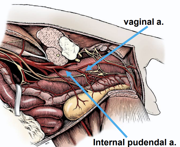
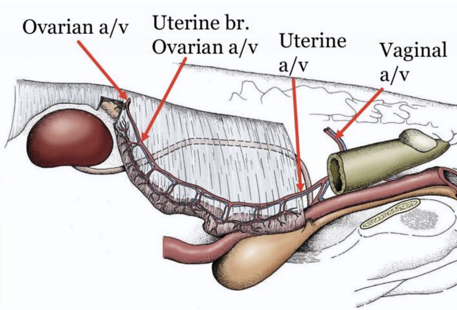
Ovarian atery
Branches directly from aorta and gives off uterine branch to ovaries
Vaginal artery
branches fron internal pudendal artery supplies vagina, vestibule, cervix. Cranial branch of uterine artery
within Broad ligament
where ovarian and vaginal artery anastomose and must be ligated in ovariohysterectomy
Repro artery pic
Uterine Veins to cauda vena cava
What vein drains the uterus
Nerve supply to female reproductive organs
caudal mesenteric plexus-sympathetic→ ovaries
Vagus nerve-parasympathetic→ ovaries
pelvic plexus-para and sympathetic→ rest of reproductive tract
pudendal nerve-motor innervation→ constrictor vestibuli and constrictor vulvae
Ovaries-Bovine
oval and small. located just cranial to pelvic inlet. Most caudally located among all species. Follicles and corpora lutea are palpable on surface
Ovaries structure-bovine
divided into outer cortex and inner medulla.
cortex dense and contains follicles and CL
medulla less compact has vessels, nerves, and lymphs, SM and CT
tunica albuginea is outermost layer of white fibrous tissue