cells
1/30
There's no tags or description
Looks like no tags are added yet.
Name | Mastery | Learn | Test | Matching | Spaced | Call with Kai |
|---|
No analytics yet
Send a link to your students to track their progress
31 Terms
cells
the functional units of the body
most cells must perform the following
maintaining its integrity and shape - dependent on plasma membrane and internal contents
obtain nutrients and form chemical building blocks - harvest energy for survival
dispose of wastes -avoid accumulation, disrupting ellular activties
some cell can also do cell division
make more cells of the same type
help maintain the tissue by providing new cells
***some cells lose their ability to divide during the development process
plasma membrane
Forms the outer limiting barrier
Separates internal contents of cell from external environment
Cilia, flagellum, microvilli- modified extension of plasma membrane
cytoplasm
Cellular contents between plasma membrane and the nucleus
Includes cytosol, organelles, and inclusions
cytosol
intracellular fluid
viscous fluid of the cytoplasm
High water content
Contains dissolved macromolecules (carbohydrates, lipids & proteins), small molecules (glucose & AAs) and ions
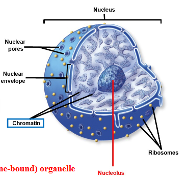
nucleus
contains the genetic material, DNA
in the chromatin
Typically only one per cell BUT
number and shape per cell depends on the cell type
erythrocytes with no nuclei
skeletal muscle cells with many (multinucleate)
red blood cells = 0
skeletal muscle cell = many
enclosed in nuclear envelope
Largest structure in the cell
externally continuous with rough ER

nuclear pores
conains channel like open passageways
•allow passage in and out of nucleus
large particles, ions, water soluble molecules
nuclear envelope
double phospholipid membrane enclosing nucleus
separates cytoplasm from nuceloplasm
externally continuous with rough ER
contains channel-like nuclear pores to let things in

nucleolus
separate (non-membrane bound) organelle
dark- staining spherical body
responsible for production of ribosomes
not present in all cells
many in nerve cells → makes many proteins
absent in sperm cells → makes no proteins
nucleoplasm
inner fluid in nucleus
membrane bound organelles
surrounded by a membrane
BECAUSE of the membrane – the activities of the organelle can proceed in an isolated environment
allows processes of organelle to go on WITHOUT disrupting other cellular processes
differ in shape, membrane, composition, enzymes = dependent on function
nucleus
endoplasmic reticulum (smooth and rough)
golgi apparatus
lysosome
peroxisomes
mitochondria (unique among organelles)
endoplasmic reticulum
Extensive interconnected membrane network
separates fluid within structure from cytosol with a continuous lumen
Extends from nuclear envelope to plasma membrane – making up ~1/2 of the membrane within a cell
synthesis = provides a place for chemical reactions
transport = moves molecules through lumen from one part of the cell to another, sequestered away from the cytosol
packaging and storage = packages and stores newly synthesize molecules
detoxification: SMOOTH ER detoxifies drugs, alcohol, and poison
structure formation: segments of ER are pinched off to form transport vesicles and peroxisomes
Rough ER
site of synthesis of proteins destined for secretion, incorporation into the plasma membrane, and as enzymes w lysosomes
Protein production by ribosomes happens here – in rough ER
Proteins inserted into membrane as they are synthesized
Original structure of protein changed – as it’s processed (additions and/or subtractions)
molecular tags – called signal sequences – determine the destination (an “address”)
Transported out in enclosed membrane sacs that pinch off from the ER membrane → termed transport vesicles
shuttle proteins from rough ER lumen to Golgi apparatus
Plentiful in cells producing much protein – such as insulin (protein) -producing cells of the pancreas
Peroxisomes produced here
smooth ER
site of lipid synthesis and carbohydrate metabolism
Continuous with rough ER
Diverse metabolic processes varying by cell
Some functions:
synthesis, transport, and storage of lipids
carbohydrate metabolism
detoxification of drugs, alcohols, and poisons
Plentiful in cells of the testes → to produce the steroid hormone testosterone (steroids are lipids)
Plentiful in liver → to detoxify alcohol, when consumed
golgi apparatus
two faces: cis face (entry and closer to ER) and trans face (exit)
synthesis: formation of proteoglycans
processing molecules: modifying and store proteins
organelle formation: synthesizes digestive enzymes for lysosomes
vesicle formation: forms secretory vesicles for delivering components of the plasma membrane and releasing content from the cell by exocytosis
Composed of several elongated, flattened saclike membranous structures (~4-5) → termed cisternae
“Warehouse” of the cell
Exhibits a DISTINCT polarity
Cis-face →closer in proximity to the ER
receiving region
Larger diameter
Trans-face → farther from ER
shipping region
Functions of Golgi
Modification, packaging, and sorting of proteins
fusion of transport vesicles from ER at cis-face
modification of molecules, e.g., addition of phosphate group
Transport of material from cis-face to trans-face – moving b/t the cisternae
Formation of secretory vesicles and lysosomes – at the trans-face
some vesicles becoming part of plasma membrane
others releasing contents outside cell – via exocytosis
Golgi extensive in cells specializing in protein secretion
endomembrane system - secretory pathway
Extensive array of membrane-bound structures → Includes ER, Golgi apparatus, vesicles, lysosomes, peroxisomes
Also includes plasma membrane and nuclear envelope
Connected directly or through vesicles moving between them
Provides means of transporting substances within cells
***Mitochondria = the only membrane-bound organelles NOT included in the endomembrane system
rough ER synthesizes proteins thar is released in a transport vesicle
vesicle from the rough ER moves to the golgi apparatus
vesicles fuses with Golgi apparatus at the cis face
proteisn are modified as they move through golgi → adding carbohydrate
modified proteins are packaged and released with secrtory vesicle from trans face
secretory vesicles
1. merge with plasma membrane to insert molecules into the plasma membrane =
2. release contents by exocytosis
3. serve as lysosomes
lysosome
lyso- meaning “dissolution”
some from soma, meaning “body”
it’s a small membraneous sac that dissolves substances
contain digestive enzymes formed by Golgi (ph 5)
Also participate in autophagy and autolysis → digestion of unneeded/unwanted substances
autophagy – digesting damaged cell components
autolysis – breaking down cellular components following cellular death
Digest contents of endocytoses vesicles
Clinical View: Lysosomal Storage Diseases
Group of heritable disorders
Characterized by accumulation of incompletely digested molecules within lysosomes
Mutation in genes that code for one of over 40 lysosomal enzymes
E.g., Tay-Sachs disease → lack enzyme needed to break down complex membrane lipids (gangliosides)
Results in accumulation of lipids within nerve cells
Cellular sign = swollen lysosomes – due to lipid accumulation
Outward signs appear as early as 6 months
Nervous system gets brunt of damage
paralysis, blindness, deafness, followed by death by age four
Lysosomal storage diseases are an extensive group of heritable disorders that are characterized by accumulation of incompletely digested biomolecules within lysosomes. Lysosomal storage diseases occur because of mutations in the genes that code for one of the more than 40 different lysosomal enzymes. Tay-Sachs disease is one example of a lysosomal storage disease. Lysosomes in affected individuals lack an enzyme needed to break down complex membrane lipids (gangliosides). As a result, these complex lipids accumulate within nerve cells.
The cellular signs of Tay-Sachs disease are swollen lysosomes due to accumulation of the lipid. Affected infants appear normal at birth, but begin to show signs of the disease by the age of 6 months. The nervous system bears the brunt of the damage. Paralysis, blindness, and deafness typically develop over a period of 1 or 2 years, followed by death, usually by the age of 4. Unfortunately, there is no treatment or cure for this fatal disease.
Peroxisomes
MOLECULES broken down by peroxisomes include – fatty acids, amino acids, and uric acid
has more than 50 different enzymes – dependent on cell type
Membrane enclaseds sacs smaller than lysosomes
pinched off vesicles form rough ER
Serves in detoxfication →
Hydrogen peroxide formed when they remove hydrogen from a molecule – hence their name
H2O2 (hydrogen peroxide) – then broken down into water and oxygen – via the catalase enzyme
Beta-oxidation = removal of two H-C units at a time from the fatty acid chain
these two units are further broken down into acetyl CoA – to be taken up by the mitochondria (for use in ATP production)
Most abundant in liver → due to detoxification properties
AND that they also participate in lipid production → important in the production of BILE
Mitochondria
Oblong shaped organelles with a double membrane
contaisn genes for producing mitochondria proteins
on a separate circular strand of DNA
engages in aerobic cellular resporiation
complete digestion of fuel molecules to synthesize ATP
Cristae = the folds of the inner membrane
These genes come from a unique, circular fragment of DNA within the mitochondria → part of supporting evidence for the hypothesis that early eukaryotic cells endocytosed small, aerobic bacteria – due to the circular DNA
interestingly, because we only get our organelles from our mother – mitochondrial DNA is something only passed on from mothers
there are certain genetic disorders that only get passed on thru mitochondrial DNA
Aerobic respiration = most efficient way to get the maximum number of ATP molecules from each molecule of glucose
ATP = the “cell’s energy currency”
Mitochondrial #’s increase – thru fission – as demands for ATP increase à such as when one exercises on a regular basis (and needs more energy)
ribosomes
non membrane bound organelles
Contain protein and ribonucleic acid (RNA)
Arranged into BOTH a large and a small subunit
Large subunit with E, P, and A sites
Made within nucleolus and assembled in cytoplasm
Bound ribosomes = attached to external surface of ER membrane – forming the rough ER
proteins for plasma membrane, export (exocytosis), or enzymes within lysosomes
destine to be incorporated into the plasma membrane, exported from the cell or housed within lysosome
Free ribosomes = suspended within cytosol
all other proteins within cell synthesized here
proteasome
located in cytosol and cell nucleus
degrade cell proteins through ATP dependent pathway
damage protein, incorrectly folded protein, proteins no longer needed
Just as with the lysosomes – proteasomes are responsible for DIGESTION of larger molecules into smaller molecules
protea – means the substance that it will break down is a PROTEIN
-somes – from soma – meaning body
it’s a membraneous sac used in protein digestion
the proteins are broken down into their building blocks → amino acids
Protein marked with ubiquitin tag for disposal
With age may be unable to normally remove proteins
centrosomes/centrioles
Usually in close proximity to nucleus
centrosomes = made of 2 centrioles
centrioles = made of microtubules
Contains pair of perpendicularly oriented, cylindrical centrioles
Surrounded by protein that is amorphous (without a distinct shape)
PRIMARY FUNCTION = organizes microtubules within the cytoskeleton
Best known for function in cell division
forms the mitotic spindle – which facilitates chromosomal movement during that process
cytoskeleton
Plays roles in:
intracellular support
organization of organelles
cell division
movement of materials
Extends through interior of cell
Anchors to proteins in plasma membrane
Formed by a framework of diverse fibrous proteins, which includes
microfilaments
intermediate filaments,
microtubules
microfilaments
Smallest components of the cytoskeleton
Actin protein monomers form two twisted actin filaments - think of twisted pearl strands
Form interlacing network on cytoplasmic side of membrane
Functions of microfilaments:
help maintain cell shape
form internal support of microvilli
separate two cells during cytokinesis
facilitate cytoplasmic streaming
participate in muscle contraction
intermediate filaments
Intermediate-sized components of the cytoskeleton
More rigid than microfilaments
Support cells structurally and stabilize cell junctions (b/t cells)
Varied protein composition between cells
e.g., keratin – found in cells of the skin, hair, and nails
microtubules
Largest components of the cytoskeleton
Hollow cylinders
Long chains of the globular protein = tubulin
Impermanent structures – elongated or shortened as needed
accomplished by addition or removal of tubulin monomers, respectively
Function to:
maintain cell shape
organize and move organelles
form components of cilia and flagella
participate in cellular vesicle transport
separate chromosomes during cell division
cilia and flagella
Movement of BOTH based on MT within their core
movement REQUIRES energy – provided by the splitting of ATP molecules
protein extneding from the cell surface
contains both cytoplasm and microtubule proteins
neclosed in plasma membrane
cilia
usually found on exposed surfaces of specific cells
usually found in large numbers
this beating of the cilia is known as the mucociliary escalator
it’s what allows you to cough up things that may have gotten into your lung
flagella
similar to cilia in sturcture
longer and usually appear alone
helps propel an entire cell
only example in humans is sperm cell
microvilli
•Microscopic extensions from surface of plasma membrane
•Much smaller than cilia
•More densely packed, lack powered movement
•Supported by microfilaments
•Form extensive plasma membrane surface
•providing increased surface area needed to absorb nutrients
•e.g., in cells of small intestine
Microfilaments – dense bundle of cross-linked actin proteins
Increased surface area – without the different layers of surface area increase in regions such as the small intestine
we would need to consume 600X more food, in order to get the nutrients we require