Looks like no one added any tags here yet for you.
What are microcytic anemias characterized by?
microcytic RBC’s, often hypochromic
typically due to defected hgb synthesis
What are the 2 categories of causes of microcytic anemias?
deficiency heme synthesis
lack of iron = IDA
defective iron utilization = sideroblasic anemia
porphyrias
deficient globin synthesis = thalassemias
Where is iron found in humans for metabolic or enzymatic functions?
hgb** majority
myoglobin
cytochrome enzymes in every cell in body
transferrin
What are the storage forms of iron? What should you think about regarding each?
ferritin = piggy bank
hemosiderin = savings account at bank
What is total body iron? How much of that is found within heme?
total = 3.7g
within heme = 2/3 so about 2.5 g of total
How does the body tightly control metabolism of iron?
95% of daily iron needs met by using recycled iron
rest is absorbed via diet (5%)
excess dietary iron excreted w/o being absorbed
internal iron CANNOT be excreted and can be toxic if too much
How can children easily take in too much iron?
eating gummy vitamins bc they taste good
About how much iron does each person lose daily?
1 mg
through sweat, loss of intestinal cells (iron stored here), urine/bile
Why do females lose more iron daily compared to others?
menstruation, pregnancy, nursing
Fe 3+
ferric iron
transferrin associated iron, storage iron
Fe 2+
ferrous iron
carries O2 = heme iron
hgb, myoglobin, cytochrome enzymes
Non-heme iron (grains and veggies) absorption
changed from Fe3+ to Fe2+ by enzymes in enterocytes
harder to absorb
** vegetarian’s/vegan’s bodies must work harder
Heme iron (meat) absorption
already in Fe2+
more easily absorbed
Where is iron absorbed in the body?
proximal portion of duodenum → then couples with transferrin in blood
goes back in Fe3+ form in order to move around the body
Where is Fe 3+ taken after it’s been absorbed?
taken to bone marrow by transferrin to make new RBC’s or to be stored as ferritin
incorporated into body cells as cytochrome enzymes
Characteristics of ferritin
water soluble
composed of ½ protein
short term storage
limited storage capacity (piggy bank)
soluble form does not stain w/ Prussian blue but will stain if clustered in siderosomes
What is the name of the protein that makes up ½ of ferritin when it is not attached to iron?
apoferritin
Distribution of ferritin
primarily intracellular
circulates between tissues and plasma (equilibrium)
What is plasma ferritin a good indicator of?
iron stores
Hemosiderin characteristics
larger aggregates than ferritin
insoluble
50% lipid, carb protein
50% iron, including denatured ferritin
yellow/brown aggregates unstained
stains blue with Prussian blue stain
Hemosiderin distribution
bone marrow - good indicator of overall stores
other tissues
Iron stores/reserves available for males and females
males = 1000mg
females = 300-500mg
When there is an increased demand for iron, which storage type is used first?
ferritin used first bc more easily accessible, then hemosiderin used
Do both ferritin and hemosiderin have to be depleted to consider someone iron deficient?
yes
With no absorption, how long would it take for a male to use up his normal reserves of iron?
8 years
How much iron do women lose each month due to menstruation?
30-40mg
What are causes of iron deficiency?
insufficient Fe in diet
increased demand
absorption problems
blood loss
Insufficient iron in diet
#1 nutritional deficiency in the WORLD
one of the leading causes of anemia in US (not #1)
Increased demand leading to iron deficiency
multiple pregnancies
periods of significant growth and development
in infants - “milk babies”
as baby’s are using less formula and breast milk, iron needs aren’t being met through normal milk at 6 months-1 year
Absorption problems leading to iron deficiency
celiac disease
defective gastric function - achlorhydria (stomach does not produce acid)
gastrectomy (bariatric surgery)
Blood loss leading to iron deficiency
#1 cause in US
females = heavy menstruation
males = GI bleeds from ulcers, colon cancer, hemorrhoids
If males have an iron deficiency, what should your first thought be until proven otherwise?
cancer
What’s another name for IDA?
sideropenic anemia
IDA PBS
micro-hypo
**RBC’s smaller than lymphocyte nucleus
IDA lab testing for bone marrow iron stores
only performed when non-invasive tests cannot diagnose
stain bone marrow aspirate specimen with Prussian blue
normal marrow has about 30-60% developing erythroblasts w/ blue iron particles in them
in IDA no stainable iron found but marrow cellularity normal-increased
Sideroblastic anemia based on bone marrow lab testing
abnormal Fe in normoblasts
What is siderocyte stain of PBS used for?
Prussian blue
other iron utilization problems, not IDA
looking for abnormal iron granules
hemosiderin granules - Pappenheimer bodies on Wright’s stain
What diseased states cause decreased serum iron concentration?
IDA
chronic infections
cancer
What diseased states cause increased serum iron concentration?
iron overloads
intravascular hemolysis
multiple transfusions (no way to get rid of)
True or False: Most serum iron is transferrin complexed?
true
IDA lab testing of TIBC
total iron binding capacity
measures transferrin capacity to carry iron
increased in IDA - body makes more transferring to capture what little iron already available
decreased in chronic diseases and cancer
How many Fe 3+ can transferrin carry?
2
What components make up TIBC?
1/3 serum iron
2/3 unbound iron binding capacity
IDA lab testing of transferring % saturation
calculation using serum iron and TIBC
% saturation = serum iron/TIBC X 100%
decreased in iron deficiency, increased in iron overload
IDA lab testing of serum ferritin
tissue ferritin in equilibrium w/ serum ferritin so good indicator of iron stores
decreased in IDA
normal to increased in ACD
increased in sideroblastic anemia and thalassemia
IDA lab testing of sTfR
serum transferrin receptor assay
looking for receptor for transferring (parking spot)
receptor expressed on all cells in proportion to Fe use
IDA = sTfR is double normal
ACD = normal values
Which is the better indicator of IDA: serum ferritin or sTfR?
sTfR
IDA lab testing of FEP or Znpp
free erythrocyte protoporphyrin
theory: RBC accumulates excess protoporphyrin bc Fe not present to bind protoporphyrin
testing via fluorescence studies
early value to chance as iron stores used
Does FEP/ZnPP elevate before or after anemia develops?
before - good tool to detect developing IDA in milk babies
What is another diseased state where FEP/ZnPP is elevated?
lead poisoning
IDA lab testing of RDW
red cell distribution width
SD of MCV divided by MCV
corresponds to level of aniso
RDW reference range
11.5-14.5%
but human eyes cannot distinguish <16%
RDW levels related to IDA and thalassemias
IDA = elevated
thalassemia = normal or slightly elevated
What are the stages of developing IDA?
iron depletion = both stores used up
iron deficient erythropoiesis = decreased serum iron, increased TIBC, may see 2 populations of cells
iron deficiency anemia = hgb decreases, progression of cell size and color
IDA vs Thalassemia
IDA
MCV decreases proportionately as anemia develops
ex. low MCV = low hgb
thalassemia
defect of globin synthesis - Fe is fine
MCV much lower for given conc. of hgb and hct compared to IDA
body compensates by making more RBC’s
Symptoms of IDA
pallor/fatigue
pica = ice/crunchy foods, clay, starch
koilonychia (spoon nails)
glossitis (smooth tongue)
Resolution of IDA
find cause and remove if possible
ulcer, GI bleed, diet, heavy menstruation
give iron - typically oral as ferrous sulfate
after transfusion
What happens to RPI after IDA treatment
initially increases and then levels off
Sideroblastic anemia
micro-hypo
iron utilization problem
Fe present and available in marrow
Fe cannot be inserted into protoporphyrin ring
Fe accumulates in mitrochondria
Fe-laden mitochondria seen on Prussian blue as “ringed sideroblasts” in marrow
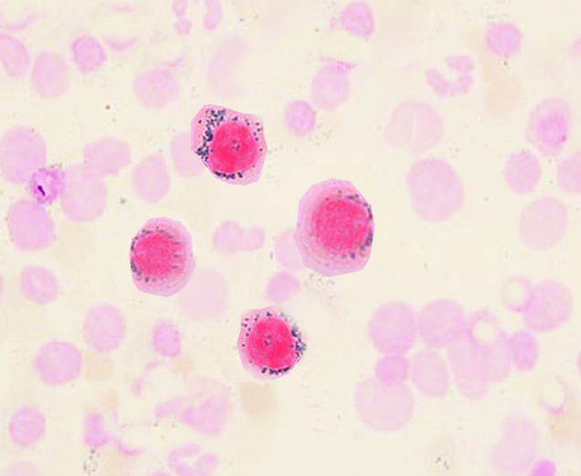
What do “ringed sideroblasts” look like?
iron (blue) in a ring around nucleus of cell
What are causes of sideroblastic anemia?
inherited enzyme defects in heme synthesis (RARE)
acquired
Acquired sideroblastic anemia
myelodysplastic syndromes and malignancies
toxic effects
alcohol abuse / some drugs
lead poisoning which blocks enzymes such as ferrochelatase causing basophilic stippling
lead accumulates in cells → aggregated ribosomes, mitochondria, siderosomes'
only seen in stained preps
PBS findings in sideroblastic anemia
dimorphic RBC population
aniso and poikylo
polychromasia
hypochromic/normochromic cells
teardrop cells
basophilic stippling
pappenheimer bodies
Other lab values associated with sideroblastic anemias
decreased RBC, hgb, hct
increased iron stores/total body iron
elevated serum iron and percent saturation of transferrin
elevated ferritin/hemosiderin
ringed sideroblasts
Treatment of sideroblastic anemias
transfusion dependent
may lead to iron overload
can be treated with iron chelator such as desferrioxamine
Anemia of chronic disease
ranges from normo-normo to micro-hypo
AKA anemia of inflammation
What are some chronic diseases that may cause ACD?
chronic infections: sub-acute bacterial endocarditis, TB
inflammatory disease
autoimmune diseases: SLE and RA
malignancies
Mechanisms that contribute to ACD
block in Fe release from macrophages to marrow in normal Fe recirculation
failure to trigger EPO release
decrease in RBC survival
marrow suppressive effect of cytokines and interleukins
Degree of anemia with ACD
usually not severe
fluctuates with activity of underlying condition
Is ACD the next most common anemia behind IDA from blood loss?
yes
Iron studies in ACD
serum iron = low
TIBC = low to normal
% saturation = low to normal
serum ferritin = normal to increased**
ZnPP = increased
sTfR = normal
Hemosiderosis
increased deposits of hemosiderin in phagocytic system
reticulo-endothelial system
macrophages very full
Examples of Hemosiderosis
multiple transfusions
sideroblastic anemia
high iron in diet
increased absorption with chronic alcoholism
Hemochromatosis
more severe form of iron overload
common genetic abnormality
involves deficiency of hepcidin
european ancestry
Where does the iron deposit in hemochromatosis?
reticuloendothelial system and soft organs
pancreas = diabetes
skin = melanin-like pigment
liver = enlarged and decreased function
gonads = hypogonadism (females = ovaries)
How to treat hemochromatosis
with iron chelator drugs and phlebotomy to remove excess cells and iron