Lab Chapter 10: Arteries
1/40
There's no tags or description
Looks like no tags are added yet.
Name | Mastery | Learn | Test | Matching | Spaced | Call with Kai |
|---|
No analytics yet
Send a link to your students to track their progress
41 Terms
What are the layers of blood vessels?
3 layers called tunics: tunica intima or interna, tunica media, tunica externa
Tunica Intima
Innermost layer (layer closest to the lumen)
Made of simple squamous epithelium (which function is diffusion)
Tunica Media
Middle, muscle layer
Contraction causes vasoconstriction: narrows lumen (lumen gets smaller)
Relaxation causes vasodilation: widens lumen (lumen gets bigger)
Tunica externa
Outermost layer
It anchors vessel to surrounding structures
Why don’t arteries have valves?
Unlike veins, arteries lack this structure bc they are high pressure vessels, so there is no tendency for backflow to occur.
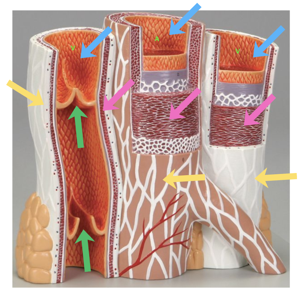
The picture shows an arteries surrounded by 2 veins. Identify what each arrow is pointing to? (Blue, pink, green, yellow)
Blue: Tunica intima
Pink: Tunica media
Yellow: Tunica externa
Green: Venous valves

Compare the tunics of arteries vs veins
Arteries
Have thicker tunica media and narrower lumen than veins
Veins
Have thicker tunica externa and larger lumen than arteries. Their tunica media is very thin.
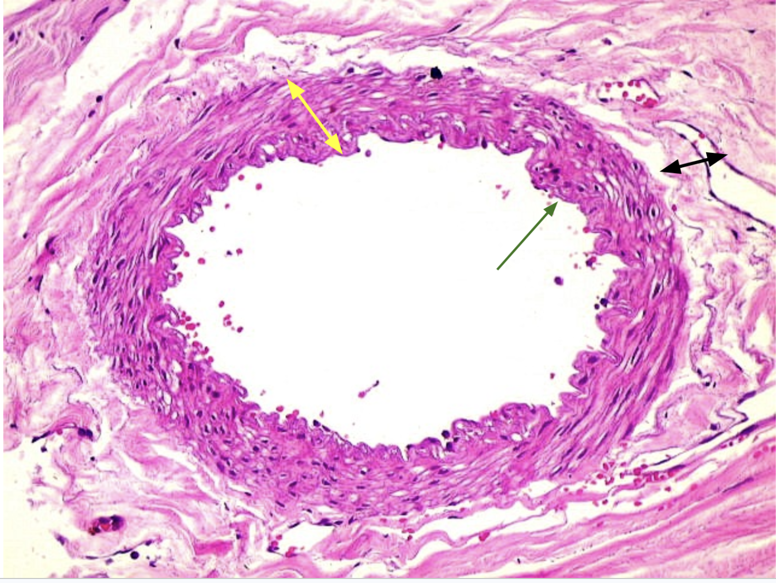
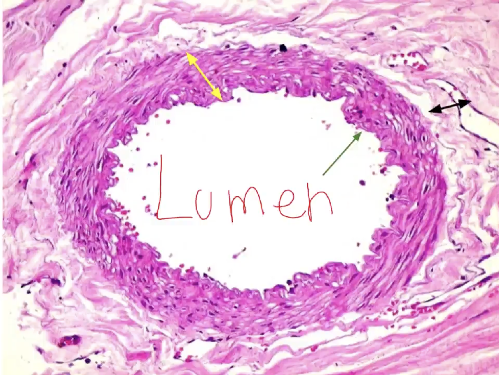
Is this a vein or an artery? Identify the layers labeled by the arrows
The middle hole of the vessel is called what?
- Artery (arteries have a narrower lumen and look tighter than veins, due to their thick tunica media)
- Green: Tunica intima/interna
- Yellow: Tunica media
- Black: Tunica externa
Middle is called the lumen

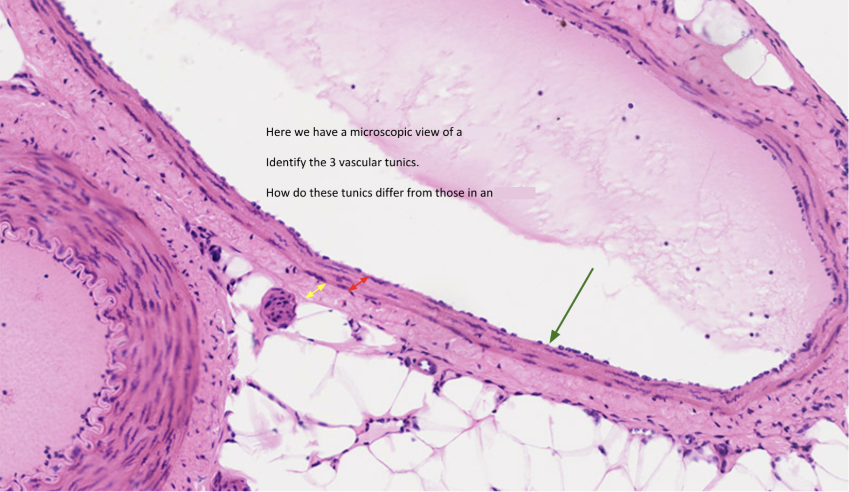
Is this a vein or an artery? Identify the layers labeled by the arrows
- Vein (notice the large lumen and thin tunica media. The tunica externa is thick)
- Green: Tunica interna
- Red: Tunica media
- Yellow: Tunica externa
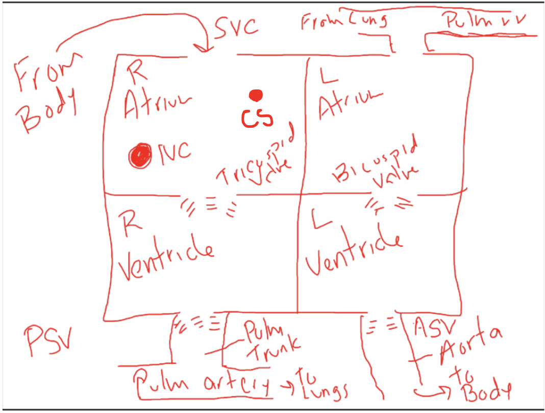
Explain process of blood flow in the heart? (Recall)
Right atrium: deoxygenated blood
Left atrium: oxygenated blood

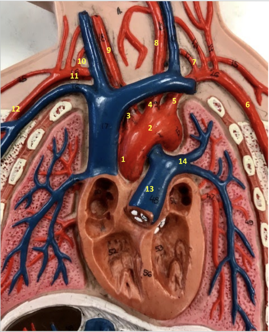
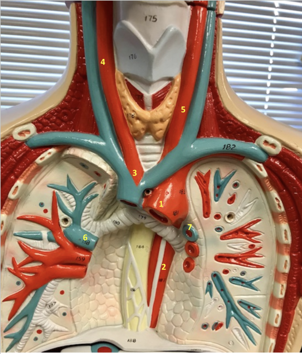
Label each part. What is the function of 3,4,5?
1. Ascending aorta
2. Aortic arch
3. Brachiocephalic artery
Supplies blood to the right shoulder and arm, and right halves of the neck and head. Notice how it branches off into 11 & 9, which goes to the shoulder (11) and neck (9).
4. Left common carotid artery
Supplies blood to the left halves of the neck and head
5. Left subclavian artery
Supplies blood to the left shoulder and arm.
6. Left axillary artery
7. Left vertebral artery
8. Left common carotid artery
9. Right common carotid artery
10. Right vertebral artery
11. Right subclavian artery
12. Right axillary artery
13. Pulmonary trunk
14. Left pulmonary artery
What are the two arteries that supply the brain with blood?
Vertebral arteries and internal carotid arteries
Vertebral arteries travel straight through vertebrae directly to brain
Internal carotid arteries enter directly into the skull to the brain

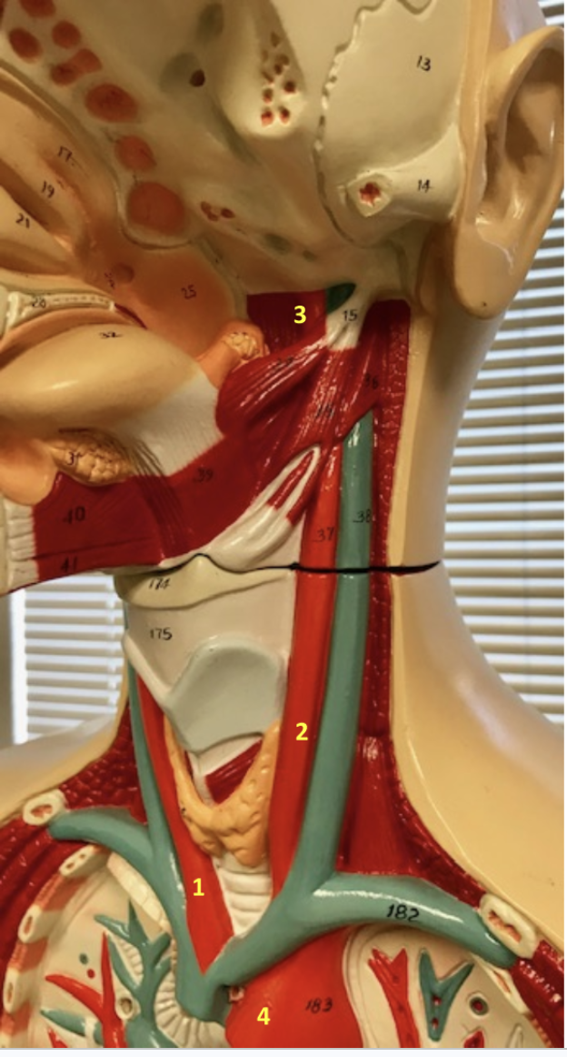
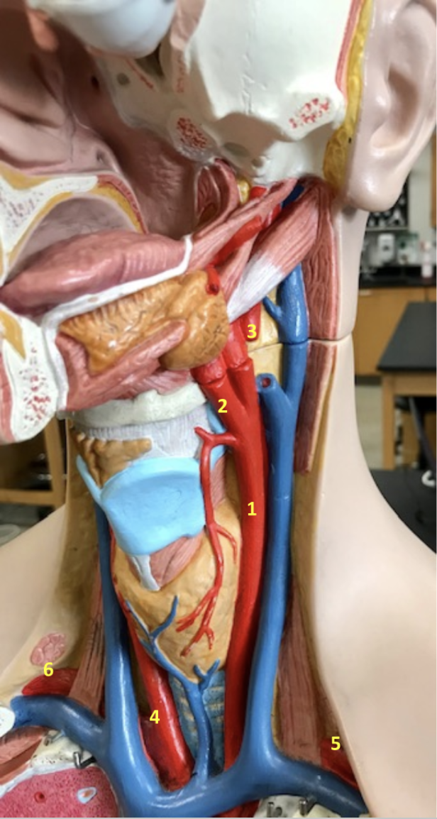
Identify each. What is the function of #5?
Neck/Head
1. Right subclavian artery
2. Right vertebral artery
3. Right common carotid artery
4. Left common carotid artery
5. Left external carotid artery: Supply blood to neck, esophagus, larynx, jaw, and face (external structures of left halves of head and neck).
6. Left facial artery
7. Left vertebral artery
8. Left subclavian artery

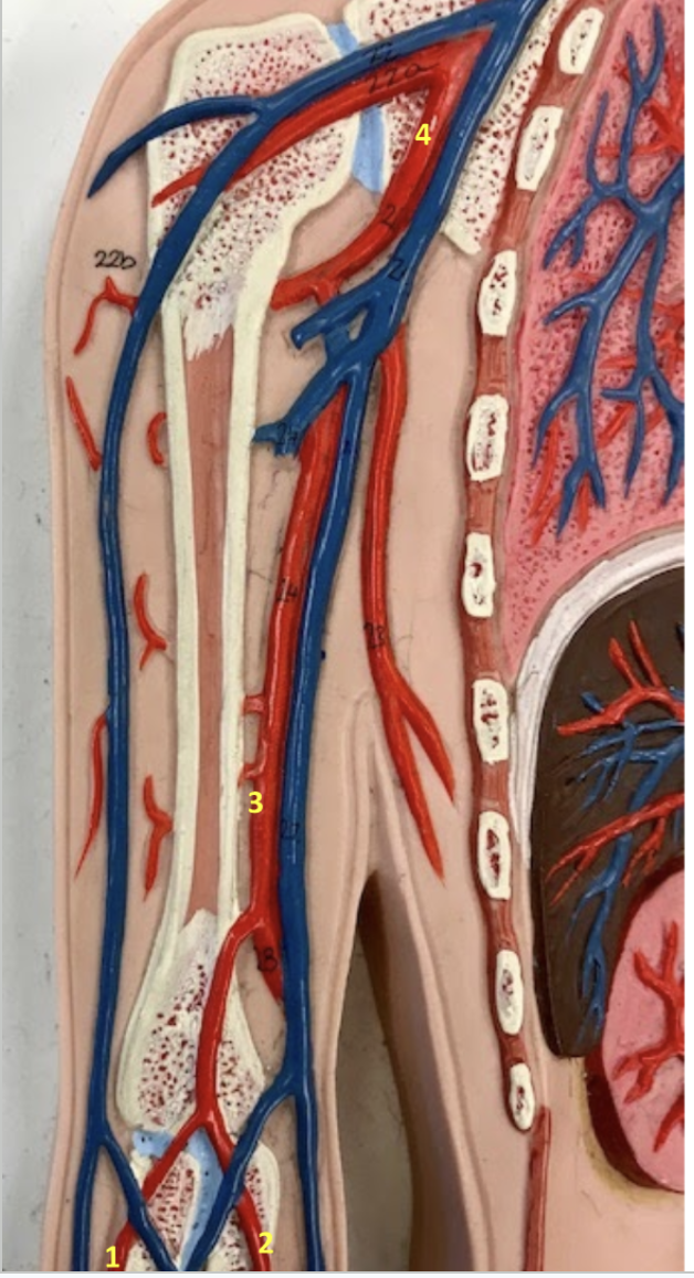
Identify each (scroll down)
Arm
1.Right radial artery (on thumb side)
2. Right ulnar artery (on pinky side)
3. Right brachial artery
4. Right axillary artery

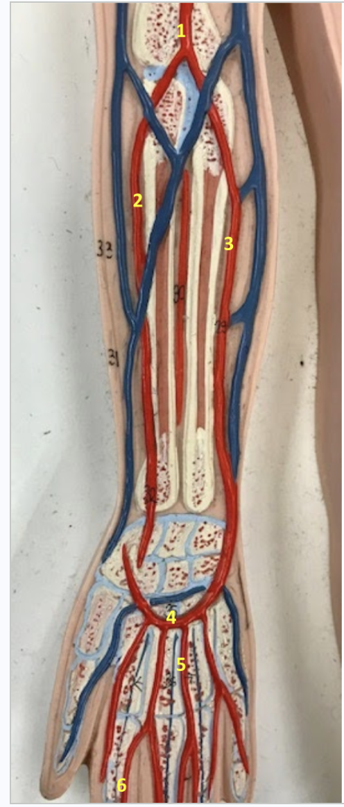
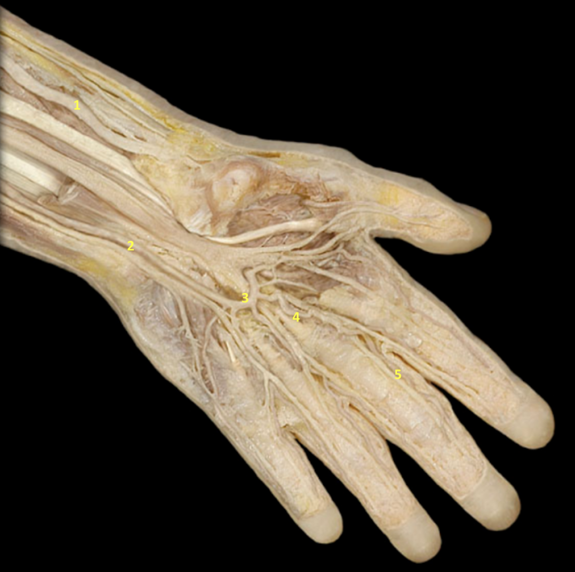
Identify each (scroll down). Function of 1, 2&3?
Forearm/Hand
1. Right brachial artery: Supply blood to upper arm
2. Right radial artery: Supply blood to the forearm
3. Right ulnar artery: Supply blood to the forearm
4. Right superficial palmar arch
5. Right metacarpal artery
6. Right digital artery


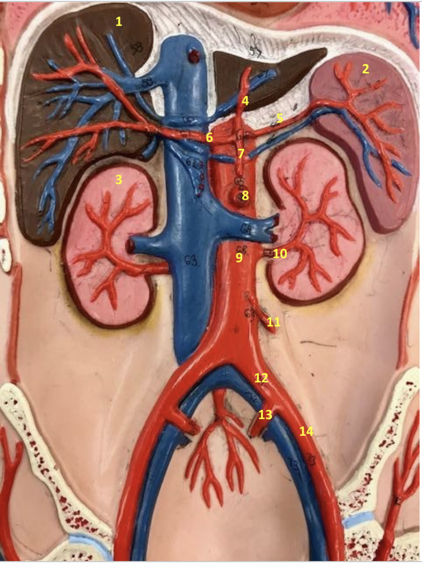
Identify each. Function of 10, 13?
Abdomen
1. Liver
2. Spleen
3. Right kidney
4. Left gastric artery
5. Splenic artery
6. Common hepatic artery
7. Celiac artery
8. Superior mesenteric artery
9. Abdominal aorta
10. Left renal artery
Supplies blood to the kidney, so does the right renal artery
11. Inferior mesenteric artery
12. Left common iliac artery
13. Left internal iliac artery
Supply blood to the pelvic walls and pelvic organs
14. Left external iliac artery
What are the 3 main branches extending from the abdominal aorta? and function?
Celiac trunk/artery #7: Splits into 3 arteries, which supply the spleen (splenic artery #5), stomach (left gastric artery #4), and liver (common hepatic artery #6) with blood.
Superior mesenteric artery #8: Supplies blood to majority of small intestine and large intestine
Inferior mesenteric artery #11: Supplies blood to distal large intestine

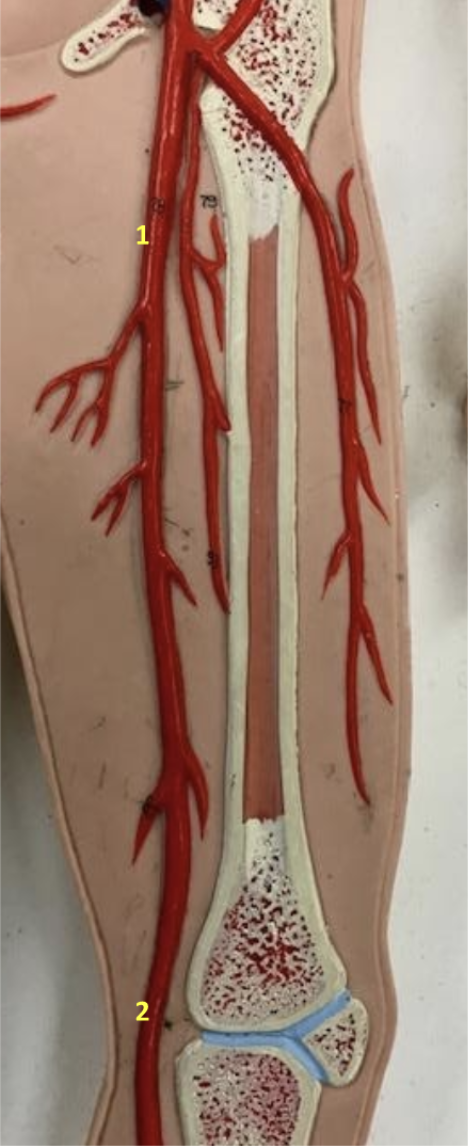
Identify (Scroll)
Thigh
1. Left femoral artery
2. Left popliteal artery


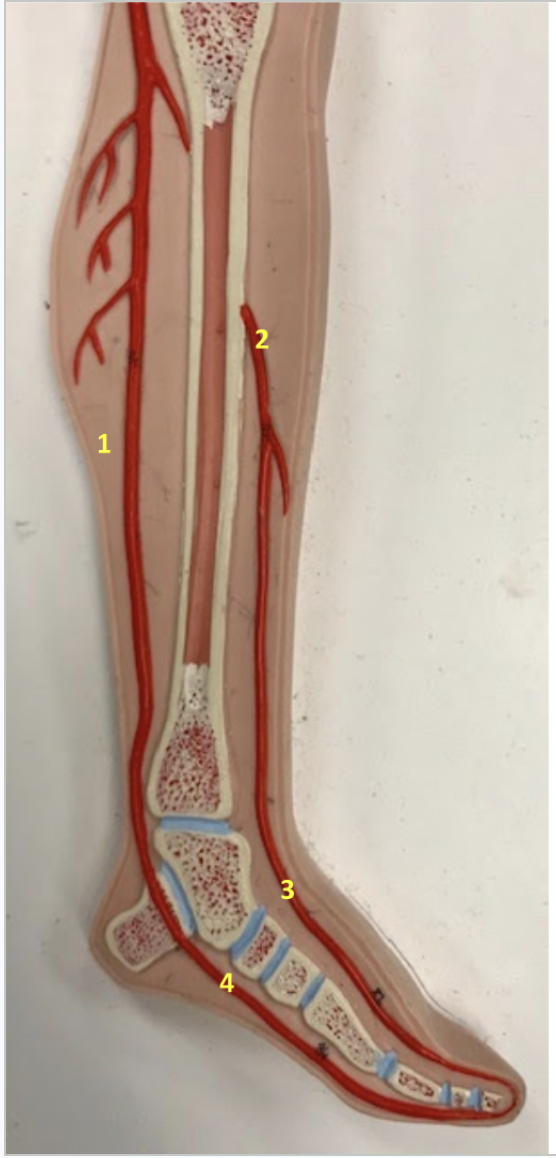
Identify (Scroll). Function of 3,4
Leg/Foot
1. Left posterior tibial artery
2. Left anterior tibial artery
3. Left dorsalis pedis artery: Supplies blood to the ankle and dorsal foot
4. Left medial plantar arch: Supplies blood to the plantar foot.
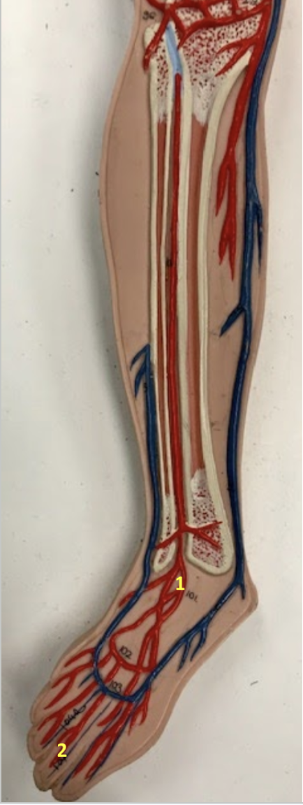
Identify (Scroll). Function of both
Leg/Foot
1. Right dorsalis pedis artery: Supplies blood to the ankle and dorsal foot
2. Right digital artery: Supplies blood to the toes


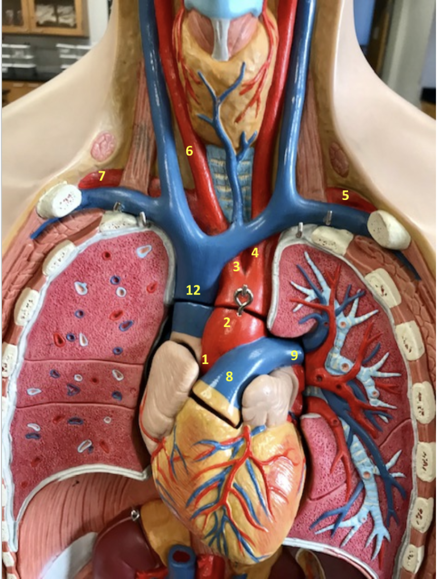
Identify each part
Thorax/Neck
1. Aortic arch
2. Descending thoracic aorta
3. Brachiocephalic artery
4. Right common carotid artery
5. Left common carotid artery
6. Right pulmonary artery
7. Left pulmonary artery

Identify each part (Scroll). Function of 3. It enters into what part of the body?
Thorax/Neck
1.Right common carotid artery
2.Left common carotid artery
3.Left internal carotid artery: Supplies brain with blood. enters directly into skull
4. Aortic arch

Identify
Abdomen/Pelvis
1. Abdominal aorta
2. Splenic artery
3. Superior mesenteric artery
4. Left common iliac artery
5. Left internal iliac artery
6. Left external iliac artery

Identify
Thorax/Neck
1. Ascending aorta
2. Aortic arch
3. Brachiocephalic artery
4. Left common carotid artery
5. Left subclavian artery
6. Right common carotid artery
7. Right subclavian artery
8. Pulmonary trunk
9. Left pulmonary artery
Identify each (Scroll)
Thorax/Neck
1. Left common carotid artery
2. Left external carotid artery
3. Left internal carotid artery
4. Right common carotid artery
5. Left subclavian artery
6. Right subclavian artery


Identify each
Abdomen/Pelvis
1. Celiac artery
2. Superior mesenteric artery
3. Right renal artery
4. Abdominal aorta
5. Right gonadal artery
6. Inferior mesenteric artery
7. Right common iliac artery
8. Left internal iliac artery
9.Left external iliac artery
10.Left femoral artery
11. Splenic artery
Identify each
Thorax/Neck
1. Ascending aorta
2. Aortic arch
3.Right common carotid artery
4.Left common carotid artery
5. Left subclavian artery
6.Pulmonary trunk
7. Right pulmonary artery


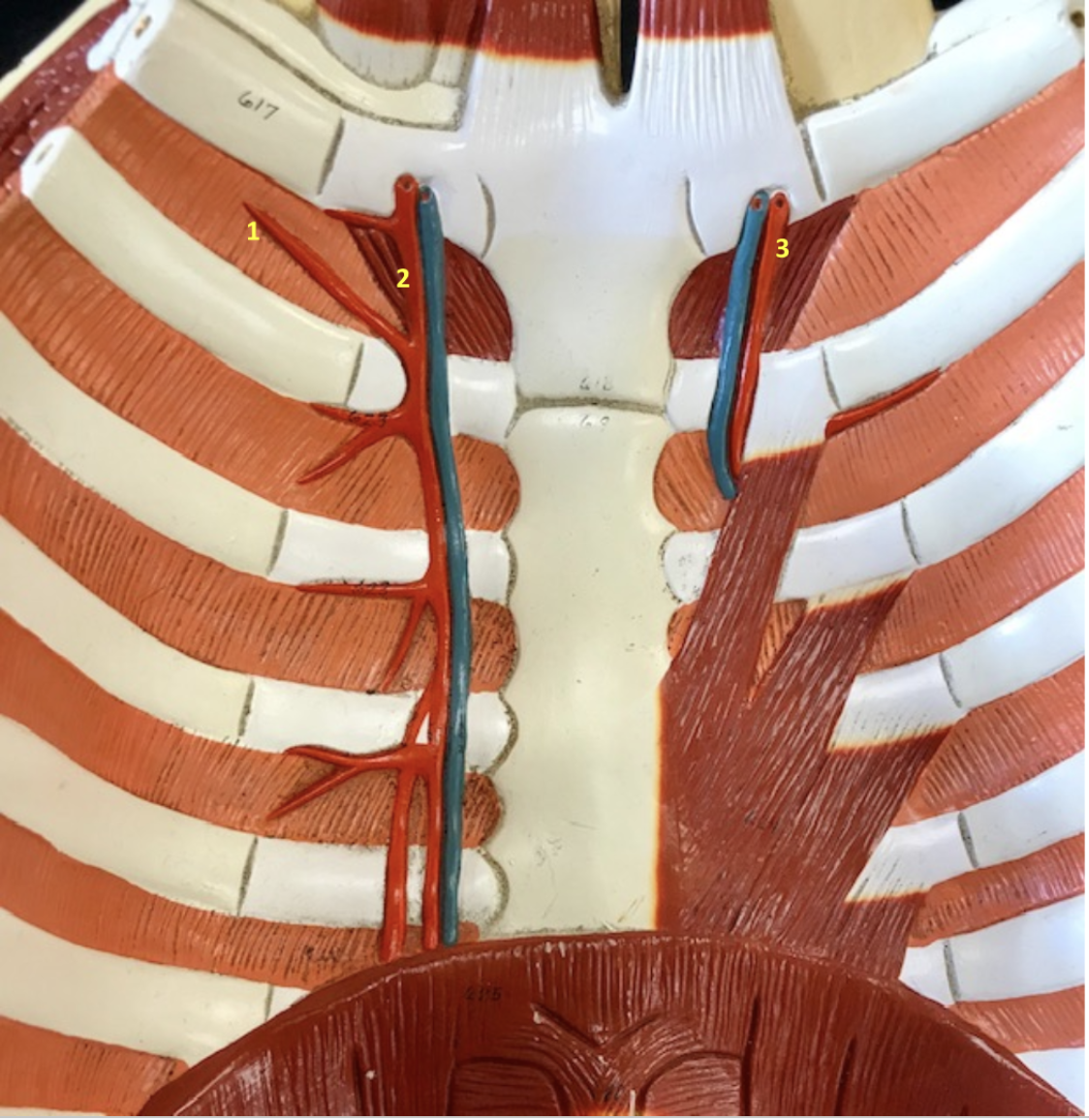
Identify
Thoracic Wall - Internal Surface
1. Left anterior intercostal artery
2. Left internal thoracic artery
3. Right internal thoracic artery
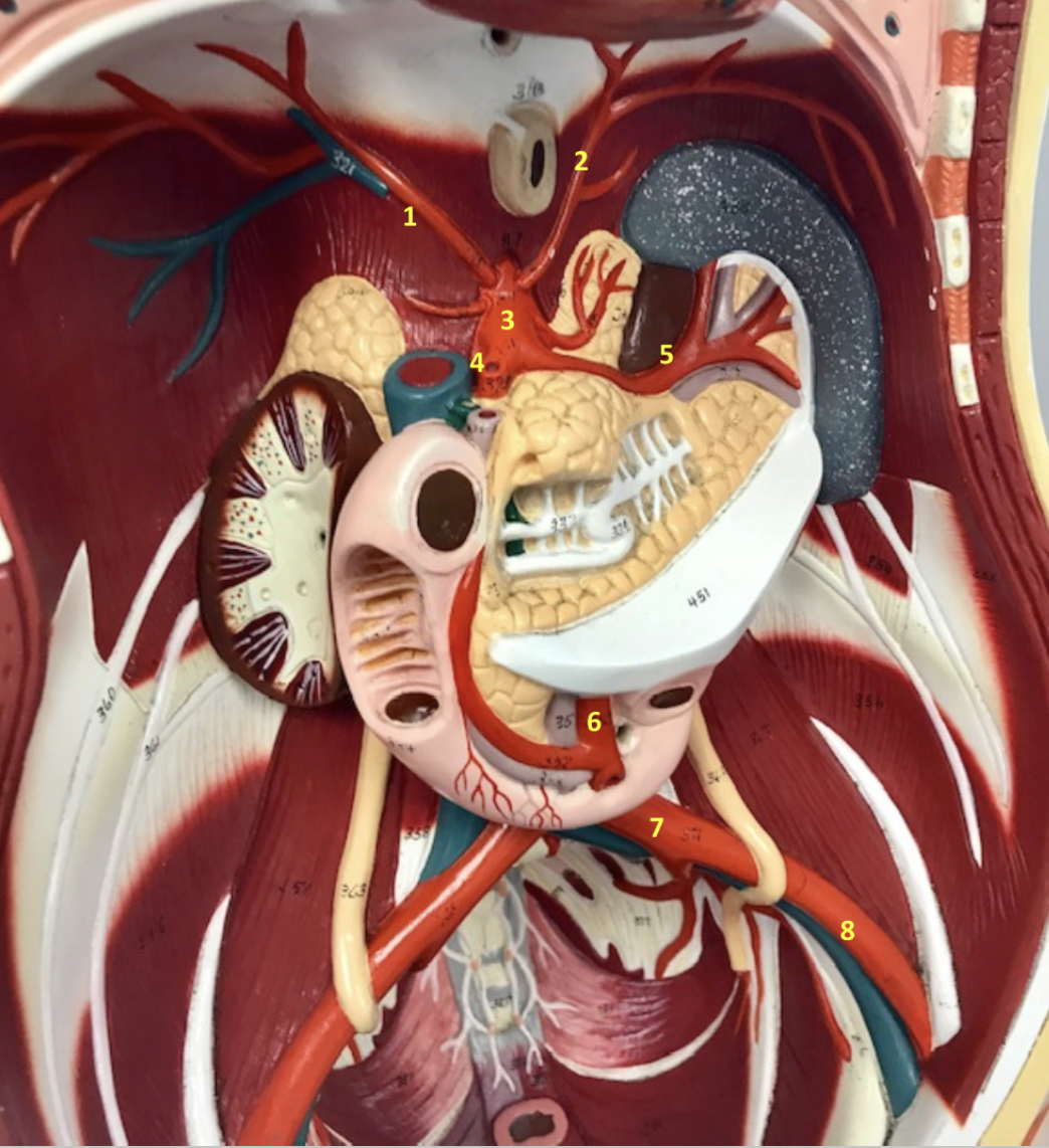
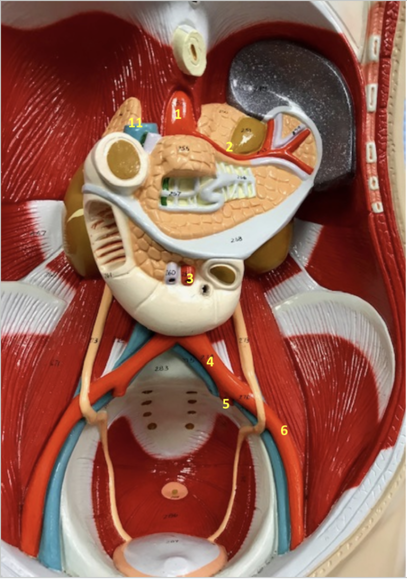
Identify each. Function of 1 &2?
Abdomen
1.Right inferior phrenic artery: Supply diaphragm with blood
2. Left inferior phrenic artery: Supply diaphragm with blood
3. Abdominal aorta
4. Celiac artery
5. Splenic artery
6.Superior mesenteric artery
7.Left common iliac artery
8. Left external iliac artery


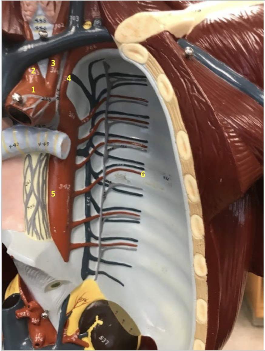
Identify each
Thorax
1.Aortic arch
2. Brachiocephalic artery
3. Left common carotid artery
4. Left subclavian artery
5. Descending thoracic aorta
6. Posterior intercostal artery
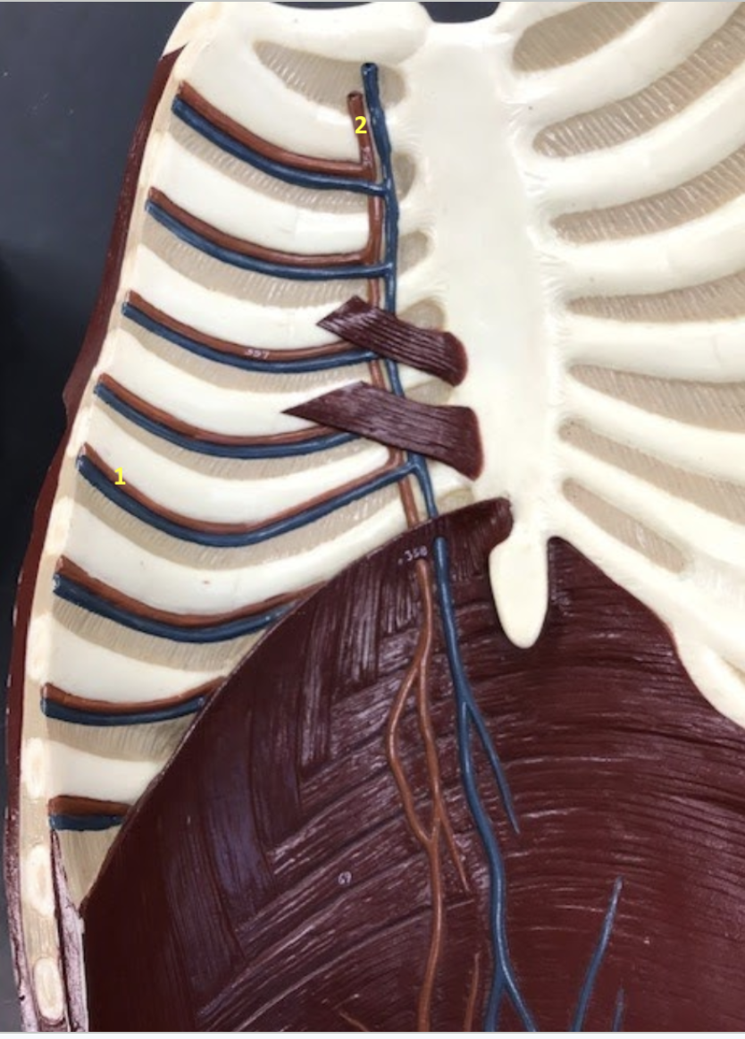
Identify each
Thoracic Wall - Internal surface
1. Anterior intercostal artery
2. Internal thoracic artery

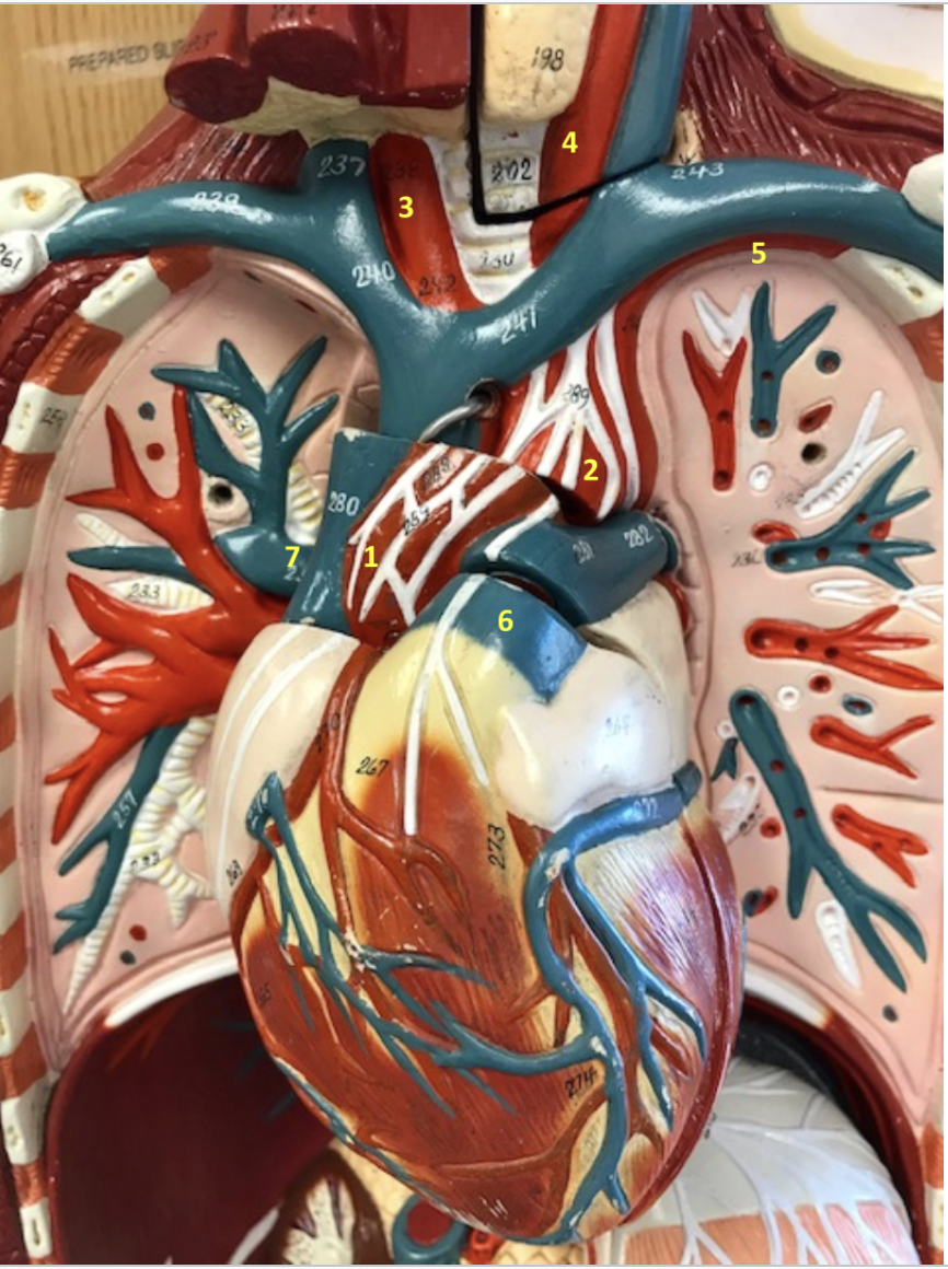
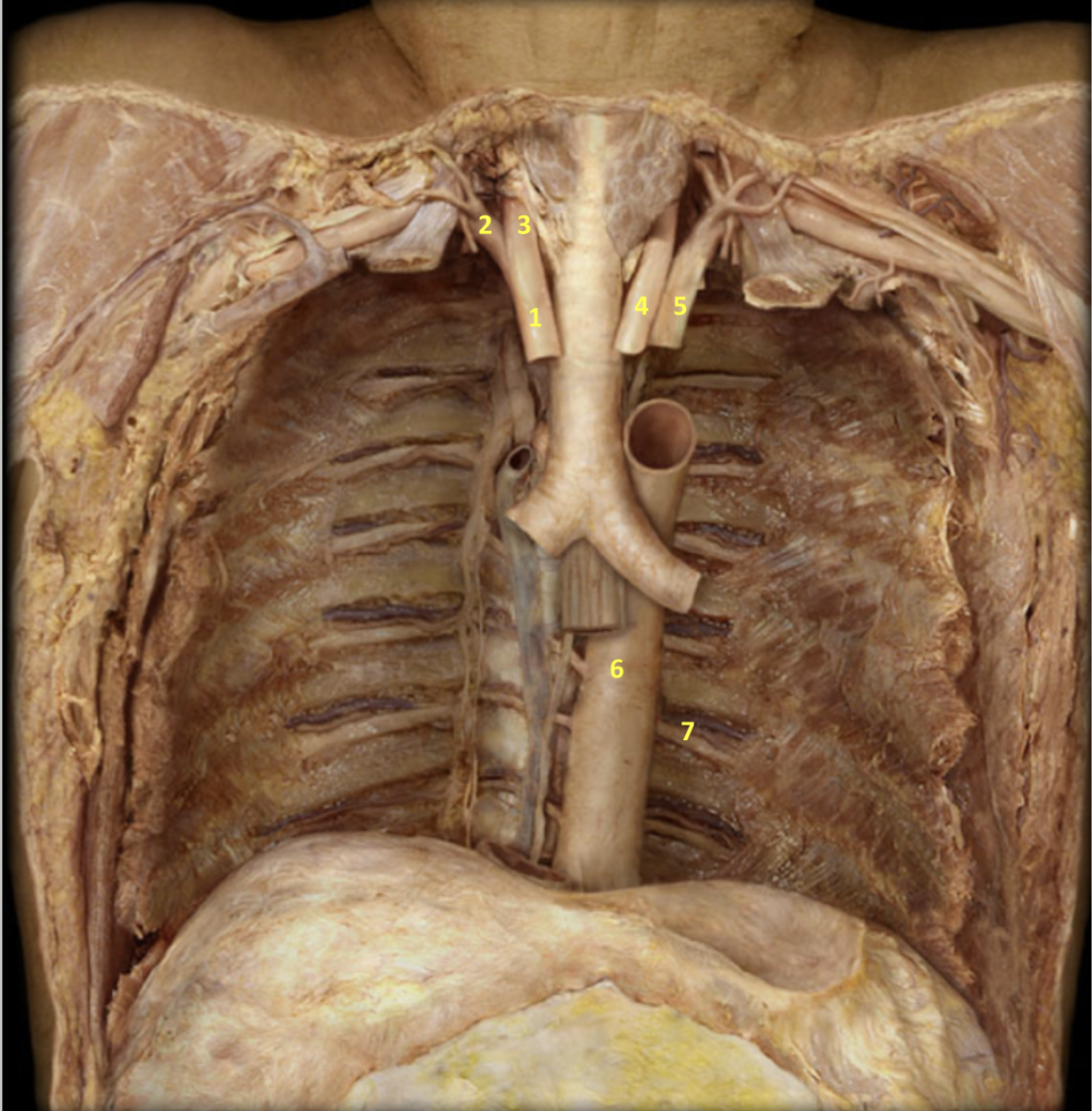
Identify
Thorax - Deep Dissection
1. Ascending aorta
2. Aortic arch
3. Brachiocephalic artery
4. Left common carotid artery
5. Left subclavian artery
6. Pulmonary trunk
7. Left pulmonary artery


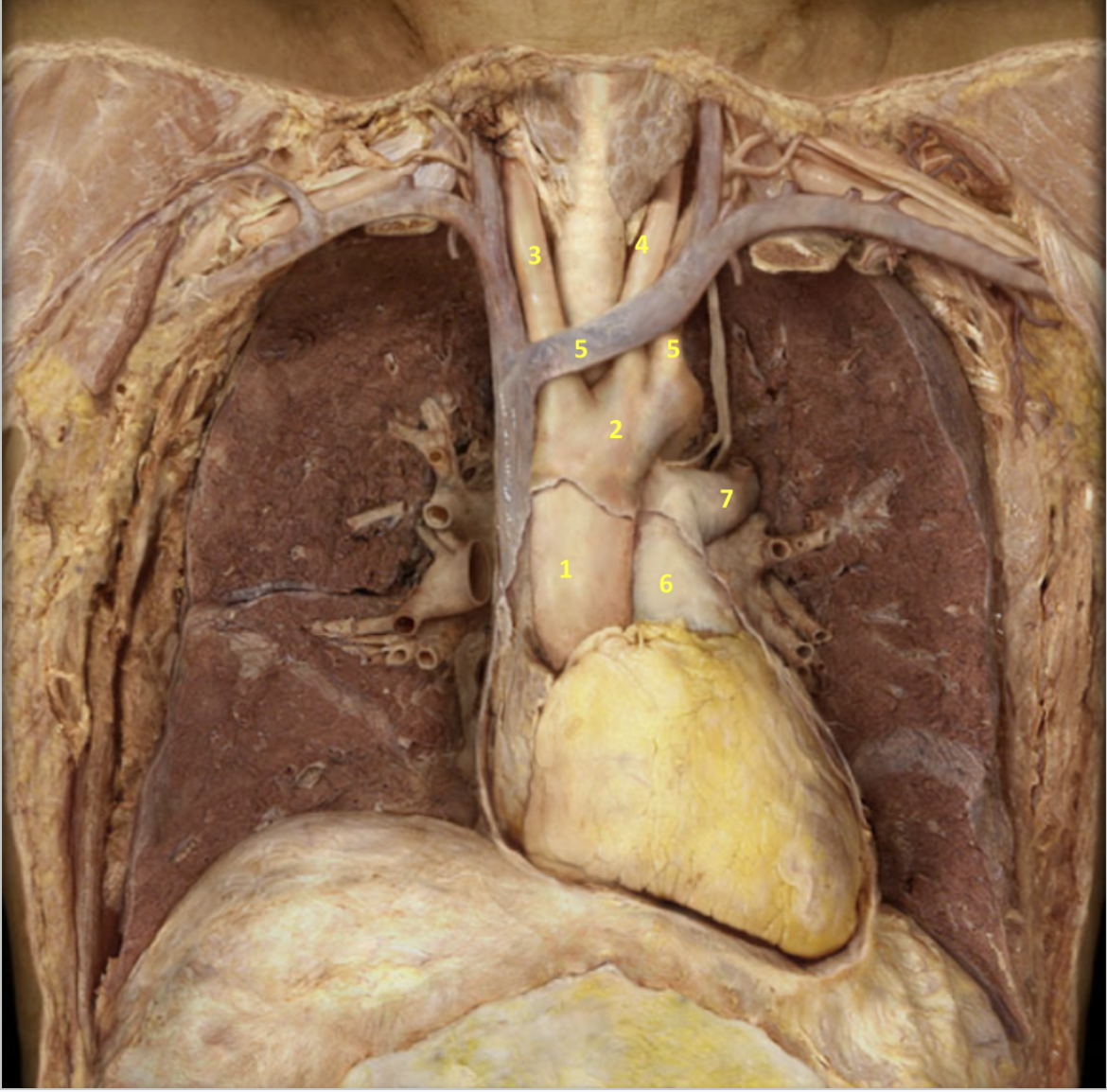
Identify
Thorax - Deeper Dissection
1. Brachiocephalic artery
2. Right subclavian artery
3. Right common carotid artery
4. Left common carotid artery
5. Left subclavian artery
6. Descending thoracic aorta
7. Posterior intercostal artery
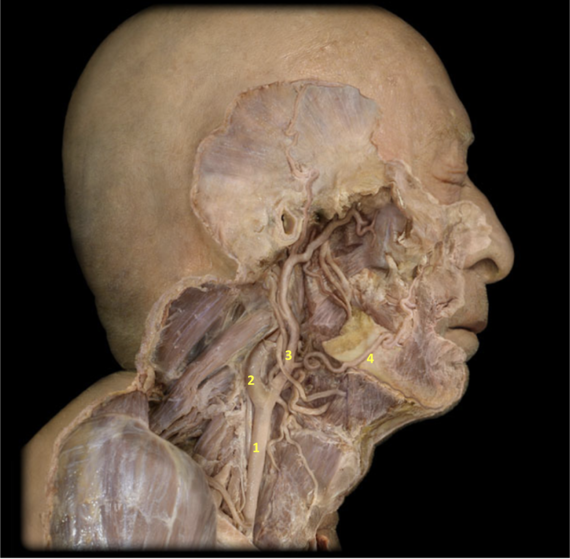
Identify each
Lateral Neck and Face - Deeper Dissection
1. Right common carotid artery
2. Right internal carotid artery
3. Right external carotid artery
4. Right facial artery


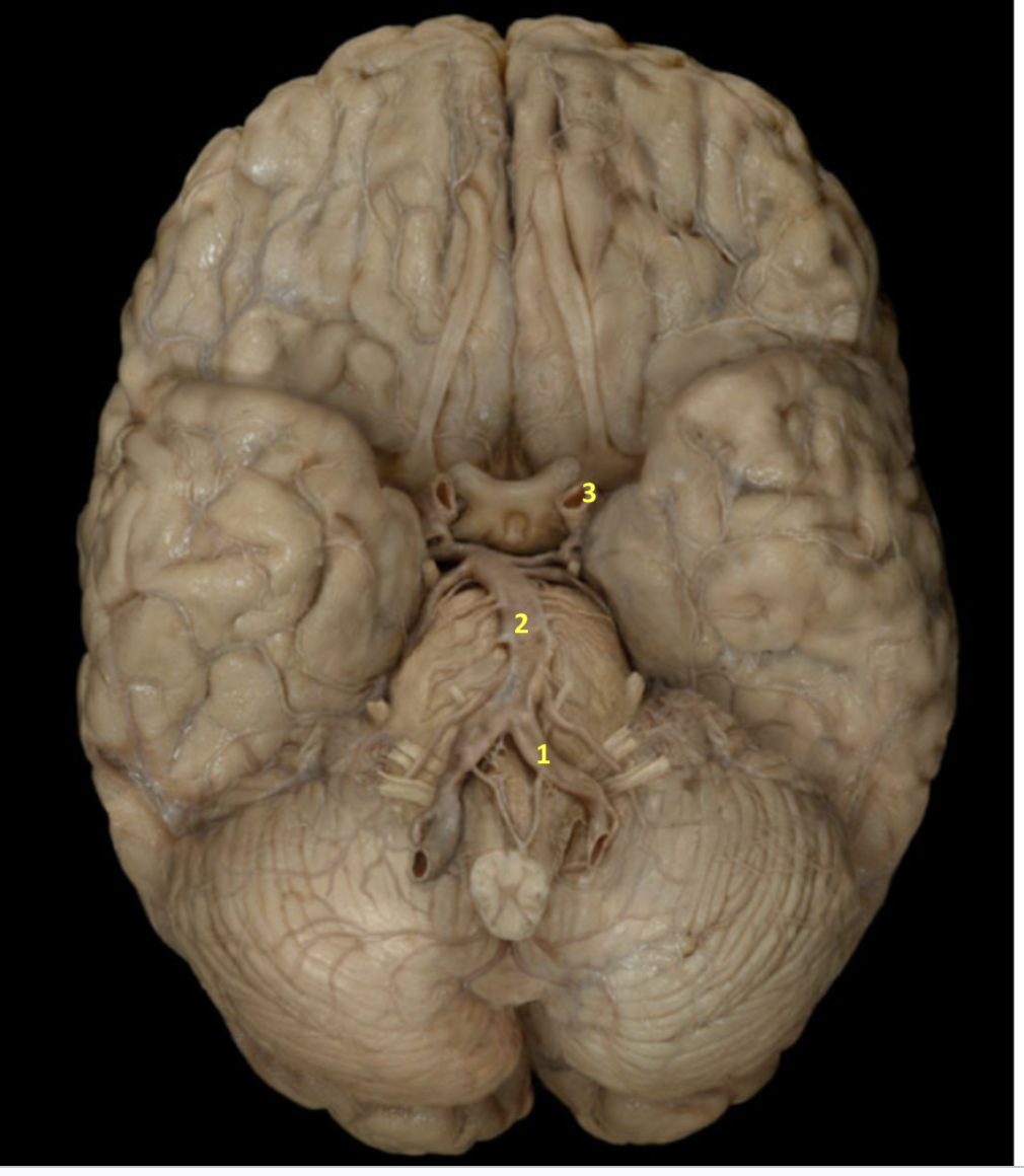
Identify
Inferior View of the Brain
1. Left vertebral artery
2. Basilar artery
3. Left internal carotid artery

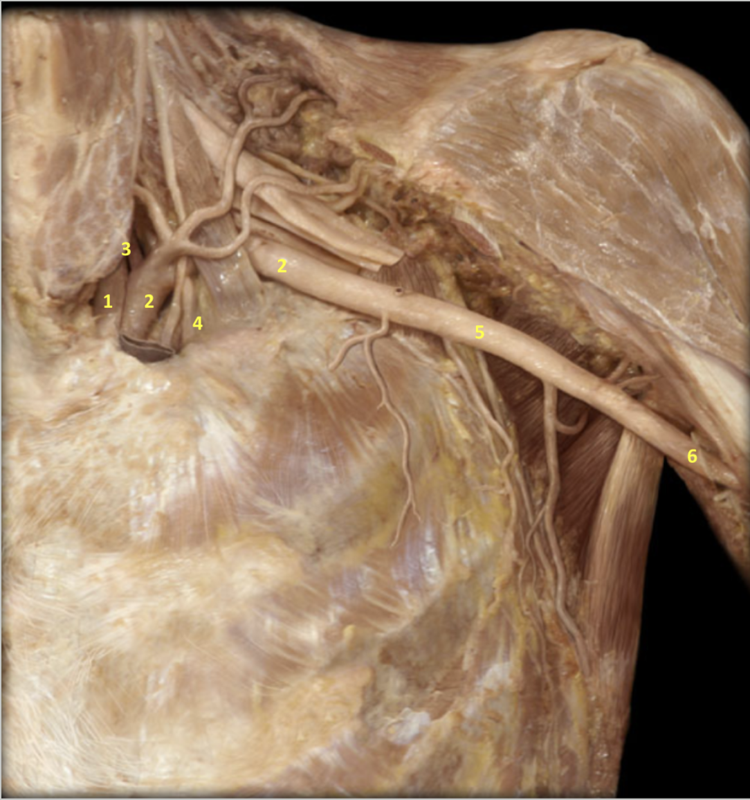
This is a deep shoulder dissection. Identify each
Shoulder - Deeper Dissection
1. Left common carotid artery
2. Left subclavian artery
3. Left vertebral artery
4. Left internal thoracic artery
5. Left axillary artery
6. Left brachial artery
Identify
Hand - Deep Dissection
1. Left radial artery
2. Left ulnar artery
3. Left palmar arch
4. Left metacarpal artery
5. Left digital artery


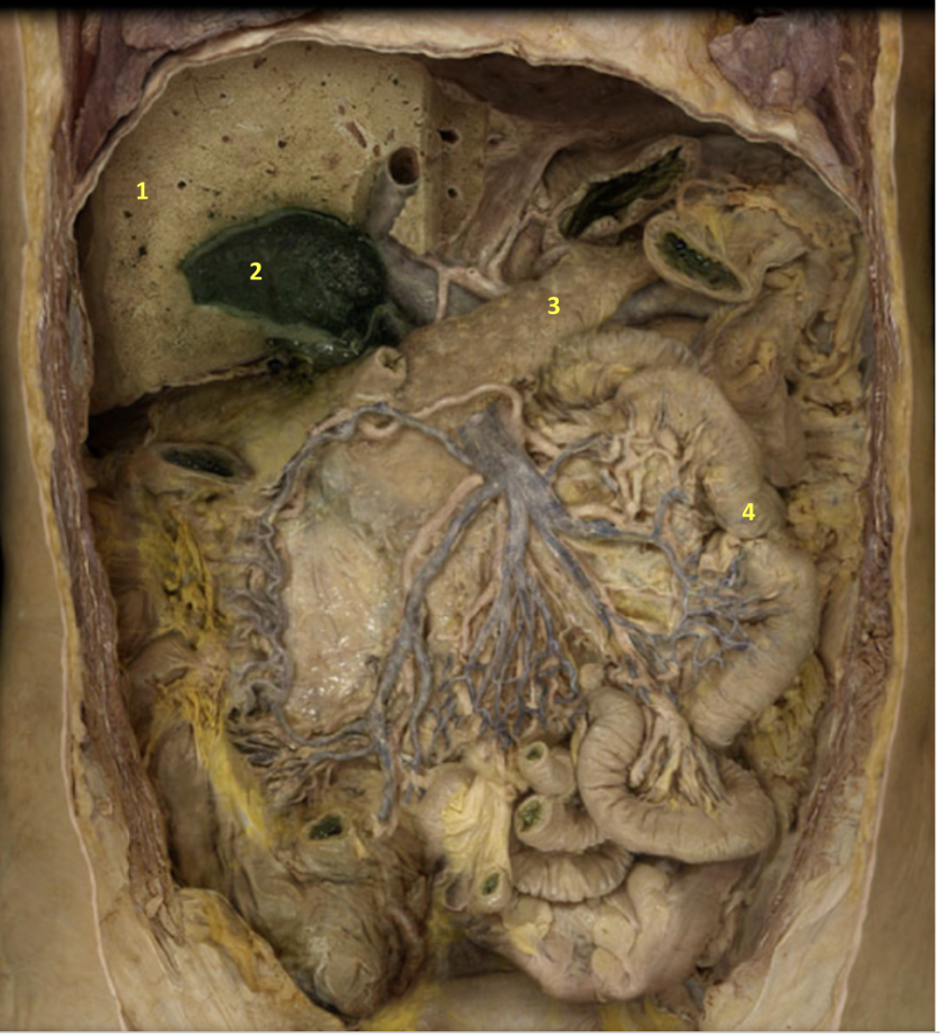
Identify each
Abdomen - Deep Dissection
1. Liver
2. Gallbladder
3. Pancreas
4. Small intestine

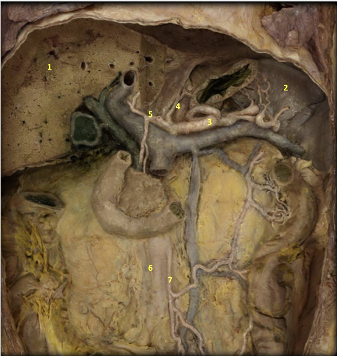
Identify
1. Liver
2. Spleen
3. Splenic artery
4. Left gastric artery
5. Common hepatic artery
6. Abdominal aorta
7. Inferior mesenteric artery

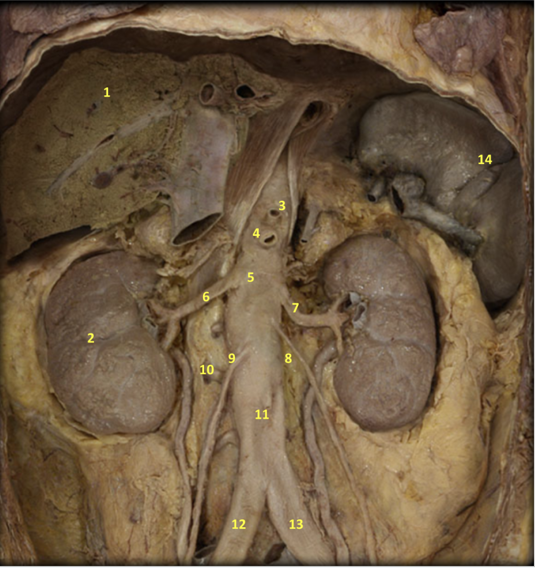
Identify
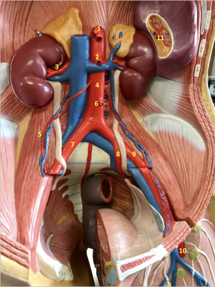
Abdomen - Even Even Even Deeper Dissection
1. Liver
2. Kidney
3. Celiac artery
4. Superior mesenteric artery
5. Abdominal aorta
6. Right renal artery
7. Left renal artery
8. Left gonadal artery
9. Right gonadal artery
10. Left lumbar artery
11. Inferior mesenteric artery
12. Right common iliac artery
13. Left common iliac artery
14. spleen
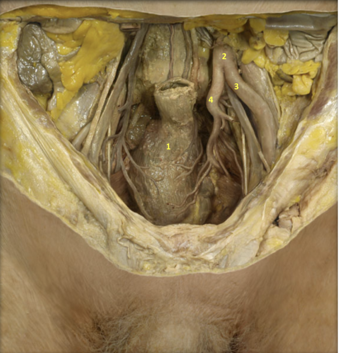
Identify
Male Pelvis - Deep Dissection
1. Rectum
2. Left common iliac artery
3. Left external iliac artery
4. Left internal iliac artery


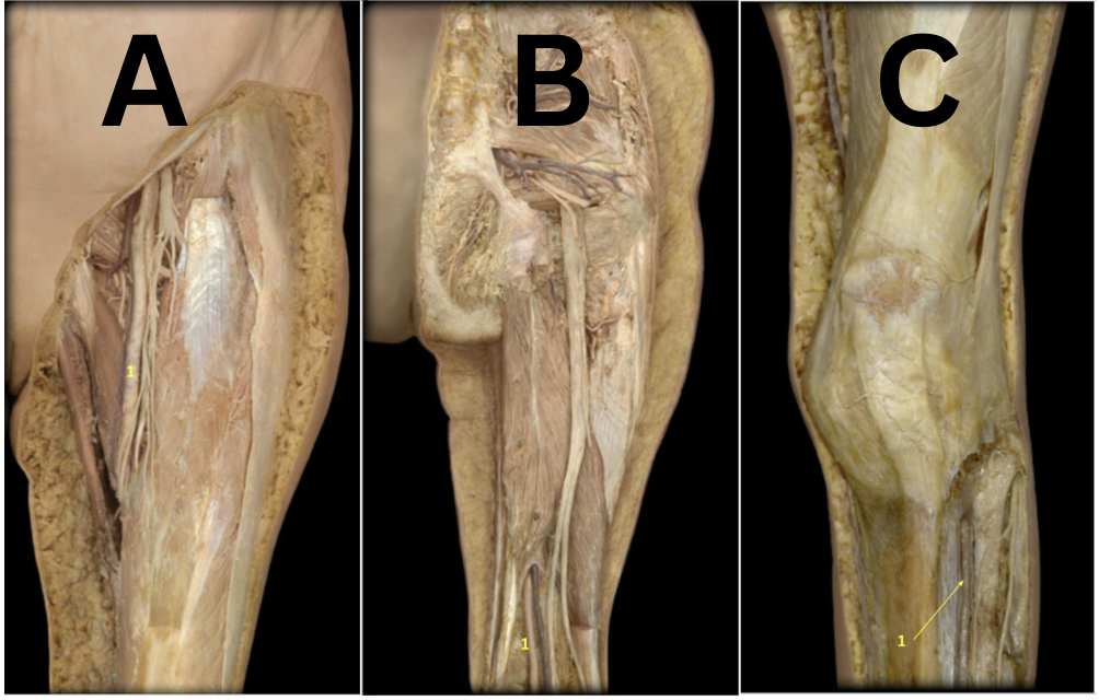
Identify #1 in each picture
A: Left femoral artery
B: Left popliteal artery
C: Left anterior tibial artery
Trace the pathway of blood from the left atrium to the right eye
Left atrium > bicupsid valve > left ventricle > aortic semilunar valve > ascending aorta > aortic arch > brachiocephalic artery > right common carotid artery > right ophthalmic artery
Trace the pathway of blood from the left atrium to the top of the right foot
Left atrium > bicupsid valve > left ventricle > aortic semilunar valve > ascending aorta > aortic arch > descending thoracic aorta > descending abdominal aorta > right common iliac artery > right external iliac artery > right femoral artery > right popliteal artery > right anterior tibial artery > right dorsalis pedis artery