Neuropsychology Test 2
1/533
Earn XP
Description and Tags
Chapters 8,9,13,14 (need to add 15 and 16)
Name | Mastery | Learn | Test | Matching | Spaced | Call with Kai |
|---|
No analytics yet
Send a link to your students to track their progress
534 Terms
Sensation and perception are related
But they are also distinct
Sensation
Changes in the sensory system in response to the environment
perception
interpretation of the changes to the environment
Sensory receptors are sensitive to a particular stimulation in the environment
sensory receptors only respond to that specific part of the environment
In the case of vision, people who are color deficient have a different experience than
people who are not color blind
In the auditory system, humans can detect sound between
20 and 20,000 Hz, whereas some animals can hear up to 120,000 Hz
Most common form of color deficiency
red-green deficient
The long wavelength cone is encoded by a gene on the X chromosome, so a mutation in this gene is more likely to impact
males than females, making this red-green deficiency more common in men than in women
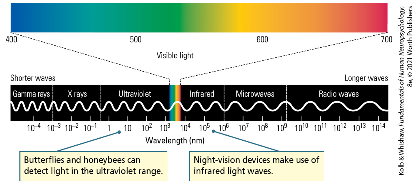
Electromagnetic Spectrum
The human eye sees only a slice of electromagnetic energy from about 400 nanometers (violet) to 700 nanometers (red).
Butterflies and honeybees can detect light in the
ultraviolet range
Night vision devices make use of
infrared light waves
A nanometer (nm) is
one-billionth of a meter
Receptors in each system are specialized to
convert a particular stimulus into electrochemical signals in the nervous system
Vision uses photoreceptors to
convert light
Audition uses vibration frequency to
convert sound
Somatosensory systems use mechanical pressure to
convert touch
Taste and olfaction use molecular shape to
activate specific receptors
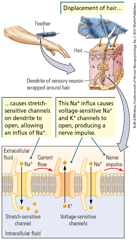
Stretch-Sensitive Channels
Tactile stimulation of a hair activates stretch-sensitive Na+ channels, which then activate voltage-sensitive channels to produce an action potential.
Stretch-Sensitive Channels Process
1) displacement of hair 2) causes stretch sensitive channels on dendrite to open, allowing an influx of Na+ 3) Na+ influx causes voltage-sensitive Na+ and K+ channels to open, producing a nerve impluse
Receptive Fields Locate
Sensory Events
Every receptor has a
receptive fieldre
The receptive field defines
what part of the external world the receptor responds to
In vision, a particular photoreceptor receives information from
a particular part of the image in front of you
For somatosensation, different receptors convey information about
touches to a specific part of the body
Receptive fields for taste, smell, and audition are less clear.
For the auditory system, those are likely the specific frequencies of sounds. For taste and smell, the receptive fields may be the physical properties (shapes) of the molecules the receptors respond to.
Receptors Identify
Constancy and Change
Some receptors remain stimulated for as long as the stimulus is present
others respond only when there is a change in the stimulus
Rapidly adapting receptors detect the change, and then adapt to
the stimulation by returning to their baseline firing rate
Slowly adapting receptors respond to the stimulus and keep responding
for as long as the stimulus is present
Receptors Distinguish
self from other
Exteroceptive receptors respond to
events outside of your body
Interoceptive receptors respond to
stimuli inside our body
Information from both types of receptors (exteroceptive and interoceptive)
is combined to create our understanding of the world
Imagine looking at the scene while you are walking along. Your exteroceptors in your eyes see the world around you, but that image on your retina is constantly changing as you move
The interoceptors account for your movements and essentially remove it from what you see in the world, so the world appears to remain stable, even though the stimulation to the photoreceptors in the eye is constantly changing
Receptor density determines
Sensitivity
The sensitivity of the system is related to
how closely together the receptors are
If the receptors are closer together, you can differentiate between
a stimulus that activates one receptor and a stimulus that activates a nearby receptor
If receptors are wide apart and have large receptive fields
stimulation anywhere in that receptive field will be perceived in the same way
Two-point sensitivity measures
how closely two stimuli can be placed and still identified as different
The tight clustering of cone cells with small receptive fields in the fovea give us
detailed color vision at the center of our visual field. The rod cells found in the rest of the retina provide less detail
Neural relays determine
motor responses
Sensory information is relayed to the brain through how many steps?
3-4 steps
The signal can be modified or behavior can be evoked at
any of the relay points
Reflexive movements away from painful stimuli are coordinated at the level of?
The spinal cord
Understanding what and where the pain is requires
requires involvement of the brainstem and cortex
Some auditory and visual processing take place in the brainstem
whereas recognizing patterns requires the cortex
Descending signals from the cortex can alter how likely it is
for sensory information to be passed on at each relay point
When engaged in an activity, such as a sporting event, you might not notice an injury.
It may be after the play or after the game that you notice a bruise or a sprain.
Central Organization of the Sensory Systems
All sensory information, regardless of the system, is encoded as action potentials
The nervous system and brain segregate sensory information into
different pathways
Different populations of neurons might encode
qualitative difference such as red vs. green
The frequency of action potentials might encode
quantitative differences such as a louder or quieter sound
Different types of sensory information are directed to
different parts of the cortex
People with synesthesia seem to have increased connections between different sensory areas
the number 1 may always be tinged blue or the number 2 may be orange. A chef with synesthesia reports that tastes are associated with shapes, so he describes one dish as “spikey” or another dish as “round”.
Visual Subsystems
Each pathway from eye to brain, numbered 1 through 7 here, traces a sensory subsystem that culminates in a neural visual center.
Frontal eye fields
eye movements
suprachiasmatic nucleus
daily rhythms (sleep, feeding, etc)
Pretectum
changes in pupil size in response to light
pineal gland
long-term circadian rhythms
superior colliculus
head orienting
accessory optic nucleus
eye movement to compensate for head movement
visual cortex
pattern perception, depth perception, color vision
visual subsystems 1-7
frontal eye fields, suprachiasmatic nucleus, pretectum, pineal gland, superior colliculus, accessory optic nucleus, visual cortex
Sensory systems represent
the external world within the brain
For each sense, there are multiple
topocographic maps
Greater number of representations is associated with
greater behavioral complexity
The primary sensory area initially processes the information
Secondary (and higher) areas perform more elaborate processing or focus on specific aspects of the stimulus
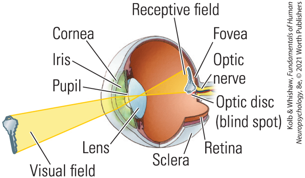
Anatomy of the Eye
Light reflected from an object is focused by the lens on the fovea of the retina
Light enters the eye through X and X and is focused on these X at the back of the eye
Light enters the eye through the cornea and lens and is focused on the photoreceptors at the back of the eye
Passing through the lens, the resulting
image is
upside down and
left-right reversed on the retina
On most of the retina, light must pass through
several layers of translucent cells to reach the photoreceptors
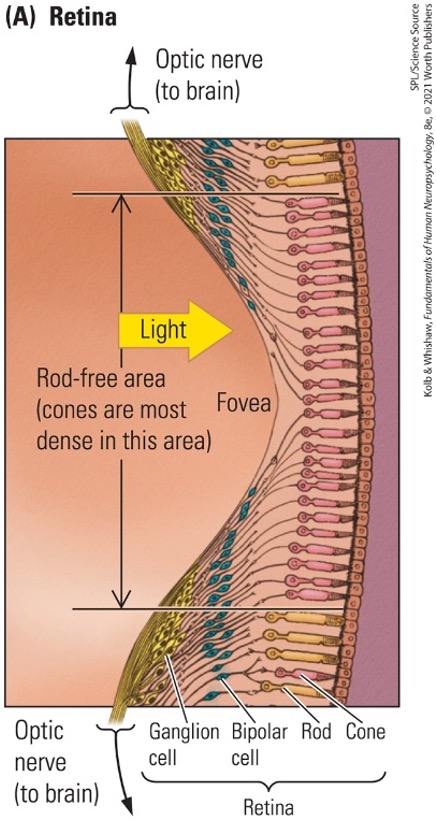
In the fovea, these overlying cells are shifted aside to
allow light more direct access without as much scattering
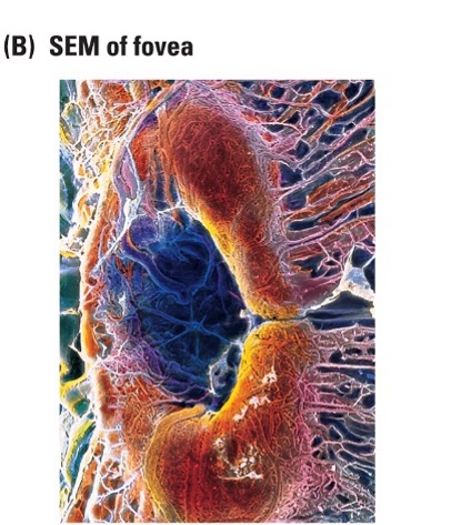
The fovea
(A) The fovea of the retina features densely packed cones. The relay cells to which the receptors connect are pushed to one side to reduce the interference to light making its way to the cones.
The retina contains two types of photoreceptors
rods and cones
Rods
Sensitive to broad spectrum of light
Sensitive to low levels of light
Found throughout the retina
Cones
Short, middle, and long wavelength cones are sensitive to specific wavelengths of light, which we perceive as color
Sensitive to higher intensities of light
Found in the retina
All photoreceptors synapse on
bipolar cells and ultimately onto retinal ganglion cells, which form the optic nerve
In bright light, looking directly at an object will provide
the highest-resolution image
Under dim light conditions, such as when looking at the stars, looking directly at an object can make it
disappear because the cones are not as sensitive in low-light conditions. To see dim stars, you are better looking off to the side of the star and seeing the star of interest with your peripheral (rod-based) vision.
The axons of the retinal ganglion cells come together to form
form the optic nerve, which exits the eye through the “blind spot”
the optic nerves from the two eyes combine
At the optic chiasm
Information from the right half of each retina, which perceives the left side of the visual field, projects to
the right hemisphere of the brain
Information from the left half of each retina, which perceives the right visual field, projects to
the left hemisphere of the brain
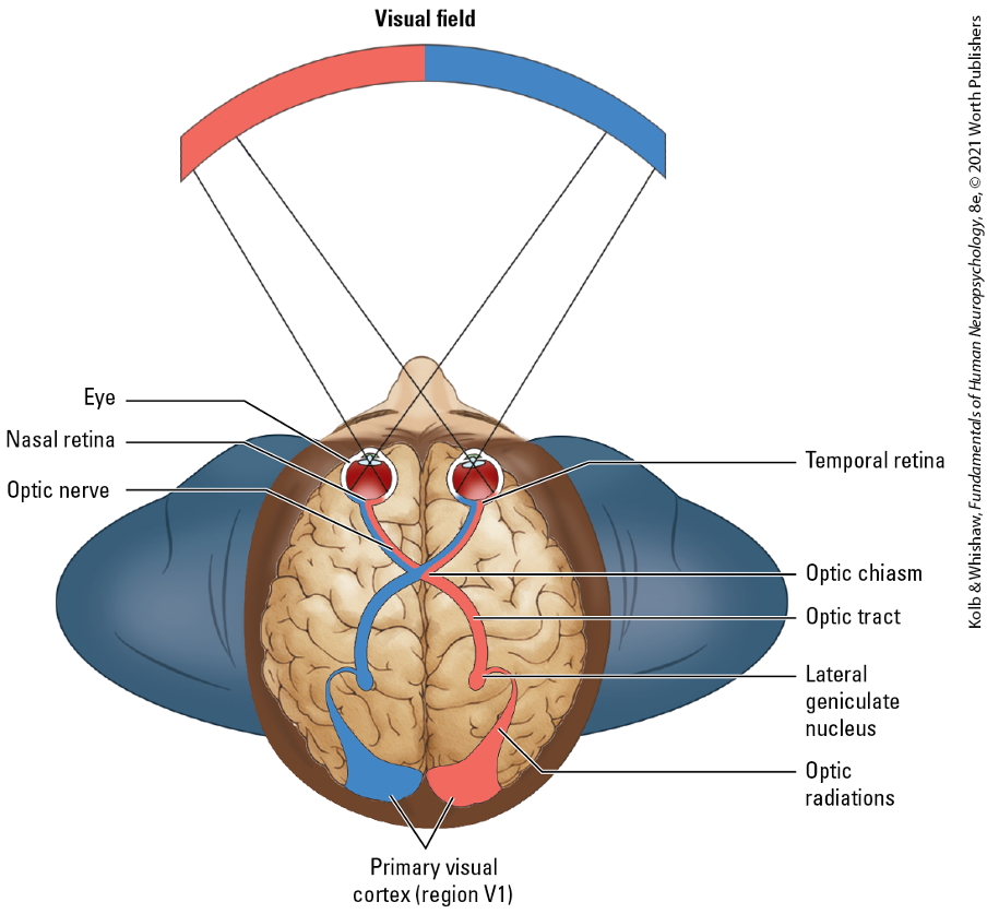
Crossing the Optic Chiasm
Dorsal view of the visual pathways from each eye to region V1 in each occipital hemisphere. Information from the right (blue) side of the visual field falls on the left halves of the retinas and ends up in the left hemisphere. Information from the left (red) side of the visual field falls on the right halves of the retinas and travels to the right hemisphere. In animals such as humans, with eyes at the front of the head, about half of the optic fibers cross at the optic chiasm.
The main visual pathway leads from the eye to
the lateral geniculate nucleus (LGN) of the thalamus to the primary visual cortex
Main visual pathway is also called the
striate cortex
The LGN is divided into six layers, and the topographic mapping of the information on the retina is
maintained in the LGN
Ipsilateral eye projects to
layers 2, 3, and 5
Contralateral eye projects to
layers 1, 4, and 6
LGN projects to layer IV of the primary visual cortex, with input from
the left eye alternating with input from the right eye, resulting in a striped appearance
The geniculostriate pathway is the name for the primary visual pathway
Damage to this pathway results in impairments in the ability to recognize form, color, or motion
Geniculostriate Pathway
Next pathway relays through the lateral geniculate nucleus (LGN) of the thalamus
In the Geniculostriate Pathway, LGN projects to
the primary visual cortex or striate cortex or V1
V1contains a retinoptic map of the visual field
Map is upside down, inverted, and reversed
Geniculostriate Pathway takes part in
pattern recognition and conscious visual functions
Tectopulvinar Pathway
Optic nerve leaves the eye and projects to the superior colliculus (optic tectum)
In the Tectopulvinar Pathway, the optic nerve projection reaches
visual areas in the temporal and parietal lobes through relays in the lateral posterior-pulvinar complex of the thalamus. It also detects stimuli and helps orient us to stimuli.
This pathway is sufficient to locate objects in space, but does not provide as much detailed information about the object viewed
Tectopulvinar Pathway
Sound is made up of
pressure variations in the air
audition can be used for
sound localization, echolocation, spoken communication, and music