module 15: sensory pathways II: hearing, chemical senses, touch
1/23
There's no tags or description
Looks like no tags are added yet.
Name | Mastery | Learn | Test | Matching | Spaced | Call with Kai |
|---|
No analytics yet
Send a link to your students to track their progress
24 Terms
Olfactory Sensory Neurons
Neurons in the nasal cavity that detect odorants and transmit signals to the olfactory bulb.
Olfactory Bulb
Brain structure that processes olfactory information from sensory neurons.
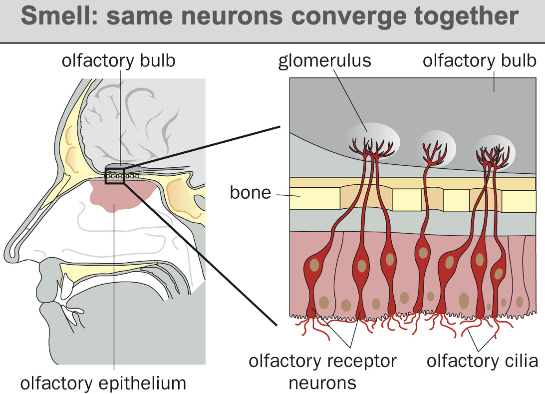
process of smell
odorants pass through the nostrils and bind to olfactory receptors on the dendrites of olfactory sensory neurones
OSNs located in olfactory epithelium
olfactory neurones converge onto glomeruli in the olfactory bulb, which is part of the brain
signals translated as scent
GPCRs in Olfaction (humans)
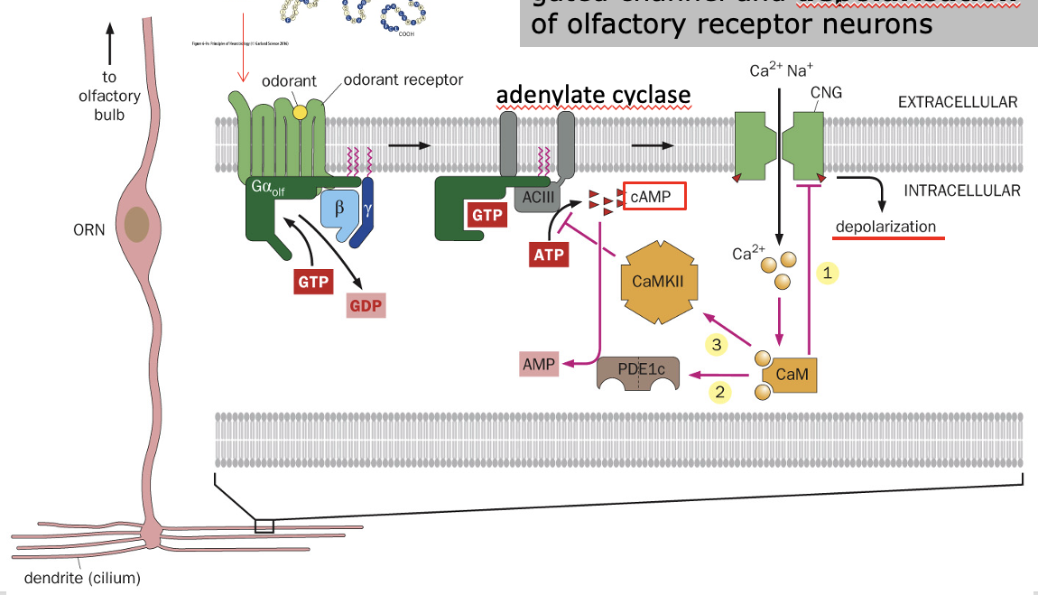
Olfactory receptors = G-protein-coupled receptors - initiate signal transduction upon odorant binding.
odorant binds to receptor on olfactory neurone, activating the olfactory GPCR
alpha subunit of GPCR exchanges GDP for GTP
Activates adenylate cyclase (ACIII)- converts ATP → cAMP
cAMP opens cyclic nucleotide-gated cation channels
Channels allow Ca²⁺ and Na⁺ entry
this depolarization of olfactory receptor neuron (ORN) → Action Potential
Insect Olfactory System
• Not GPCR-based
• Uses ion channels that open directly upon odorant binding
Olfactory Signal Transduction- Comparison to Vision
• Similarity to Vision:
• Both use GPCRs
• Vision: Rhodopsin GPCR - Ligand = Light
• Olfaction: Olfactory GPCRs - Ligand = Odorant
• differences to Vision
• Olfaction: Depolarization of ORN
• Vision: Light causes hyperpolarization of rods/cones
olfaction and vision circuit comparison
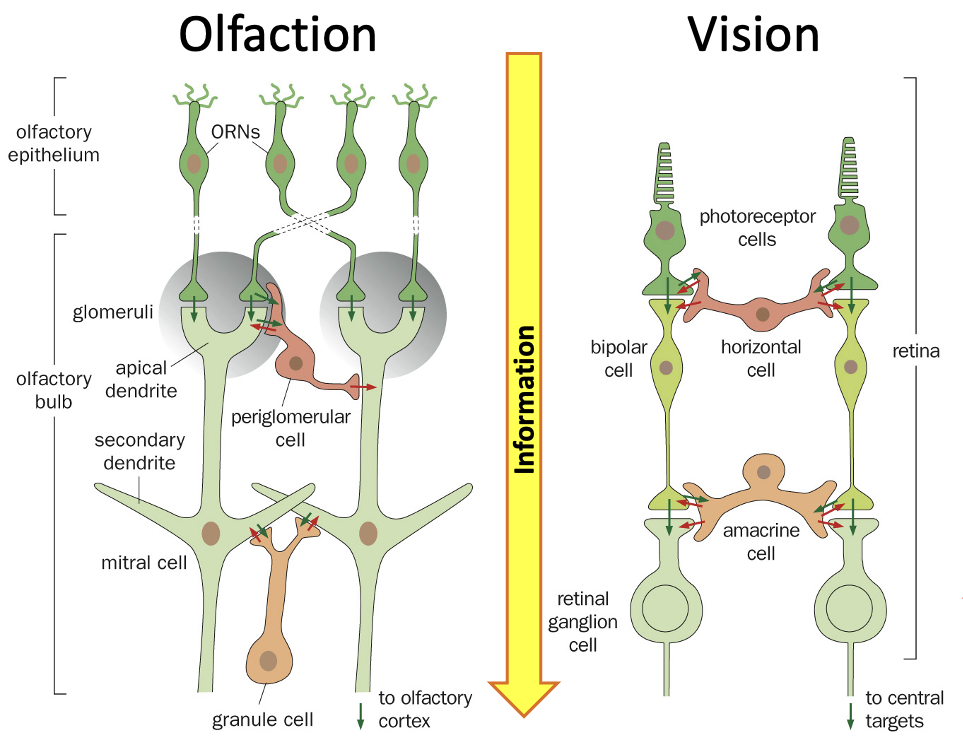
1. Pathway: Smell vs. Vision
• Vision: Light travels through photoreceptors → bipolar cells → retinal ganglion cells → brain
• Smell: Odor signals go from olfactory receptor neurons (ORN) → mitral/tufted cells → cortex (brain)
2. Role of Interneurons (Helper Cells)
• Between these layers, there are helper cells called periglomerular & granule cells.
• These cells inhibit (reduce) activity using a chemical called GABA.
• This helps sharpen the smell signals, so your brain can tell similar smells apart.
3. Difference from Vision: No “Image,” Just Smell Patterns
• In vision, you have a spatial receptive field → each part of the retina responds to a different part of the image.
• In smell, you don’t have a “smell map” like an image. Instead, you have an “odor receptive field”, meaning neurons respond to specific odor patterns.
• The inhibitory interneurons help by adjusting the intensity of the smell so you can detect odors even if they are very weak or very strong.
Combinatorial Coding in Olfaction
Each odorant binds to multiple receptors and each receptor responds to multiple odorants allowing for a vast range of detectable smells.
thousands of different smells but we don’t have thousands of different receptors— how do we detect and tell them apart?- combinatorial coding:
One receptor can detect many different odorants (but with different strengths).
One odorant can activate many different receptors (not just one).
The brain figures out which smell it is based on the unique combination of receptors that get activated.
Example
• Imagine you have three receptors (A, B, and C):
• Lemon might activate A + B strongly and C weakly.
• Vanilla might activate B + C but not A.
• Smoke might activate A + C but not B.
• The brain “reads” these different patterns to identify the smell
Five Basic Tastes related to survival
Bitter = avoid poisons
Sweet = sugar & carbohydrate (attractive).
Umami = l-amino acids (monosodium glutamate) (attractive)
Salty = Na+ (low salt= attractive, high= repellant)
Sour = acids/H+ (repellant)
taste receptor cells
not neurones but are neuroepithelial cells
can regenerate
release neurotransmitters when tastings bind- activate ends of gustatory nerves (taste nerves).
The gustatory nerves carry the taste information to the brain, where it is processed, allowing you to recognize different flavors.
taste buds
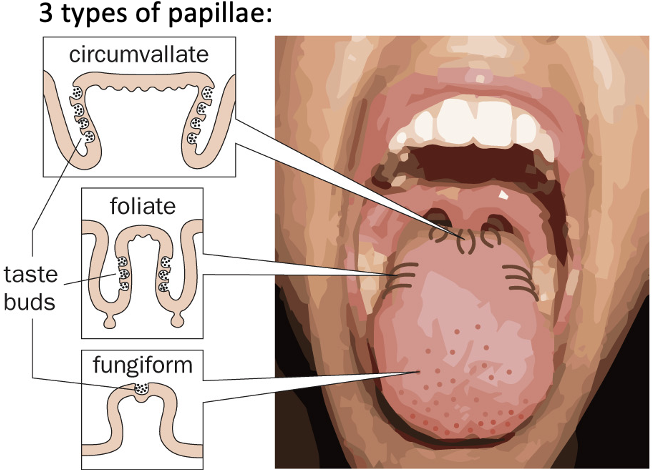
clusters of taste receptor cells form taste buds
localised to several kinds of papillae that are located in different areas of the tongue
Papillae
Structures on the tongue containing taste buds- detect different flavours when eating
papillae types
circumvallate papillae
Large, dome-shaped papillae at the back of the tongue
arranged in a V-shape,
contain many taste buds.
foliate papillae
On the sides of the tongue,
appear as small ridges
contain taste buds
fungiform papillae
Mushroom-shaped
found on the front and sides of the tongue,
contain taste buds.
GPCRs in Taste
G-protein-coupled receptors (eg T1R1 T1R2 T1R3) detect diff taste types
sour - OPTOP1: single proton channel, not a T receptor
bitter - T2R family
umami - T1R1 + T1R3
sweet - T1R2 + T1R3
sour (in mice) - ENaC (epithelial sodium channel)
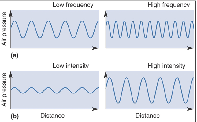
hearing: air pressure waves
we detect sound as variations in air pressure
normal hearing: 20-20,000 Hz
lower frequency waves = lower pitch
lower intensity waves = quieter
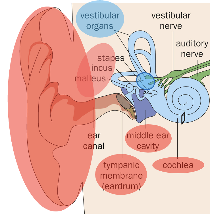
auditory system
consists of external ear, middle ear and inner ear
middle ear- bones:
malleus - First bone, receives vibrations from eardrum
incus - Passes vibrations from malleus to stapes
stapes - Sends vibrations to the inner ear
inner ear = cochlea - converts sound vibrations into nerve signals
so system detects sound
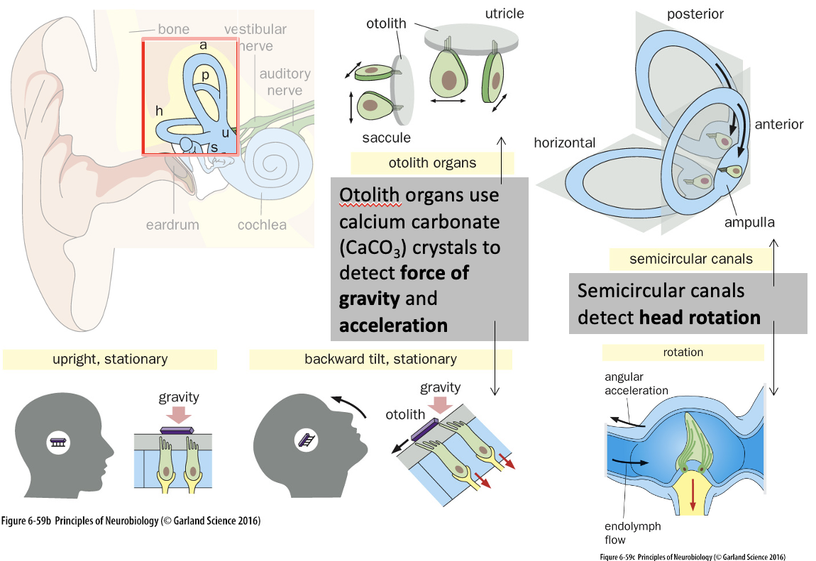
vestibular system
consists of semicircular canals and otolith organs
semicircular canals - Detect rotational movements
posterior - Tilting motion (ear to shoulder)
horizontal - Side-to-side motion (shaking “no”)
anterior - Nodding motion (up/down)
otolith organs - Detect linear acceleration & gravity using calcium carbonate crystals
utricle - Horizontal motion (e.g. moving forward in a car)
saccule - Vertical motion (e.g. moving in an elevator)
so system detect gravity, acceleration and head rotation - help maintain balance & spatial awareness!
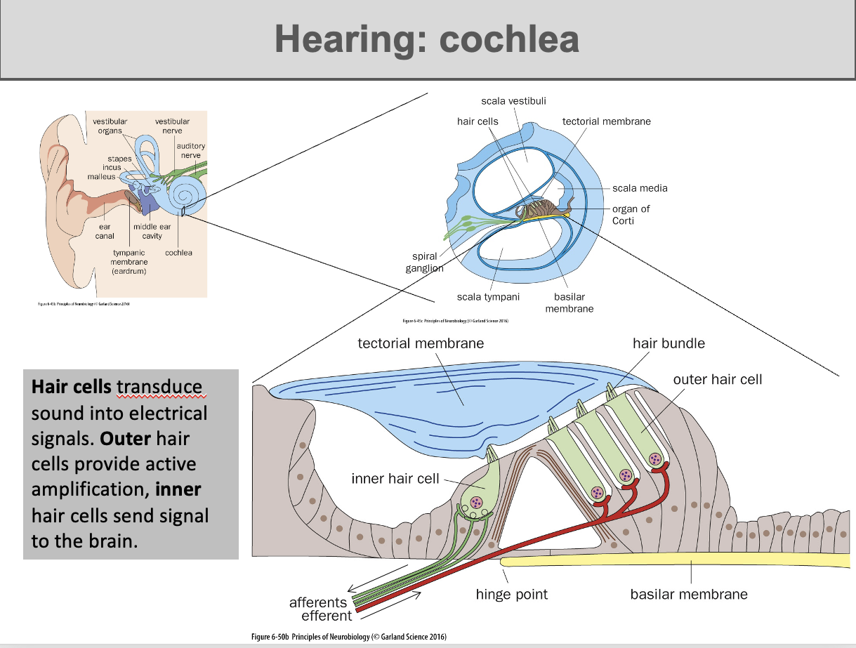
Cochlea + hearing
cochlea = Spiral-shaped fluid-filled structure in the inner ear responsible for sound transduction (converting mechanical sound waves into electrical signals that the brain can interpret as sound)
key structures
Basilar membrane → Moves with fluid vibrations, causing hair cells to bend.
Tectorial membrane → Overlays hair cells and interacts with them during sound detection.
Organ of Corti → Contains hair cells responsible for hearing
contain hair cells
Inner Hair Cells → Send sound signals to the brain.
Outer Hair Cells → Amplify sound by adjusting vibrations of the basilar membrane (using prestin- allows these cells to change their shape)
Process: Sound waves move fluid → Hair cells bend → Electrical signals sent to the brain!
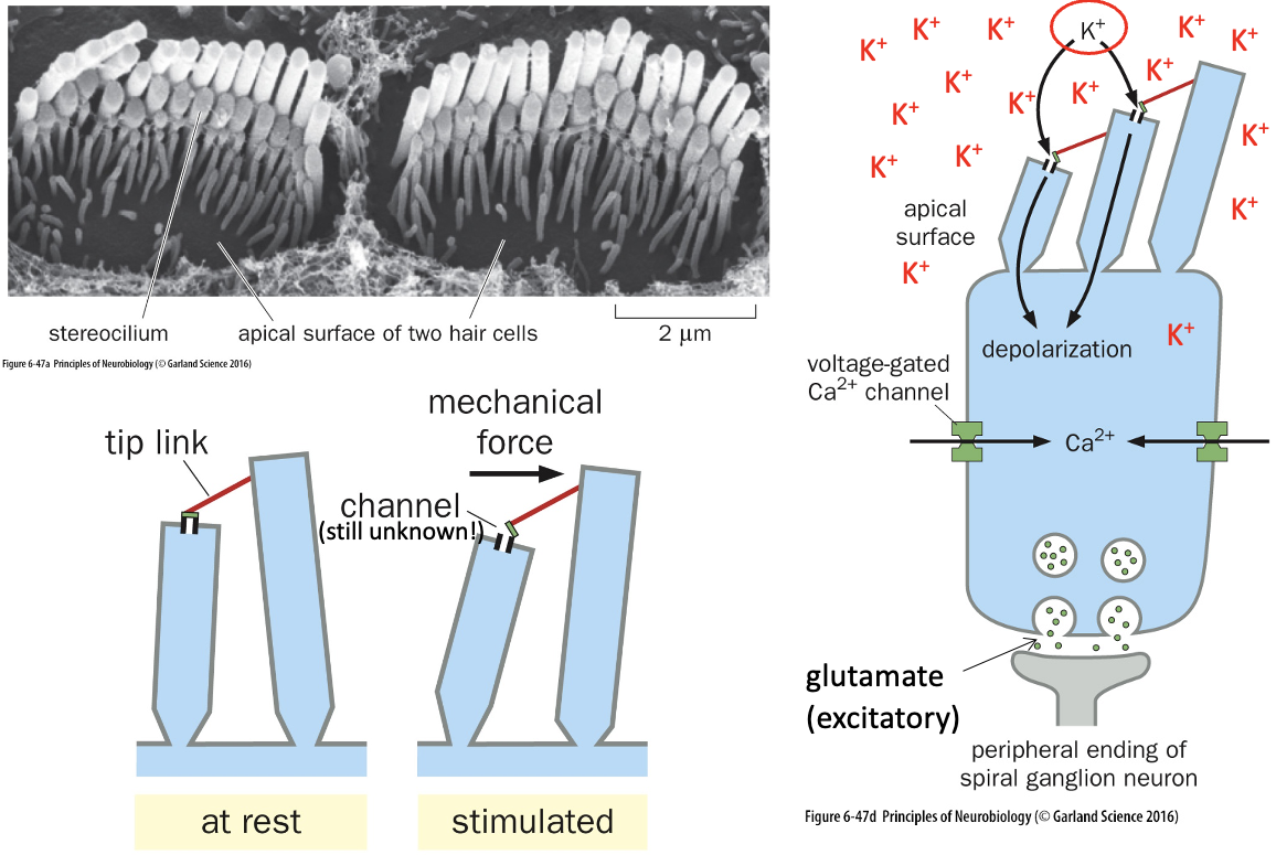
Hair Cell Mechanotransduction
Function: Converts mechanical sound vibrations into electrical signals (mechanotransduction).
Key Features
Stereocilia → Arranged like a staircase on hair cells.
Tip Links → Connect stereocilia and help open ion channels.
Unknown Ion Channel → Pulled open by mechanical force, allowing K⁺ (potassium) to enter
Fast Depolarization Process
1⃣ K⁺ influx causes depolarization.
2⃣ Hair cells release glutamate, an excitatory neurotransmitter.
3⃣ Spiral ganglion neurons receive signals and transmit them to the brain.
Result: Rapid sound detection through direct mechanical transduction
Tonotopic Representation
cochlea = tonotopically organized - different regions detect different frequencies.
Basilar Membrane Properties
Base → Narrow & stiff → Detects high frequencies (e.g., 20,000 Hz).
Apex → Wide & flexible → Detects low frequencies (e.g., 20 Hz).
Result: Different hair cells respond to specific sound frequencies, allowing the brain to distinguish pitch
Interaural Time Difference
comparing the difference in time it takes for a sound to reach each ear
used to detect position of sound source
mechanosensation
converting mechanical stimuli into neuronal impulses
mechanoreceptors
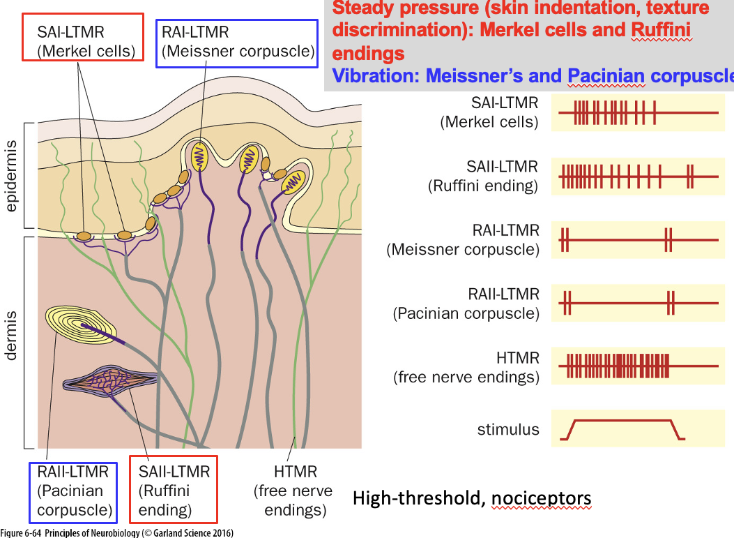
we detect pressure, texture, and vibration through specialized mechanoreceptors in the skin.
Functions of diff receptors
Slow-Adapting (Steady Pressure & Texture Discrimination):
Merkel Cells (SAI-LTMR) → Detect fine details & sustained pressure.
Ruffini Endings (SAII-LTMR) → Detect skin stretch.
Rapidly-Adapting (Vibration & Light Touch):
Meissner Corpuscles (RAI-LTMR) → Detect light touch & low-frequency vibration.
Pacinian Corpuscles (RAII-LTMR) → Detect deep pressure & high-frequency vibration.
High-Threshold Mechanoreceptors (HTMR):
Free Nerve Endings → Detect pain (nociception) and extreme stimuli.
have myelinated nerve fibers for fast conduction of sensory signals
Piezo Channels
Ion channels in Merkel cells
responsible for detecting mechanical force.