Micro Exam 2
1/104
There's no tags or description
Looks like no tags are added yet.
Name | Mastery | Learn | Test | Matching | Spaced | Call with Kai |
|---|
No analytics yet
Send a link to your students to track their progress
105 Terms
Examples of mycobacterium diseases
TB & leprosy
Gram stain & morphology for M. tuberculosis?
Doesn’t stain. Bacilli. Need acid fast stain, doesn't become decolorized. Similar to Gram + w thicker peptidoglycan layer, also w waxy coat & liparabinomannam. Cultures very slowly.
Gram stain & morphology for M. leprae?
Neither + or -. Bacilli. Doesn’t culture at all.
Reservoir M. tuberculosis
Human
Transmission M. tuberculosis? At risk?
Aerosol, low infectious dose. Immuocompromised at risk (old, young, AIDS, Humira)
Reservoir M. leprae
Human
Transmission M. leprae?
Skin breaks & respiratory. Not very infectious, low incidence. From armadillos
Virulence factors of M. tuberculosis
Inhibits production of ROS
Siderophores compete for Fe resources
Protection by waxy coat
Evades killing/lysis after phagocytosis by macrophages (phagolysosome fusion). Body forms granulomas to contain infected cells.
Virulence factors of M. leprae
Avoids killing like M. tuberculosis to survive in the body long term
Infects only Schwann cells (needed for myelination of periphery) → nerve damage
Clinical symptoms of M. tuberculosis? Phases/types?
Pulmonary
Primary, latent, secondary phases
Chronic cough, hemoptysis, fever, malaise, difficulty breathing, cachexia
Extrapulmonary spread
Spinal TB: degradation of cartilage between vertebrae, collapse of spinal column
Military TB: Spreads through blood, white nodules in lungs
Clinical symptoms of M. leprae? Phases/types?
Leprosy/Hansen’s Disease
Tuberculoid leprosy: ring-shaped, pale, flat, scaly, numb, red-ringed lesions. Disease is contained within granulomas.
Lepromatous leprosy: permanent damage to skin, nerves, limbs, eyes causes crippled hands
Compare & contrast M. tuberculosis & M. leprae.
Neither fit Gram characteristics / traditional morphology & culture categories
Reservoir is in humans
Both transmit via respiratory aerosol
TB is more infectious than leprosy
M. leprae also spread via skin contact
Both have systemic symptoms & symptoms that worsen over time
TB is primarily pulmonary, leprosy is primarily skin-based
long-term treatment with Abx
What is the Mantoux test? How does it work?
Diagnosis for TB. Inject small amount of tuberculin (protein extract) into skin. If previously exposed (infected) will form a wheal ~48-72h later due to delayed type hypersensitivity (type 4 overreaction of immune system). Caused by memory T cells activating macrophages.
Ways to confirm TB diagnosis?
Clinical symptoms, chest radiograph, sputum culture & stain
How is leprosy diagnosed?
Observation of skin lesions: usually numb and patchy
Biopsy of skin → acid fast rods, dermal nerve inflammation, neuromuscular granulomas present
Doesn’t culture at all, requirements unknown
Obtain travel history

What is Hansen’s disease?
leprosy
How is TB treated?
Long term treatment with a multi-drug cocktail for 6-9 months to prevent Abx resistance. Waxy coat protects bacteria.
What is the BCG vaccine?
Named after founder, ses attenuated strain of Mycobacterium bovis from a cow to protect against TB. Used in regions w high TB rates, not in U.S. to better monitor rare infections that do occur.
What are IGRAs?
Interferon gamma release assays: Mix blood sample with TB antigens, leukocytes release IFNy which is measured. Can be used in addition to BCG vaccine.
How is leprosy treated? Prevented?
Treatment w multi-drug cocktail until lesions disappear. No form of prevention.
What is NTM?
Non-tuberculous mycobacteria are organisms in the environment, opportunistic. Outbreaks in tattoo parlors.
What kind of virus is Dengue virus?
Flaviviridae genus & RNA virus
Where is the reservoir for DENV? Transmission method?
Reservoir: Humans
Transmission: Aedes aegypti mosquito species vector, lives in warmer climates like Africa → global warming increasing prevalence.
How does a hematocrit test work?
Centrifuge blood
It separates into plasma on top and RBCs on the bottom.
Determine the % volume of RBCs to plasma. Should be ~50%.
Is DENV an obligate or facultative pathogen?
Obligate: needs host cell to survive & replicate
Initial pathogenesis of dengue?
Virus infects immune & endothelial cells
Infected cells release cytokines
Plasma leakage, causes inc. hematocrit (diff ratio)
Platelets decline (thrombocytopenia)
Less bone marrow produced
Fragile vascular system from decreased platelet function
Clinical features of dengue? Phases/types?
Febrile phase: virus replication → fever, headache, vomiting
Critical phase: inc. hematocrit, dec. platelets, risk for:
Dengue hemorrhagic fever: bleeding in urine, stool, vomit, skin, nose, gums
Dengue shock syndrome: plasma leakage causes hypovolemic shock bc heart can’t pump blood
Recovery phase: Fluids reabsorbed (no leakage), regeneration of platelets, Ab titer inc.
Primary dengue: Infection w 1st serotype
Secondary dengue: Infection with a different serotype (there are 4). Abs from the primary infection can’t neutralize 2nd serotype. They bind to the 2nd strain partially, allow it to bind to receptors and into the cell. The virus doesnt neutralize in the endoscope and replicates.
How does neutralization work? (Its blocked in dengue)
Multiple Abs bind to virus.
Virus cant bind to receptors and enter the cell.
Virus can’t escape the vacuole once endocytosed.
What is ADE?
Antibody dependent enhancement: Abs from primary infection enhance entry of secondary serotype. Causes worse infection.
Treatment for DENV?
Supportive care such as IV fluids & blood transfusion
How should dengue be diagnosed?
Look for IgM (first Abs produced) and IgG (produced within days of infection) via serology
PCR
NS1 presence: an antigen, part of viral protein
Use these to distinguish between primary and secondary. Be able to use examples of primary and secondary infection serology and patient samples at diff timepoints to diagnose with primary or secondary.
Describe analytical sensitivity/specificity & clinical sensitivity/specificity.
Analytical sensitivity: Smallest amount of Ag that can be detected.
You won’t test positive for COVID until a few days after infection.
Analytical specificity: If the test can distinguish between strains
The COVID test can differentiate between SARS-CoV2 and common cold viruses.
Clinical sensitivity: % of people who have infection that actually test positive
Can you use the test to determine if a patient has disease?
Does high analytical sensitivity = high clinical sensitivity? B. pertussis
Clinical specificity: % of people who don’t have disease that actually test negative
A COVID test that has false positive from a cold virus
Neisseria gonorrheae Gram stain? Morphology?
Gram - diplococci, within neutrophils
Neisseria gonorrheae reservoir and transmission?
Reservoir: asymptomatic infections
Transmission: contact of mucosal surfaces (oral, anal, vaginal)
Virulence factors of Neisseria gonhorreae
Siacilic acid coat mimics host molecules, allows for evasion.
Uses antigenic variation
Describe antigenic variation
Group of pathogens. One has a variation, eg in spike protein. Abs attack everything but the variant. The variant can now proliferate, expand, and spread. (Think COVID)
How do clinical features of Neisseria gonorrheae differ in males and females? General clinical features? Spread?
Presents as urethritis in males. Also urethritis in females, may progress to pelvic inflammatory disease when it spreads to uterus, ovaries, fallopian tubes. Can be spread to child during birth. Clinical features reported by case study patient: chills, malaise, painful urination, discharge, tenderness
Disseminated gonococcal infection: infection spreads to blood, may cause joint pain (septic arthritis)
How is gonorrhea diagnosed
Gram stain & culture of urethral discharge and/or synovial fluid (if septic arthritis), should see bacteria within neutrophils
NAAT
How should gonorrhea be treated and prevented?
Treatment: Abx, test for other STIs
Prevention: Use condoms. Vaccine can’t be made due to antigenic variation
Gram stain & morphology of Neisseria meningitidis
Gram - diplococci (see coccus in name)
Describe the reservoir and transmission of neisseria meningitidis
Reservoir is nasopharyngeal carriage. Transmitted via respiratory → close quarters, mass gatherings have higher risk from prolonged exposure. More common in young people
Virulence factors of neisseria meningitidis
Antiphagocytic capsule
Antigenic variation
Lots of endotoxin produced → risk of capillaries leaking leading to sepsis and other systemic problems
Describe the clinical symptoms of Neisseria meningitidis
Spreads quickly, systemic effects in young people:
Sepsis from endotoxin
Headache & nuchal rigidity from meningitis
Bleeding in the dermis (skin petechiae)
Disseminated intravascular coagulation: If clots obstruct flow, tissues have lack of oxygen & nutrients (ischemia), hemorrhage
What is disseminated intravascular coagulation & what is it caused by?
If clots obstruct flow, tissues have lack of oxygen & nutrients (ischemia), hemorrhage. Can be a severe consequence of Neisseria meningitidis
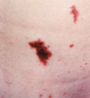
How is Neisseria meningitidis diagnosed & treated?
Diagnosis: Meningitis & petechiae (see image). Gram stain of CSF will confirm.
Treatment: Abx. Prophylactic treatment for close contacts
What is the meningococcal vaccine, who is it given to?
Abs for Neisseria meningitidis. Given to age 25 and below & at risk groups.
Gonococcus refers to
Neisseria gonorrheae
Meningococcus refers to
Neisseria meningitis
Major difference between gonococcus and meningococcus
Gonococcus: Stays relatively local (urethra)
Meningococcus: Spreads quickly, evades, sepsis, meningitis
Gram stain, culture, & morphology of Treponema pallidum?
Does not Gram stain, need silver stain. Spirochete. Does not culture (fastidious)
What disease does Treponema pallidum cause
Syphilis
What is the reservoir and transmission method for Treponema pallidum
Reservoir: Humans
Transmitted via mucous membrane contact & lesions. Mostly MSM demographic. Increasing in the US.
Describe the phases of syphilis and clinical features:
Primary syphilis
Chancre at infection site
Weeks later
Disappears untreated
Secondary syphilis
Months later
From untreated chancre
Spread = systemic features: fever, malaise, lymphadenopathy
Rash on palms & soles (maculopapular)
Wart-like genital lesions (condyloma lata)
Resolves untreated
Latent syphilis
Secondary syphilis has resolved, asymptomatic
Seroreactive: Abs present
Can spread to child
30% of cases progress from here
Tertiary syphilis
Decades later
Granuloma formation = tissue damage: skin, liver, bone, testes, aortic aneurysm
Dementia, ataxia, vision loss, seizure
Virulence factors of Treponema pallidum (syphilis)
“Stealth pathogen”: balance between the immune system & pathogen allows it to stay present for decades
How is syphilis diagnosed
Clinical skin signs
Skin scraping under silver stain shows spirochetes
Treponemal test: Abs present for life
Nontreponemal test: Looks for related markers of cell damage
Treponemal test vs Nontreponemal test
Treponemal: Abs against pathogen present for life
Nontreponomal test: Looks for biomarkers from cell damage
How can syphilis be treated & prevented
Treatment: Penicillin will cure primary or secondary. Tertiary can be treated but any damage done is permanent.
Prevention: Condom use. No vaccine bc stealth pathogen.
Chlamydia trachomatis Gram stain & morphology
Similar to Gram -, minimal peptidoglycan. Round.
Reservoir & transmission of Chlamydia trachomatis?
Reservoir is humans, obligate intracellular (can’t live outside). Transmitted via mucous membranes (genital, anal, oral). Can pass from mother to child during birth (perinatal) as conjunctivitis. More common in women.
How are clinical features of chlamydia trachomatis different between males & females?
Males: Infection of urethra & epididymis → painful urination, cloudy discharge, testicle tenderness
Females: Infection of cervix epithelium & urethra → painful urination, vaginal discharge, bleeding. Also can ascend to become PID
Pelvic inflammatory disease
See gonorrhea also! Infection ascends to uterus, ovary, fallopian tube; can be transmitted during childbirth. Can also cause infertility & ectopic pregnancy (implants outside uterus)
What is trachoma, what is it caused by?
Eye infection
Chlamydia trachomatis
More common in crowded places, poverty, poor hygiene (not U.S.)
Virulence factors of chlamydia trachomatis? How do they contribute to disease development?
Adheres to urinary epithelium (elementary bodies). Gets endocytosed. Prevents phagolysosome fusion. Becomes reticulate bodies to replicate, then exocytosis
What are elementary bodies? What is their complement?
Elementary: Present outside the vacuole, used to infect.
Reticulate bodies: within the vacuoles, grow and replicate after endocytosis of elementary bodies
How is chlamydia trachomatis diagnosed
NOT gram stain: too small, lack peptidoglycan
NAATs
Case study used endocervical swab to collect sample. Also ultrasound.
How is chlamydia trachomatis treated
Abx besides beta lactase since there is no peptidoglycan to act against.
How can chlamydia be prevented
Use of condoms. To prevent trachoma, practice hygiene.
Gram & morphology of all pathotypes of E. coli?
Gram - rods
Reservoir & transmission of ETEC
Reservoir: infected humans
Transmission: fecal oral w high infectious dose
Commonly causes traveler’s diarrhea, high risk of infection in Asia, Africa, Middle East, Mexico, South America
Virulence factors / pathogenesis of traveler’s diarrhea?
Adhesion to micropili. Enterotoxin disrupts ion transport in GI epithelium, causing net secretion of fluid & watery diarrhea
What clinical features does ETEC cause
Gastroenteritis: refers to diarrhea & vomiting.
Infectious traveler’s diarrhea (secretory/watery).
Also nausea, vomiting, fever, delayed 1-2 days from exposure
What is traveler’s diarrhea usually caused by? What kind of diarrhea is it?
ETEC. Secretory/watery
How is ETEC infection diagnosed, treated, and prevented
Diagnosed by presence of watery diarrhea (no clinical test).
Self limiting, basic oral rehydration can be used.
Prevent by eating only well cooked food & drinking bottled water in high risk areas
Differences between ETEC and STEC
STEC has lower infectious dose
STEC more severe clinical features, larger outbreaks
STEC stand for
Shiga toxin producing E.coli
Reservoir & transmission of STEC
Reservoir: animals
Transmission: fecal oral via food, low infectious dose
Major serotype causing STEC?
O157:H7
Virulence factors & pathogenesis of STEC
Adhesion to GI epithelium.
Type 3 secretion system to inject into cells
Shiga toxin - AB5, acquired by horizontal gene transfer from Shigella dysenteriae. Endocytosed, escapes to cytoplasm, binds rRNA & blocks protein synthesis, cell dies
What clinical conditions can STEC cause?
Inflammatory/Bloody Diarrhea: Shiga toxin damage to intestine epithelium, causes loss of fluid, epithelial cell death → Capillaries exposed → bleeding
Hemolytic Uremic Syndrome (HUS): Shiga toxin damages EC’s. Causes clots in small capillaries. Damages kidneys. Thrombocytopenia. RBS fragment in capillaries (hemolytic anemia). Also damages filter in kidneys, clogs them. Causes electrolyte imbalance → uremia, hematuria. Can lead to acute renal failure.
Other general symptoms: Fatigue, fever, headache, low abdominal pain, pale, blood in stool, albumnuria
How is STEC infection diagnosed
Stool culture & look for blood
Detection of Stx
Urinalysis
CBC: should be more leukocytes
NAAT
How is STEC treated & prevented
Treated w oral rehydration. Rarely w Abx, might stress bacteria to produce more toxin.
Prevention: Handwashing, cooking food hot enough
What do commensal strains of E. coli cause? Why?
Opportunistic infections occur from bacteria in the gut. Usually cause UTIs: rectal bacteria adheres to perineum. Mechanical disruption, bacteria enters urethra. Also transmitted via birth.
Motility: they have flagella
Exotoxin released with growth, damages epithelium, inflammation & hemorrhage
Define UTIs and the diff types
Infection causes frequent, burning, cloudy urination.
Lower UTI: Urethra & bladder, vaginitis, proastatis
Upper UTI: Kidney
What are “fecal coliforms”?
Bacteria from mammal intestines, commonly contaminate water. Screening for E. coli.
Difference between bacterial vaginosis & urinary tract infection
Vaginosis is imbalance of normal microbiome
UTI is ascending infection, usually E. coli, males & females
How are UTIs treated & prevented
Given UTIs empirically, unless “complicated” (underlying problem, men & at risk women) then urine culture for specifics.
Drink lots of water to flush bacteria.
What are the 3 types of clinical features E. coli can cause?
UTIs, neonatal meningitis, diarrhea (see diff types & examples)
Why would E. coli cause neonatal meningitis
Exposure to pathogenic strain during delivery
Gram & morphology of Enterobacterales?
Gram - rods
Reservoir & transmission of Shigella species?
Infected humans
Fecal oral, very low infectious dose (10-100)
Virulence factors of Shigella species?
Resistant to stomach acid
Facultative intracellular pathogen. Uses M cells in colon to inject VFs by T3SS. Allows bacterial uptake.
Intracellular motility via actin polymerization
Shiga toxin: destroys epithelium, exposes lamina propia
2 Shigella species we are learning about? Differences?
dysenteriae & sonnei, sonnei is more common in US. Dysenteriae more likely to cause bloody diarrhea.
How are M cells involved in shigellosis
They are epithelial cells that regularly sample the GI for Ags, give to lymphoid follicle/mucosa associated lymphoid tissue. Shigella species use them to enter epithelium.
Shigellosis clinical features
Dysentery (bloody diarrhea) days after exposure w fever, cramps. Diarrhea has mucus from shedding of GI mucus lining. Also risk for hemolytic uremic syndrome from Shiga toxins.
How is shigellosis diagnosed, treated, prevented?
Diagnosis: Stool culture. Special differential & selective media (Hektoen enteric agar). Case study: Shiga toxin test, CBC (hemolysis & thrombocytopenia), Urinalysis for albuminuria
Treatment: Oral rehydration, avoid Abx → more Shiga toxin production. If sick enough (fever >103, hypovolemia, too much diarrhea, hospitalization, etc)
Prevention: Sanitation to prevent fecal water/food contamination
What media is used for Shigella species culture
Hektoen enteric agar
Salmonella typhimurium virulence factors
Sensitive to acid, need to ingest more for effects
Uses M cell transcytosis (but NO actin polymerization)
Inhibits phagolysosome fusion by blocking hydrolytic enzymes, macrophage cant kill
How is Salmonella diagnosed, treated, prevented? Differences between typhirium & typhi?
Diagnosis: stool & blood culture
Differences between Shigella & Salmonella?
Clinical features of S. typhimurium
Gastroenteritis, can be mild or severe.
Diarrhea
Fever
Cramping
Usually not bloody