EKG rhythms
1/31
There's no tags or description
Looks like no tags are added yet.
Name | Mastery | Learn | Test | Matching | Spaced | Call with Kai |
|---|
No analytics yet
Send a link to your students to track their progress
32 Terms
Sinus Rythms
Electricity originates in the SA node
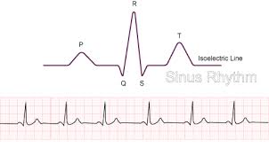
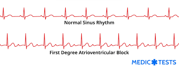
Normal Sinus Rhytm (NSR)
P wave: Normal
QRS: Normal
PR interval: Normal
Reg/Irg: Regular
Rate: 60-100 (normal)
Other:
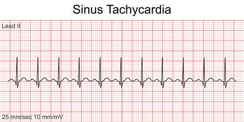
Sinus Tachycardia
P wave: Normal
QRS: Normal
PR interval: Normal
Reg/Irg: Regular
Rate: over 100
Other: Causes could be caffeine, drugs, hyperthyroid, etc
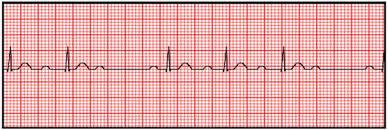
Sinus BradyCardia
P wave:Normal
QRS:Normal
PR interval:Normal
Reg/Irg:regular
Rate: below 60
Other: Age, medications, drugs, heart disease, athlete, etc
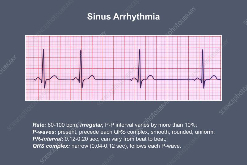
Sinus Arrythmia
P wave:Normal
QRS:Normal
PR interval:Normal
Reg/Irg: Occasional Irregular
Rate: Persons underlying heart rate
Other:(Bc of breathing/ preassure from vagal nerve)
Normal Sinus Rythms with occasional (irregular)
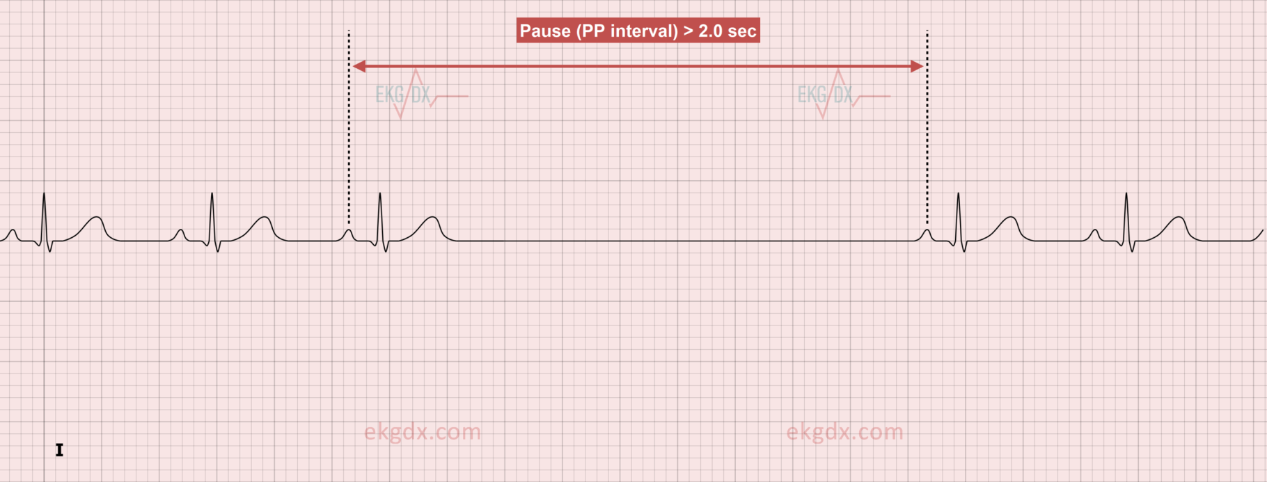
Sinus Pause
Normal Sinus Rhythm with a pause.
2.5 seconds or longer
If a 6 second pause =Medical emergency
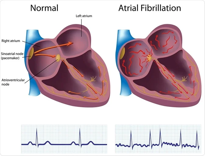
Atrial Rhythms
Orginates somewhere other than the SA node (in atrial)
Ventricles are not affected (QRS is always normal)
Atrial Flutter ( Saw tooth)
P wave: Flutter Waves/sharp
QRS: Normal
PR interval:Cant determain (Mulitple P waves)
P:QRS: varies 2:1 3:1 4:1 (Always 1 QRS)
Reg/Irg: Regular
Rate: Varies with the P:QRS rate
Other: T waves are hidden
AV node bocks the extra electrical defib
Automatcity Bc electricity is coming from the wall of atrial cells
Only lets so many electrical impulses through at a time
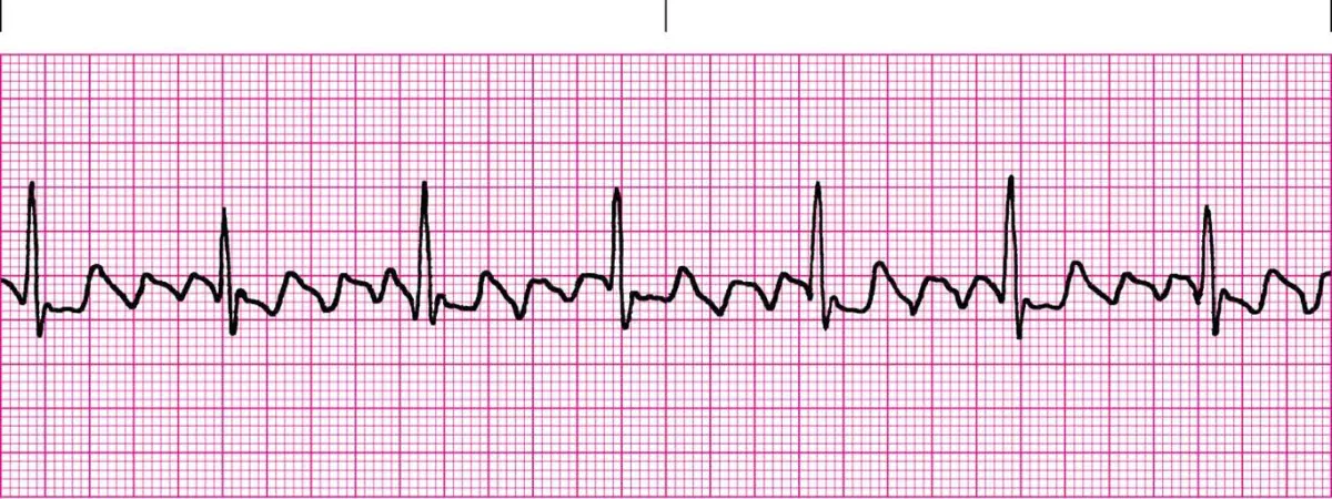
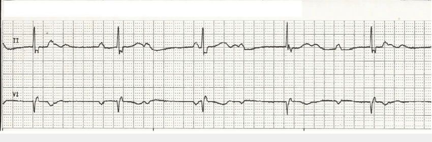
Atrial Fibrillation (A-FIB)
P wave: Fibrillatory waves
QRS: Normal
PR interval: Cant determain
P:QRS:Cant determain
Reg/Irg: irregular
Rate:60-100bpm
Other: Decreased cardiac output
scribbles
chaos in atria
Blood begins to pool= risk for a blood clot
(problem in the atria) they don't coordinate in beat
AV node lets some of the electricity through more than atrial flutter
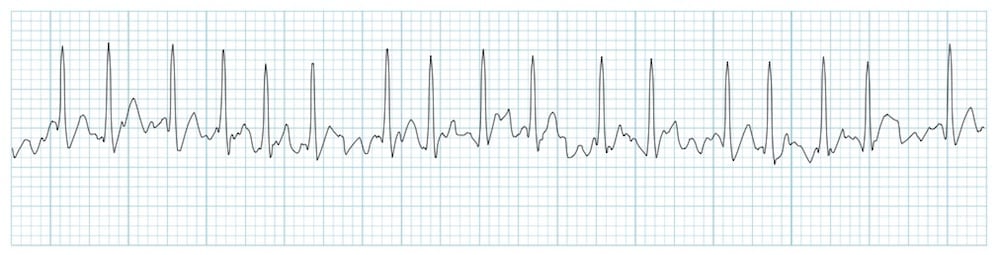
Atrial Fibrillation w Rapid Ventricular Reponse (AFIB w/ RVR)
P wave: Fibrillatory Waves
QRS: Normal
PR interval: Cant determine
P:QRS: Cant determine
Reg/Irg: Irregular
Rate: Greater than 100
Other: Lets more impulses through
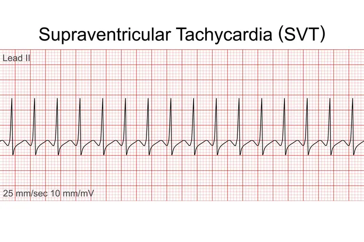
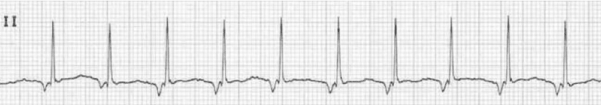
SupraVentricular Tachycardia (SVT)
P wave: P waves hidden in T waves
QRS: Normal
PR interval: Cant determine
P:QRS: Cant determine
Reg/Irg: Regular
Rate: 150 or greater
Other: Orginates in atria, AV junction, or SA node
Lets every impulse through
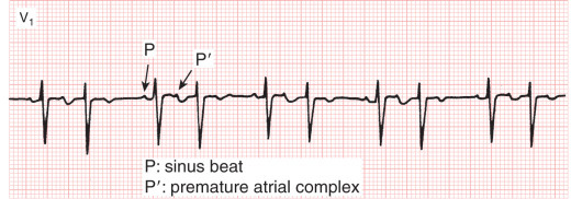
Premature Atrial Contraction (PAC)
EVerything normal except its (Irregular)
Singular, P wave is distorted =comes from atria
Occurs because of an early electrical impulse occurs other from a location in the atria other than the SA node
Looks like a thumb and on top of something - before the next normal sinus P wave should appear.
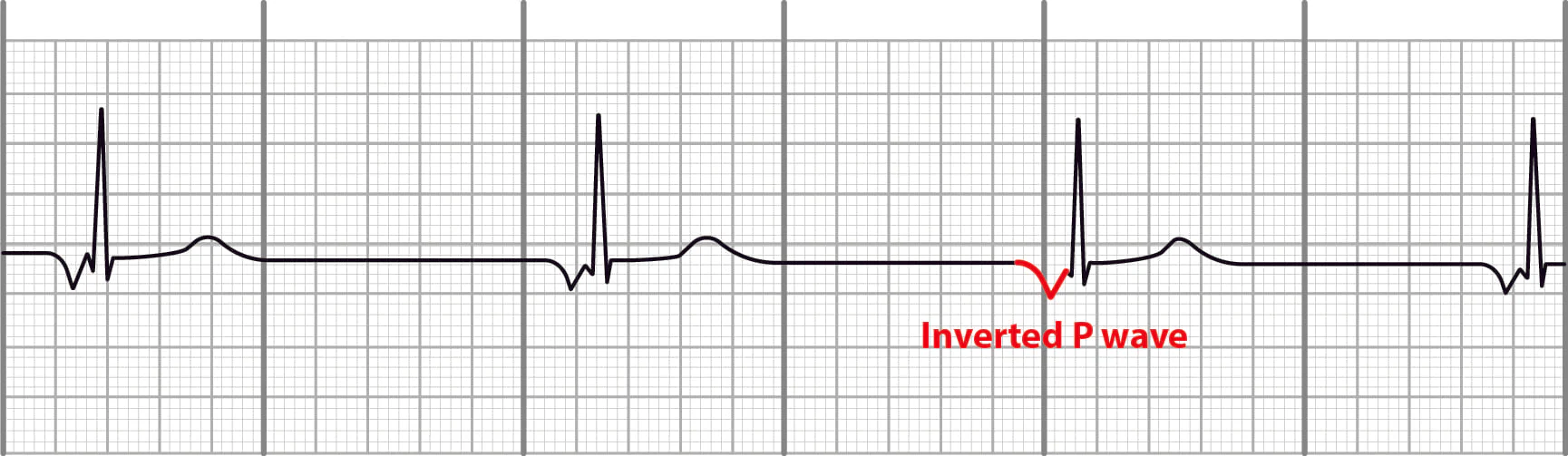
Junctional Ryhtms
AV node is the pacemaker
The p Wave is always either Inverted before QRS, Absent, Or inverted After QRS
The difference between junctional Ryhtms is the rate
Intrinsic Rate: 60-40
Junctional Brady Cardia
P wave: Inverted before QRS, Absent, Inverted After QRS
QRS: Normal
PR interval: N/A
P:QRS: N/A
Reg/Irg: Regular
Rate: 60-40
Other:
Junctional Escape
P wave: Inverted before QRS, Absent, Inverted After QRS
QRS: Normal
PR interval: N/A
P:QRS: N/A
Reg/Irg: Regular
Rate: less than 40 BPM
Other:Whole Ryhthm will be junctional
Acclerated Junctional
P wave:Inverted before QRS, Absent, Inverted After QRS
QRS: Normal
PR interval: N/A
P:QRS: N/A
Reg/Irg: Regular
Rate: 60-100Bpm
Other:
Junctional Tachycardia
P wave:Inverted before QRS, Absent, Inverted After QRS
QRS: Normal
PR interval: N/A
P:QRS: N/A
Reg/Irg:Regular
Rate:Greater than 100 bpm
Other:
Heart Block Rhythms (AV Block)
-Problem in the AV node or the conduction pathways just below it (the His–Purkinje system).
electrical signals traveling from the atria (top chambers) to the ventricles (bottom chambers) are slowed down or completely blocked.
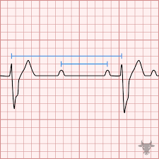
First Degree AV Block
P wave:Normal
QRS: Normal
PR interval: Yes but its long
P:QRS: 1:1 ratio
Reg/Irg:Regular
Rate:Any
Other: Everything is normal except there is a long PR interval
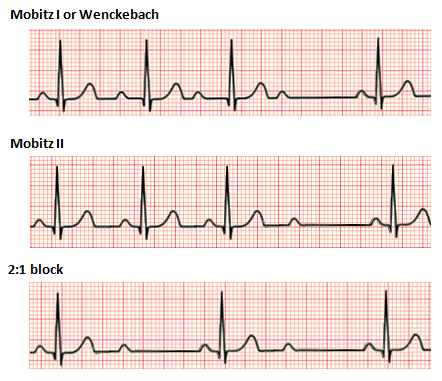
Second Degree AV Block (Mobitz 1 or Wenkebach)
P wave:Normal
QRS: Normal
PR interval: Progressivley longer units and QRS drops
P:QRS Dropped beats but not always
Reg/Irg: irregular
Rate:Any
Other: Long, Longer, LONGERRR, Drop, Now you have a wenkebach
Second Degree AV Block (Mobitz 2)
P wave:Normal
QRS: Normal
PR interval: Yes.. but when QRS dropped no
P:QRSDropped QRS but not always
Reg/Irg: Regular or Irregular
Rate:Any
Other:Intermittent dropped QRS
no PR interval lengthening
Third Degree AV Block (Complete Block)
P wave:Normal
QRS: Normal
PR interval: none
P:QRS none
Reg/Irg: regular or irregular
Rate:Any
Other: -
P/QRS independent of eachother
-Alls Ps will march together
-All QRS will march together
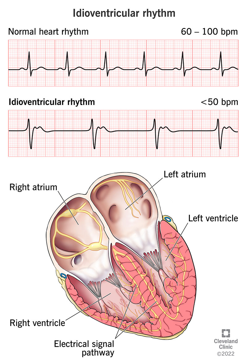
Ventricular Ryhtms
Orginates in the Ventricles
Always WIDE QRS= ventricles receiving electrical impulses by themselves= EMERGENCY ACTION
Idioventricular (AGONAL)
P wave:No
QRS: Bizzare and Wide
PR interval: none
P:QRS none
Reg/Irg:Regular
Rate: intrinsic Rate 20-40 and accelerated 40-100
Other: NOT SHOCKABLE
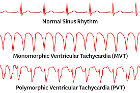
Ventricular Tachycardia (V-TACH) Monomorphic
P wave:No
QRS: Wide, Upside down
PR interval: none
P:QRS none
Reg/Irg:N/A
Rate:N/A
Other: QRS is wide and similar in apperance
Shockable if person doesn't have a pulse and is unconscious, Not shock able if person has a pulse and is awake BUT, prepare for a code blue but iniate a rapid
Ventricular Tachycardia (VTACH) Polymorphic
P wave:No
QRS: Wide, Multifocal ballet like
PR interval: none
P:QRS none
Reg/Irg: N/A
Rate:N/A
Other:”Twisting of Points” torsades de pointes
-QRS can vary in size and shape
No pulse = shock
pulse=dont shock
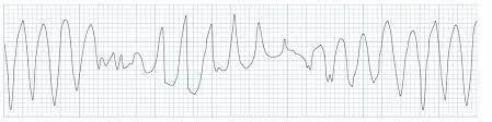
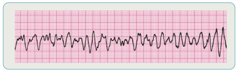
Ventricular Fibrillation (VFIB)
P wave:No
QRS: Fibilitory Waves
PR interval: none
P:QRS none
Reg/Irg: N/A
Rate:N/A
Other: EMERGENCY IMMEDIATE ACTION=CODE BLUE
ALWAYS SHOCK (never a pulse)
Premature Ventricular Contraction (PVC)
Charateristics: QRS Wide and fat (lost p wave)
Concerns:More than 6 a min is not a perfusing beat
Not a Ryhtms just an abnormal beat
Bigeminy- every other beat
Trigenimy- every 3 bats
quadrigeminy- every 4 beats
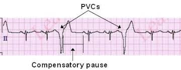
couplet- together
run of vtach- 3 PVC together
Atrial Pacer
spike before the p wave (Atrial)
Ventricular pacer
before the QRS (Ventricular)
atriocentricular pacer
2 spikes (atria and ventricles)
Asystole
no electrical rythm
Do not shock ( START CPR)