L1: Synaptic plasticity
1/69
There's no tags or description
Looks like no tags are added yet.
Name | Mastery | Learn | Test | Matching | Spaced | Call with Kai |
|---|
No analytics yet
Send a link to your students to track their progress
70 Terms
How are synapses dynamic
With repeated use→ show short-term or long term changes
Synaptic plasticity
What does synaptic plasticity allow
allows nuerons to store information
in response to different degrees of experience
Changes can either
increase synaptic efficacy →potentiation
Decrease synaptic efficacy→ depression
Short term vs long term plasticity
Short term
timescale of ms→ several mins
contribute to computations in neural circuits
Long term
mins→ hours→ years
hypothesised to be the basis of learning and memory
What happens when a presynaptic neuron is stimulated experimentally/physiolgically
action potential travels the length of the axon
until it reaches the presynaptic terminal
Ca enters via voltage-gated calcium channels
leads to local increase in Ca concentrations
Release Neurotransmitters in synaptic cleft
glutamate in excitatory
binds to AMPA ionotropic receptor
entry of Na+ into postsynaptic neuron
generates current→ EPSC
The mean amplitude/size (m) of the EPSC of the postsynaptic cell is equal to
p*n*q
p→ probability of release
q→ quantal size→ i.e postsynaptic receptor response to single discrete (one quantal) of vesicle transmitter release
n→ number of release sites→ readily releasable vesciles
SYNAPTIC TRANSMISSION→ WITHOUT PLASTICITY
Due to no plasticity yet…
after some time another AP arrives at the presynaptic terminal
BUT
we get the SAME mean amplitude/size (m) of EPSC
However, there is residual Ca ions left at presynaptic terminal
Why are there residual Ca ions left at the presynaptic terminal
result of the arrival of an AP
When does residual go back to baseline?
after 200 ms
What happens if another AP arrives within the 200ms?
leads to greater Ca concentration increase in presynaptic neuron
as levels build up on the residual level already left over
increases probability of release (p) of glutamate
→ amplitude (m) of the second EPSC
→ PAIRED PULSE FACILITATION
What does the interval between the two stimuli determine?
the extent of increase of the second EPSC
The longer the interval between the two stimuli→ the lower the amplitude of the second EPSC generated
because there will be less residual calcium available for the arrival of the second AP at the presynaptic terminal
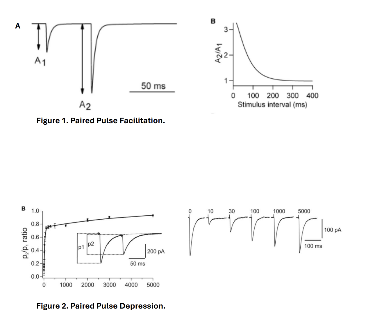
Paired pulse depression
arrival of the second AP to be shortly after the first AP
however
the second stimulus results in a smaller EPSC amplitude in the postsynaptic neuron
Why is there smaller EPSC amplitude in the postsynaptic neurone?
decrease in readily available vesicle pool
i.e number of release sites (n)
The longer the interval between the two stimuli…
the less the size of the attenuated second EPSC
Why sometimes we get depression due to arrival of a paired pulse and another time facilitation?
if the first synaptic transmission results in larger response (bigger EPSC)
→ the direction of the change/plasticity will be towards decrease
PAIRED PULSE DEPRESSION
If the first synaptic trasmission gives a smaller response
there is a tendency to facilitate that synapse to become stronger
therefore: direction of plasticity will depend on the strngth of the synpatic transmission prior tot he second pulse arriving
Facilitation and depression coexist?
can co-exist at the same synapses
with history of activity influencing the direction of plasticity
High release probability favours (p)…
depression
i.e is there was originally alot of release from before
it is less likely that in the second time around there will be enough NT to release
if there hasn’t been enough time to get NT vesicles back?
Low (p) favours
Facilitation
Therefore this shows an example of plasticity
the strength of the synaptic transmission can be dynamically altered
Computations and short term synaptic plasticity
short term plastisicty takes place over period of milliseconds to seconds
not long lasting
→ they take part in synaptic computations
Example of this: High pass and low pass filtering
High pass filtering
presynaptic neurone stimulated at a high frequency
then this frequency is not filtered
its is passed to the post synaptic neuron
→ post synaptic neuron also fires at the same frequency
This is due to the mechanism of…
Paired pulse facilitation:
As presynaptic neurone fires action potentials at a high frequency
the EPSC on post synaptic neurone increases in size
results in higher chance of action potential generation as the membrane depolarises to its threshold for firing
allows the postsynaptic neurone to fire at the sam frequency as the presynapatic neuron
What happens if the presynaptic neuron is activated at a lower frequency
EPSCs do not increase
→ no facilitation
→ less chance of action potential generation/firing of the post synaptic neurone
i.e lower frequencies are filtered out
What does this help to make sure?
only the presynaptic neurone (and hence synapases) that are very active pass on the info
to a post-synaptic neurone
→ the less active synapses to be filted out
OVERALL: high pass filtering
Low pass filtering
→ Converse situation
due to paired pulse depression
Because the cell has a high probability → so a low frequency stimulation will excite but a high frequency will cause low filter to pass and the high frequency to stop.
Paired pulse depression
If presynaptic neuron fires at a high frequency:
decrease in EPSC size (because not enough NT? coz it is a high p neuron?)
less chance of action potential firing as threshold for AP generation is not reached
higher frequnecies are filtered out
if presynaptic neurone is firing a a low frequency
no attenuation of the EPSC
AP can generated
(the EPSC was large to begin with during paired pulse depression)
Long term plasticity: allows neurons to store information for how long?
longer time period of hours→ days→ months→ yyears
What changes in the synapes in this plasticity
structurual changes
spines!
changes involving molecular mechanisms
→ changes in neurotransmitter release
and/or
changes invovling receptors and post-synaptic levels
Long-term potentiation (LTP)
lasting changes that result in strengthening of synaptic transmission
Long term depression (LTD)
lasting changes that lead to weakening of synaptic transmission
There are different forms of experimentally induced LTP and LTD depending on…
the brain region and transmitter receptors involved
Why has the hippocampus been extensively studied?
as well defined layers → allow experimenter to relatively easily electrically stimulate one layer whist recording from the other that receives a synapase from the stimulated layer
Hippocampus (and neocortex) are two structurates involved in learning and memory
How has the hippocampus been studied
LTP and LTD protocols:
induced in different regions of the hippocampus and cortex
including dentate gyrus, CA1 region, visual cortex and somatosensory cortex
One form of LTP
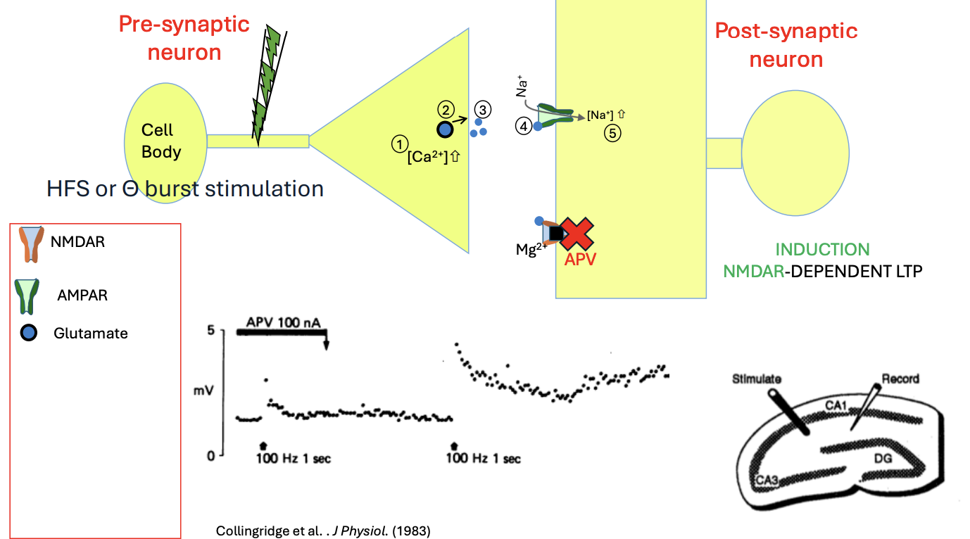
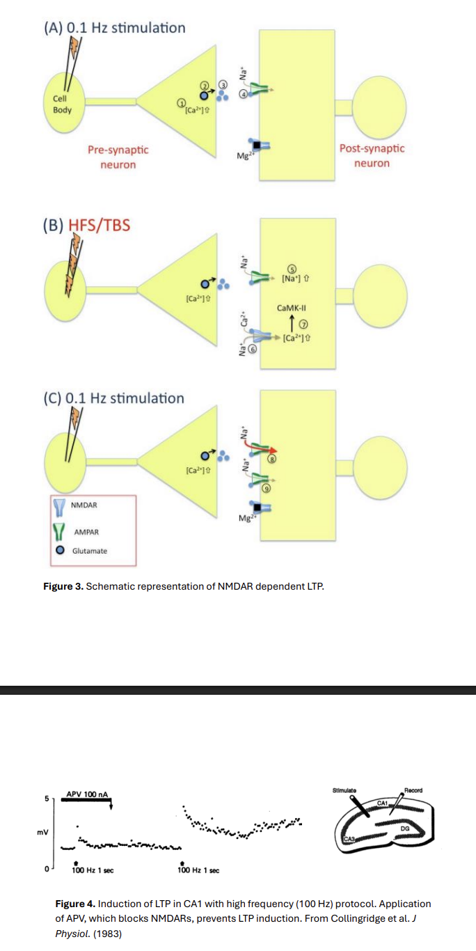
NMDAR-dependent LTP
How is NMDAR-dependent LTP induced experimentally
in different regions of the brain
CA1 or hippocampus
What does experimental stimulation cause?
Stimulate pre-synaptic neurones→ e.g Schaffer collaterals in the hippocampus
result→ arrival of AP in the presynaptic terminals of the neuron
Calcium enetry via voltage gated Ca Channels
leads to release of glutamate from presynaptic terminal
Glutamate bind to AMPARs of the postsynaptic membrane
allows Na+ influx through AMPARs
leads to slight depolarisation of the post-synaptic spine
How is this depolarisation recorded?
Via electrode
Group of CA1 neurone via an extracellular electrode
→ field Excitatory Post Synaptic Potentials
From a single neurone via intracellular electrode or using whole cell patch clamp technique
How does the experimenter stimulate the pre-synaptic Schaffer collaterals
at a constant amplitude and frequency
such as every 10 seconds
→ forms the baseline recording
What happens in baseline recording
AMPARs activated
synaptic efficacy remains the same→ each pre-synaptic stimulation there is the same size of AMPAR-mediated post-synaptic response
NMDARs→ still blocked by magnesium ions at hyperpolarised potentials
→ NO ION FLUX through NMDARs
Experimental protocol that can have a long lasting change on synaptic efficacy
activation of presynaptic neurone
at a high frequency
Types:
100 Hz High frequency stimulation (HFS)
Theta burst stimulation (TBS)→ a non-invasive brain stimulation technique that uses short, high-frequency bursts of magnetic pulses to modulate brain activity
What does this high frequency of presynaptic neuron sttimulation lead to
increased Na+ flux in post-synaptic neurone
lead to further depolarsiation of post-synaptic spine
Mg blockage of NMDARs is relieved
NMDARs allows Na+ and Ca+ entry
accumulates in the post-synaptic spine
Ca can act as a second messenger (the first messenger is glutamte)
activates specific intracellular mechanisms
What intracellular mechanisms are activated
Activation of calcium calmodulin (CaM)
activates calcium-calmodulin-dependent protein kinase II (CaMK-II)
kinase phsophorylate post-synaptic AMPA receptors at the GluA1 subunit and GluA2 subunit
increasing AMPAR conductance
CaMK-II can also be involved in…
trafficking of more AMPA receptors to the synaptic spine
Thus, subsequent to delivery of HFS or TBS, when the freuency of presynaptic stimulation is returned to pre-induction frequency…
when back to original frequency baseline
the AMPAR conductatnce is larger than prior to TBS/HFS delivery.
So synaptic efficacy increases
This is a long lasting (up to an hour) in creases
THEREFORE is LONG TERM POTENTIATION
A larger synaptic efficacy means that
there is higher probability of AP generation in the post-synaptic neurone
as a result of synaptic transmission
The induction of LTP occurs
during the short period of HFS/TBS delivery
where NMDARs are open
As LTP induction requires activity of NMDARs
this LTP is called NMDAR-dependent LTP
Evidence:
if the experimenter blocks NMDARs during HFS/LTP protocol
LLTP cannot be induced
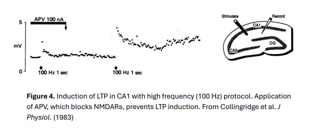
During LTP induction, NMDARs act as…
as coincident detectors
Meaning→ they are activated only when the pre-synaptic release of glutamate is coincident with post-synaptic depolarisation
What allows them to be coincident detectors
blockage of magnesium at resting potentials
Coincident detection can be better appreciated during
Pairing protocols
Pairing protocols
For induction of NMDAR-dependent LTP :
a high frequency closer to baseline levels
AT SAME TIME AS the post-synaptic neurone is depolarised by the experimenter
→This depolarisation is enough to relieve the magnesium block of NMDARs
Therefore what is required for LTP induction
NOT: high freuency stimulation of presynaptic neuron is not required to gradually depolarise the post-synaptic neuron via more soduim entry AMPAR receptor
instead
The concident of presynaptic neuron and post-synaptic neuron is again the requirement for LTP induction
What allows this coincidence of presynaptic firing and post-synpatic depolarsiaion
the magnesium blockage of NMDARs at hyperpolarised potentials
and relief→ and hence Ca2+ entry
How fast must the depolarisations be for LTP to be induced
depolarisation of post-synaptic neuron within 10ms
following the EPSP generated as a result of the presynaptic release of glutamate
in order for the LTP to be induced
what happens if the postsynaptic cell is depolarised much later than the EPSP generation
the LTP is not induced
Overall, what are the unique properties of NMDARs that form the basis of the role in inducing LTP
high calcium permeability
blockage by Magnesium at resting membrane potentials
slower kinetics of NMDA receptors compared to AMPARs
The properties of NMDARs contribute to three features of LTP
Co-operativity
Input Specificity
Associativity
Cooperativitity
if more presynaptic fibres are recruited
by increasing the intensity and not the frequency of the stimulation protocol
then more glutamate will be released
→ higher chance that the postsynaptic neurone (or neurones) will be depolarised
hence leading to removal of magnesium blockage of NMDARs
what ensures that only pathways that are highly active induce LTP→ input specificity
because depolarsiation of the postysynaptic neurones (the output)
and the magneisum relief of NMDARs only occurs at the postsynaptic spines that receive the high frequency input
→as other pathways without high frequency input and postsynpatic depolaristion do not change in synaptic efficacy
→ input specificity
Input specificicity
Associativity
follows from input specificity and cooperativity:
If there are two pathways that target the same postsynaptic neurone (or neurones)
but pathway A cannot induce LTP by itself
but pathway B can induce LTP by itself
(pathway A has smaller intensity of stimulation than pathway B)
i.e recruitetes less fibres than pathway B
→ IF high frequency stimulation is simultaenously applied to both pathways
Then LTP is induced in both pathways also
Why is this?
postsynaptic target will be depolaised sufficiently enough
to allow for opening of NMDARs
and Entry of calcium ions into the spine of pathway A
This is again the idea of coincident detection:
presynaptic glutamate release is coincident with postsynaptic depolarisation
The induction of NMDAR-dependent LTP during TBS/HFS leads to…
subsequent increase in AMPAR components on post-synaptic neurone
This increase is called→
The expression of NMDAR-dependent LTP
i.e only the induction is NMDAR dependent
Not post induction:
if the experimenter blocks NMDARs post HFS/TBS application (after LTP induction)
the synaptic efficacy remains high
NMDAR dependent LTP is an example of
Hebbian plasticity
Canadian pshycholist Donald Hebb
When an axon of Cell Ais near enough to excite a cell B and repeatedly and persistently takes part in firing it
some growth process or metabolic change takes pace in one or both cells suchas that A’s efficiency (as one of the cells firing B) is increased
In the case of NMDAR dependent LTP, the expression of this cahnge is the…
the increase in AMPARs
on the postsynaptic membrane
Overall summary of NMDAR dependent LTP (need to check this)
Apply HFS/TBS
NMDAR more likely to open Mg gate
Ca2+ into postsynaptic neuron
Causes kinase actiivty
increases no. AMPARs to membrane (expression)
and also more spines? thicker?
stronger efficacy
Order of paired protocol determines if LTP or LTD
Post synaptic second= LTP
Pre synaptic second =LTD