AQA A Level Biology: CELLS
1/51
There's no tags or description
Looks like no tags are added yet.
Name | Mastery | Learn | Test | Matching | Spaced | Call with Kai |
|---|
No analytics yet
Send a link to your students to track their progress
52 Terms
Structure of Nucleus
1. Nuclear envelope: double membrane surrounding nucleus, outer membrane continuous with the (R)ER of the cell.
2. Nuclear pores: allow the passage of larger molecules, such as mRNA, out of the nucleus.
3. Nucleoplasm: granular, jelly-like material making up the bulk of the nucleus.
4. Chromosomes: protein-bound, linear DNA.
5. Nucleolus: small spherical region(s) in nucleoplasm. Manufactures ribosomal RNA and assembles ribosomes.
Function of the Nucleus
1. Controls cell's activities - produces mRNA and tRNA - protein synthesis. Controls entry and exit of materials, and contains nuclear reactions.
2. Retains genetic material in the form of DNA and chromosomes.
3. Manufactures ribosomal RNA and ribosomes.
Structure of Mitochondria
1. Double membrane surrounding organelle - controls entry and exit of material.
2. Cristae - extensions of the inner membrane, providing a large surface area for the attachment of enzymes and other proteins during respiration.
3. Matrix - makes up the remainder - contains proteins, lipids, ribosomes and DNA (allows mitochondria to produce own proteins) and some respiratory enzymes.
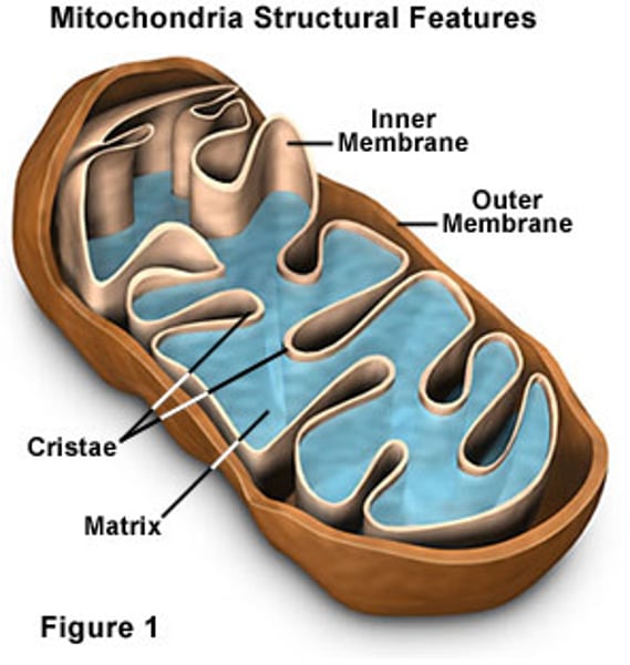
Function of the Mitochondria
1. Sites of Krebs Cycle and oxidative phosphorylation pathway in aerobic respiration - responsible for ATP production.
Structure of Chloroplasts
Found in plants and algae.
1. Chloroplast envelope - double plasma membrane, highly selective, surrounds the organelle.
2. Grana - stacks of disc-shaped thylakoid membrane.
3. Thylakoids - contain chlorophyll used in photosynthesis, can be linked by lamellae to other grana.
4. Stroma - fluid-filled matrix where Calvin Cycle takes place. Also contains starch grains.
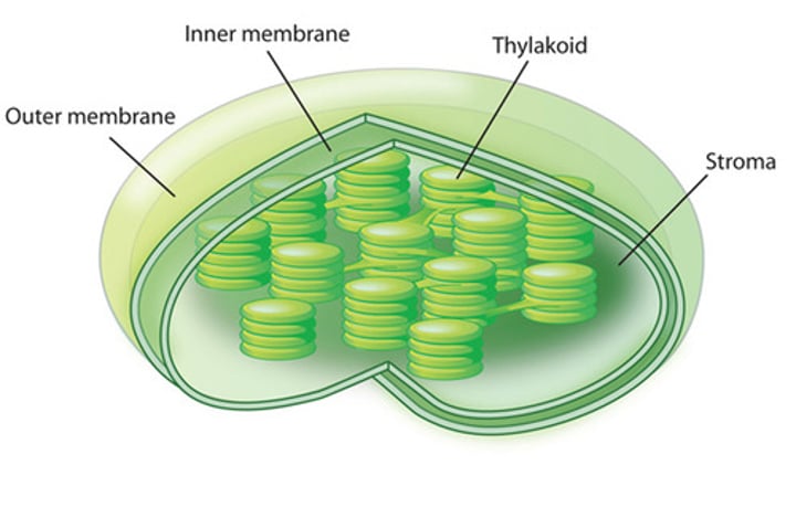
Functions of Chloroplasts
Site of Photosynthesis:
LDR in thylakoid membranes.
LIR in stroma.
1. Granal membranes provide a large SA for LDR - photosystems, e- carriers and enzymes etc.
2. Chloroplasts contain DNA and ribosomes - can quickly and easily manufacture some of the proteins needed for photosynthesis.
Structure of Endoplasmic Reticulum
1. 3D system of sheet-like membranes - continuous with the outer membrane of the nuclear double membrane.
2. Membrane contains a network of tubules and flattened sacs called cisternae.
3. RER - ribosomes on the outer surface of the membranes.
4. SER - lacks ribosomes on its surface and is often more tubular in its appearance.
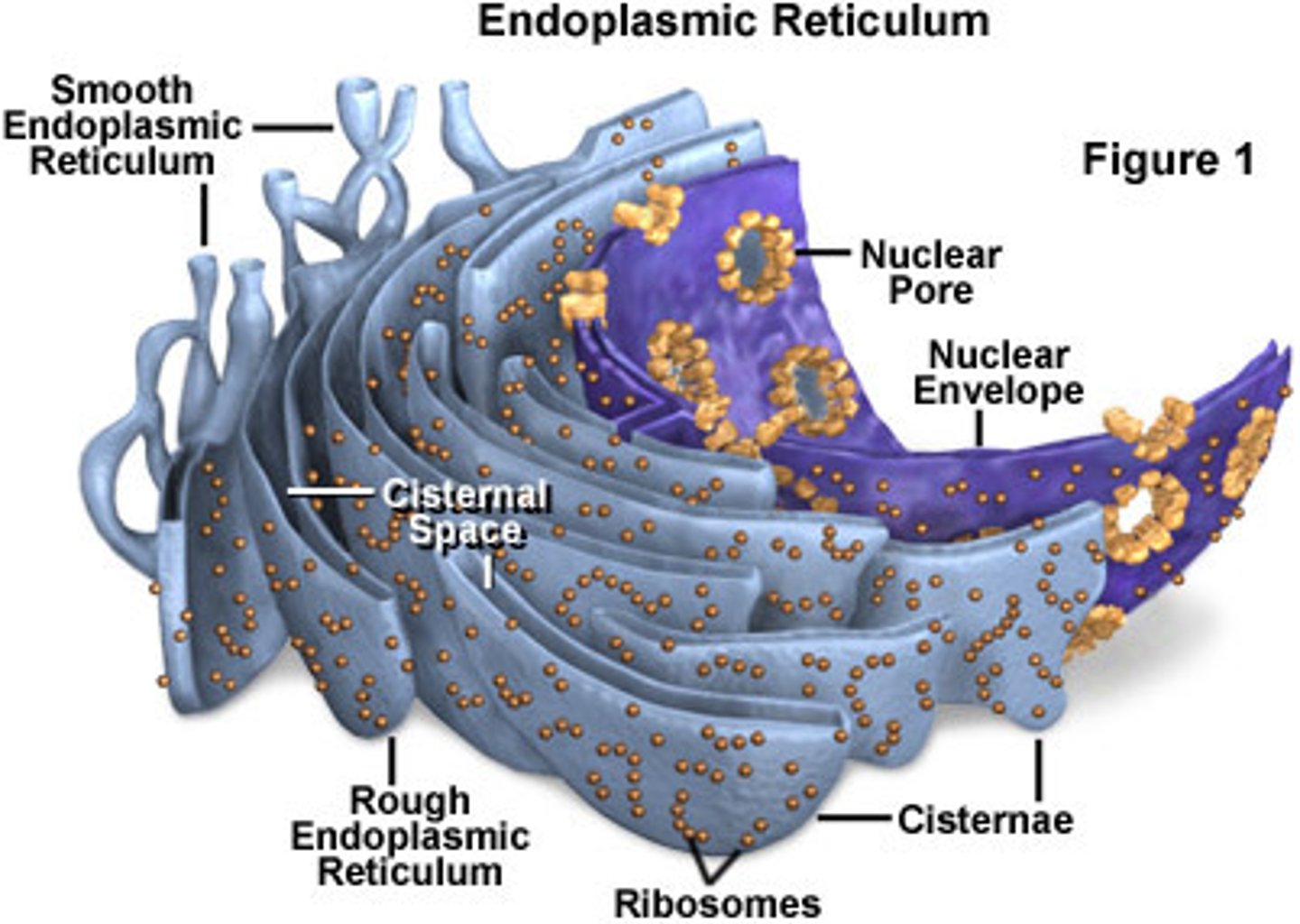
Function of Endoplasmic Reticulum
RER
1. Large SA for protein/glycoprotein synthesis.
2. Provides a pathway for material transport throughout the cell, especially for proteins.
SER
1. Synthesises, stores and transports lipids and carbohydrates.
Structure of Golgi Apparatus
1. Compact system of flattened sacs and stacked membranes (cisternae).
2. Vesicles - modified proteins and lipids transported to cell membrane where they fuse with it, and then egest contents to the outside.

Function of Golgi Apparatus
1. Form glycoproteins by adding carbs to proteins.
2. Produce secretory enzymes, such as those secreted by the pancreas - apparatus is developed in secretory cells, especially those in the small intestine.
3. Secrete carbs, such as cellulose for plant cell walls.
4. Transports, modifies and stores lipids.
5. Forms lysosomes.
Structure of Lysosomes
Golgi vesicles with proteases, lipase and lysozymes.
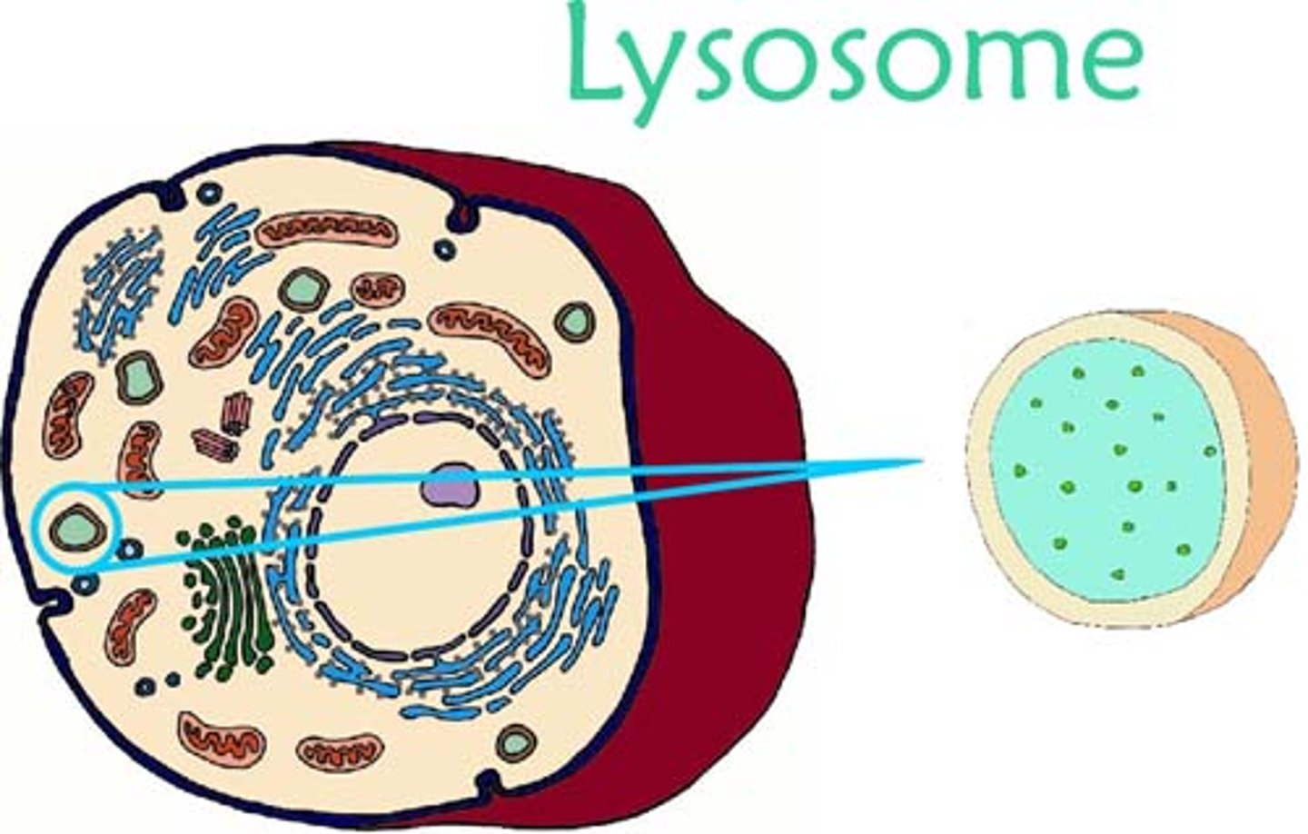
Functions of Lysosomes
1. Hydrolyse foreign material ingested by phagocytes.
2. Exocytosis of enzymes to destroy extra-cellular material.
3. Apoptosis - programmed cell death.
Autolysis - breaking down cells after death.
4. Digest worn out organelles - can recycle chemicals.
Structure of Ribosomes
1. Small cytoplasmic granules found in all cells, free-floating or associated with RER.
2. 80S - found in eukaryotic cells, slightly larger.
3. 70S - in prokaryotic cells, slightly smaller.
4. 2 Subunits - large and small - contain ribosomal RNA and proteins.
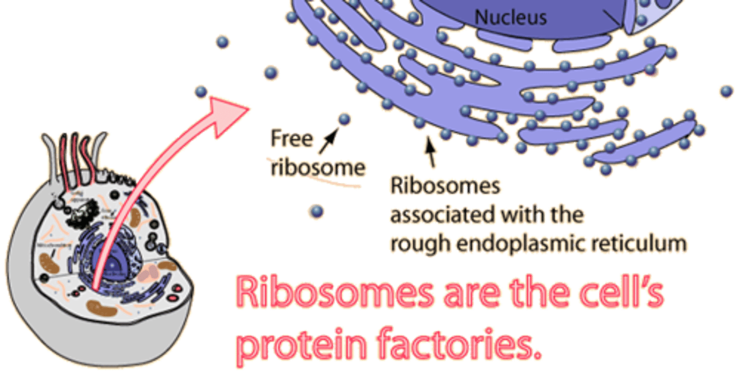
Functions of Ribosomes
Carry out translation stage of protein synthesis to produce polypeptides.
Structure of Cell Wall
Found in plants, algae and fungi.
1. Cellulose microfibrils embedded in a matrix - contribute to overall cell wall strength are considerably strong.
and other polysaccharides.
2. Middle lamella - marks the boundary between adjacent cell walls and cements adjacent cells together.
Functions of Cell Wall
1. (Cellulose) - to provide mechanical strength to prevent cell wall bursting under pressure created by osmotic entry of water.
2. To provide mechanical strength to the cell as a whole.
3. Allows water to pass along it - contributes to the movement of water through the plant.
Structure of Vacuoles
1. Fluid-filled sac bounded by a single membrane.
2. Single membrane around it called tonoplast.
3. Solution of mineral salts, sugars, amino acids, wastes and sometimes pigments such as anthocyanins.
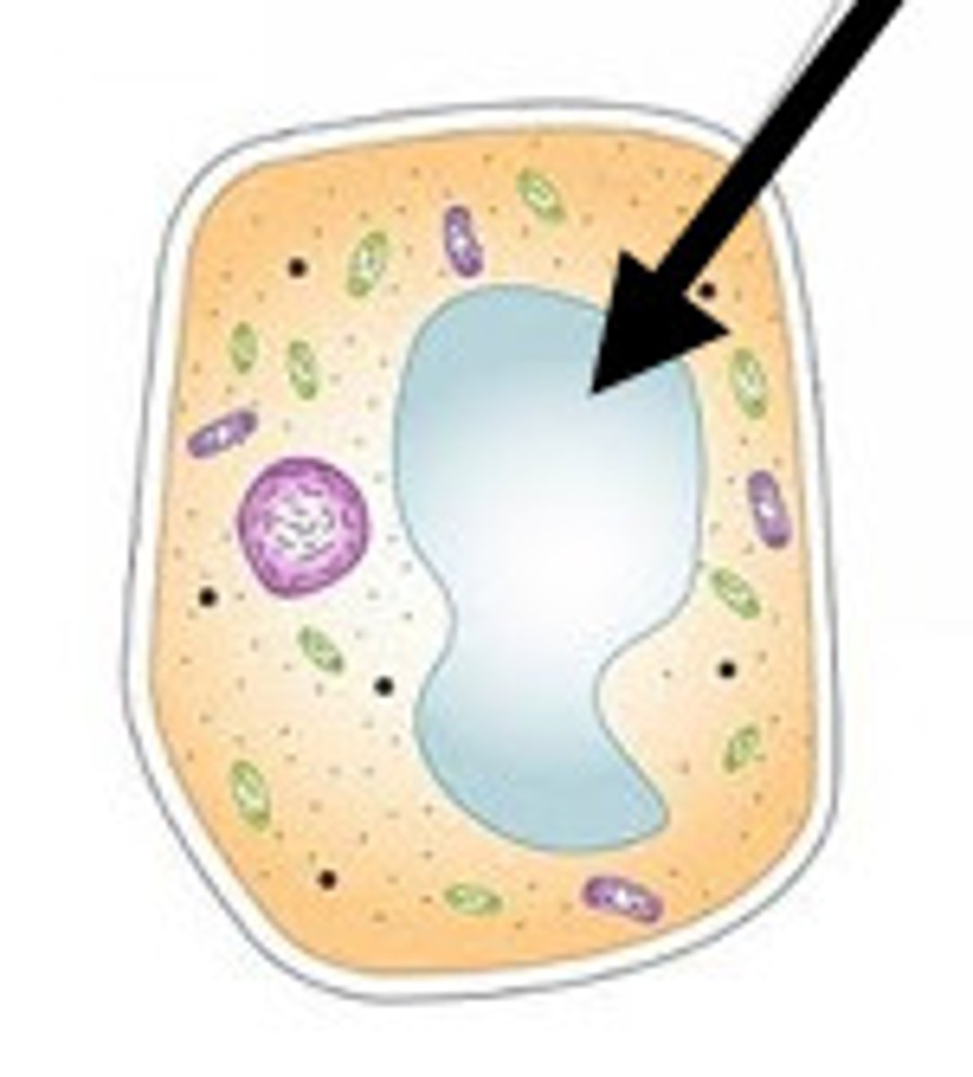
Functions of Vacuoles
1. Support herbaceous plants and herbaceous parts of woody plants by making cells turgid.
2. The sugars and amino acids can act as a temporary food source.
3. Pigments - may attract pollinating insects due to colour.
Define Tissue
Give an example
A collection of similar cells that are aggregated together and work together to perform a specific function.
Example = epithelial tissue - consists of sheets of cells, lining the surfaces of organs, often having a protective or secretory function.
Define organ.
Comment on the difference between capillaries, veins and arteries, relative to the term organ.
A combination of aggregated tissues that are co-ordinated together to perform a variety of functions, one of which is the predominant major function.
While capillaries, veins and arteries all have the same major function, i.e.e carrying blood, capillaries are not organs, unlike veins and arteries as they are made up of only one tissue - epithelium.
Define eukaryotic cell
A larger cell with a true nucleus that is bounded by a nuclear membrane/nuclear envelope.
Can you name and explain an example of a eukaryotic cell with specific adaptations?
A sperm cell:
- Many mitochondria for ATP production which to allow movement
- Undulipodium (tail) for movement towards egg
- Acrosome (specialised lysosome) to help the sperm penetrate the egg
- Diploid nucleus, as it is a gamete
Define prokaryotic cell
A smaller cell which has no true nucleus or nuclear envelope.
Can you describe how prokaryotic cells differ from eukaryotic cells?
Prokaryotic cells:
- have no nucleus
- smaller 70S ribosomes
- cytoplasm lacks membrane-bound organelles
- much smaller cell
- cell wall contains murein (a glycoprotein)
- plasmids may be found in prokaryotic cells
Eukaryotic Cells:
- have nucleus
- 80S ribosomes
- membrane-bound organelles
- larger cell
- cell wall made of cellulose or chitin
- no plasmids
Can you list other features of prokaryotic cells?
Prokaryotic cells can have:
- one or more plasmids
- a slime capsule surrounding the cell
- one or more flagella
Principles of Optical Microscopes
Simple convex glass lenses used in pairs in a compound light microscope to focus an object at a short distance by 1st lens, then magnified by 2nd lens.
Can you describe the pros and cons of optical microscopes?
Pros:
- cheap
- images in colour
- no training required
- live specimens
Cons:
- low magnification x1500
- low resolution
- 2D images
Principles of Transmission Electron Microscopes
1. Electron gun produces e- beam, focused onto the specimen by a condenser electromagnet.
2. Beam passes through a thin section of the specimen from below. Parts absorb e- and appear dark; others let e- pass through and appear bright - produces image on screen - photomicrograph.
Can you describe the pros and cons of transmission electron microscopes?
Pros:
- high resolution images
- high magnification
- visible internal structures
Cons:
- expensive
- training is required
- no colour images
- 2D images
- only thin specimens
Principles of Scanning Electron Microscopes
1. Beam of e- directed onto surface of specimen - passed back and forth across specimen.
2. e- scattered by specimen - scattering pattern analysis allows us to get a 3D image.
Can you describe the pros and cons of scanning electron microscopes?
Pros:
- 3D images
- high magnification
- high resolution
- thick specimens
Cons:
- expensive
- training is required
- no colour images
How do you prepare a slide for an optical microscope?
1. Pipette a drop of water onto the slide
2. Use tweezers to place a thin section of your specimen on top of the droplet
3. Add a drop of a stain
4. Add a cover slip - remove all air bubbles
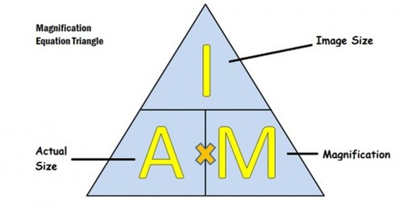
What is the difference between magnification and resolution?
MAGNIFICATION - increasing the size of an image. Up until the limit of resolution, an increase in magnification = an increase in detail.
RESOLUTION = minimum distance apart that two objects can be for them to appear as separate items.
What is the formula to calculate Magnification?
Can you describe the principles of cell fractionation and ultracentrifugation in separating cell components?
1. Homogenisation
- tissue is broken up in a cold, isotonic buffer solution to release the organelles into a solution
2. Filtration
- the homogenised cell solution is filtered through a gauze
- this separates any large cell debris
3. Ultracentrifugation
- the cell fragments are poured into a test tube and placed in a centrifuge and spun at a low speed
- a thick sediment - the pellet - is at the bottom of the tube and the fluid above is the supernatant
- the supernatant is drained into a new tube and spun again at a higher speed
- a new pellet forms and again, the supernatant is drained off and spun again at an even higher speed
- this process is repeated at higher speeds each time until all the organelles are separated out
Why is a cold, isotonic buffer needed?
COLD - to reduce enzyme activity that could break down organelles.
ISOTONIC - same water potential as tissue sample - to prevent water moving in or out of the cells by osmosis, causing lysis.
BUFFERED - to prevent changes in pH which could affect/denature enzymes.
How are the organelles separated out during centrifugation?
They are separated in order of mass and the order is usually:
- nuclei
- mitochondria
- lysosomes
- endoplasmic reticulum
- ribosomes
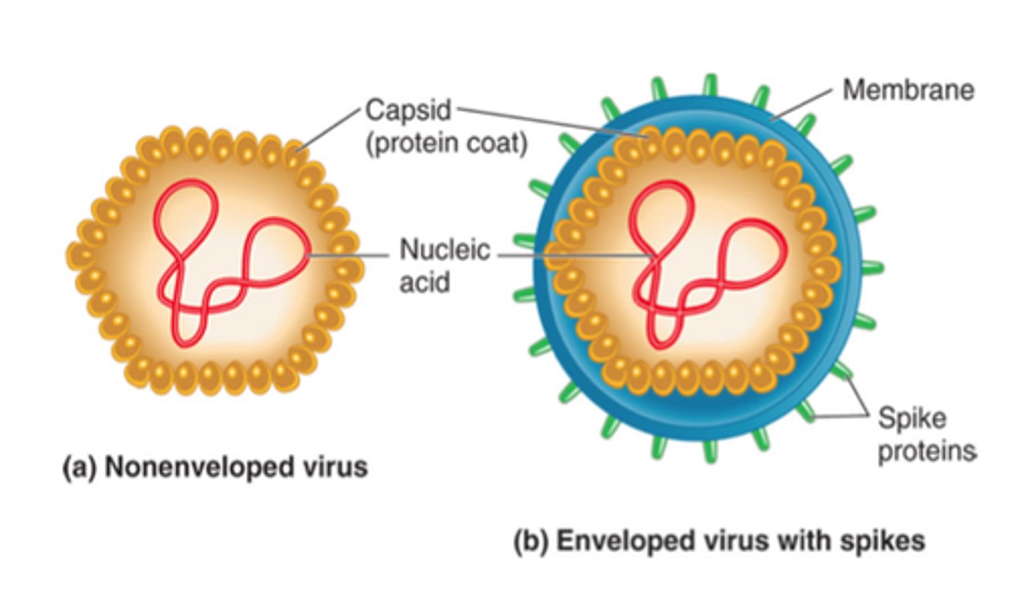
Define Virus
Acellular, non-living particles. Smaller than bacteria.
Can you explain that viruses are acellular and non-living?
Viruses are nucleic acids surrounded by protein so they are not living.
Can you describe the structure of virus particules?
- no plasma membrane, cytoplasm, ribosomes
- a core of genetic material : DNA or RNA
- capsid surrounding core
- attachment proteins around edge of capsid
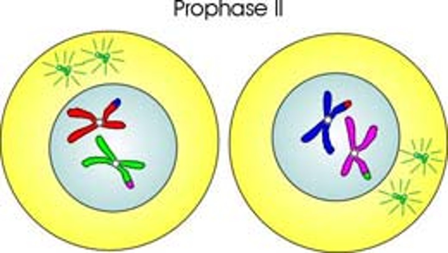
Outline the differences between mitosis and meiosis.
Mitosis results in 2 genetically identical diploid daughter cells.
Meiosis results in 4 genetically different haploid daughter cells.
List the stages of Mitosis
Interphase
Prophase
Metaphase
Anaphase
Telophase
Outline Interphase
Precedes Mitosis
1. Cell is not dividing.
2. Considerable cellular activity - replication of DNA, two copies on centromere.
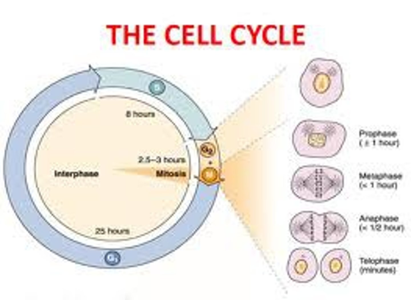
Outline Prophase
1. Chromosomes become more visible, thicken.
2. Centrioles move to opposite ends of the cell (poles).
3. Spindle fibres develop from each of the centrioles (spindle apparatus).
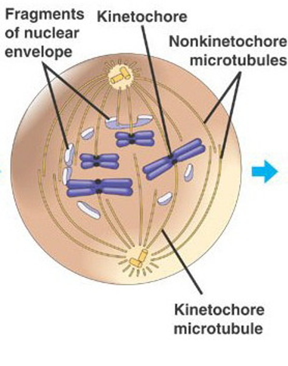
Outline Metaphase
1. Chromosomes seen to be made up of two chromatids.
2. Microtubules attach to centromere - chromosomes pulled to the cell equator where they line up.
Outline Anaphase
1. Centromeres divide, separating each pair of sister chromatids.
2. Chromatids pulled to their respective poles as spindles contract, centromeres first - v-shaped.
Outline Telophase
1. Chromosomes reach their respective poles and then uncoil, become long and thin again - chromosomes again.
2. Spindle fibres disintegrate; nuclear envelope and nucleolus reform.
3. Cytoplasm divides in cytokinesis.
Why is mitosis so important?
GROWTH - ensures that all cells growing from original cell of an organism are genetically identical.
REPAIR - Important that replacement cells produced have an identical structure and function to lost cells.
REPRODUCTION - Single-celled organisms divide by mitosis to give 2 new organisms - each new organism is genetically identical to the parent organism.
How does cancer arise? Difference between benign and malignant?
Result of gene damage controlling mitosis and cell cycle.
Mutant cells are structurally and functionally different - most mutant cells die but surviving mutant cells become tumours.
Malignant - grow rapidly, less compact, more likely to be life-threatening.
Benign - grow more slowly, more compact, less likely to be life-threatening.
How can cancer be treated?
How can cancer be treated?
Involves killing dividing cells by blocking a part of the cell cycle - cell division and therefore cancer growth ceases.
Chemo disrupts cell cycle by preventing DNA replication or by inhibiting the metaphase stage of mitosis by interfering with spindle formation.
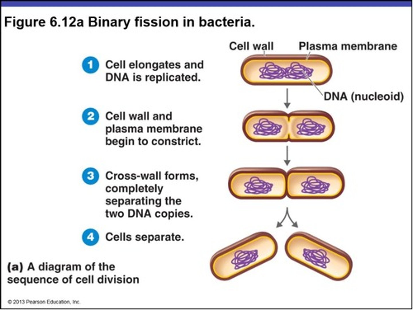
Outline the process of Binary Fission
How prokaryotic cells divide:
1. Circular DNA molecule replicates, and both copies attach to the cell membrane.
2. Plasmids also replicate.
3. Cell membrane begins to grow between the two DNA molecules and begins to pinch inwards - dividing cytoplasm in two.
4. New cell wall forms between the DNA molecules, dividing the original cell into two identical daughter cells - each with a copy of the circular DNA and a variable number of copies of the plasmids.
Outline how viruses replicate
Acellular, non-living, so do not undergo cell division.
1. Attach to host cell with the attachment proteins on their surface.
2. Inject nucleic acid into host cell.
3. Viral nucleic acid then hijacks the cell's machinery and codes for metabolic processes to produce viral components - nucleic acid, enzymes, structural proteins, which are then assembled into new viruses.