Osmoregulation (Lectures 24-26)
1/67
Earn XP
Description and Tags
BIO 225, Duan, W26
Name | Mastery | Learn | Test | Matching | Spaced | Call with Kai |
|---|
No analytics yet
Send a link to your students to track their progress
68 Terms
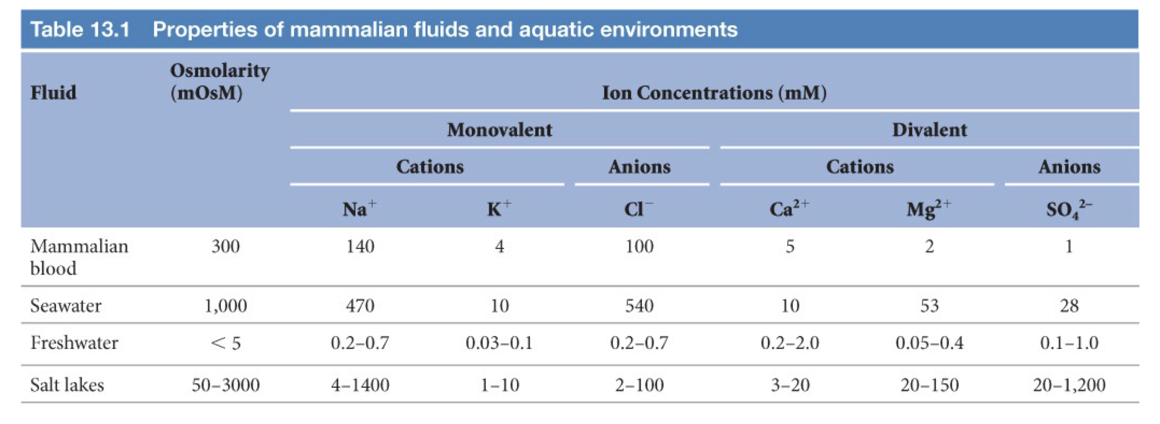
Different fluids and Aquatic Environments Properties
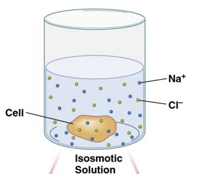
Isosmotic Solution
Solution matches the osmolarity of the cell so water does not flow net in or out, but there is still a flow of water, net flux is 0.
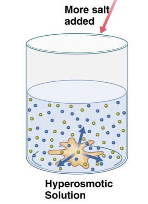
Hyperosmotic Solution
Solution has a much higher osmolarity than the cell, so water leaves the cell to try and balance these solute concentrations
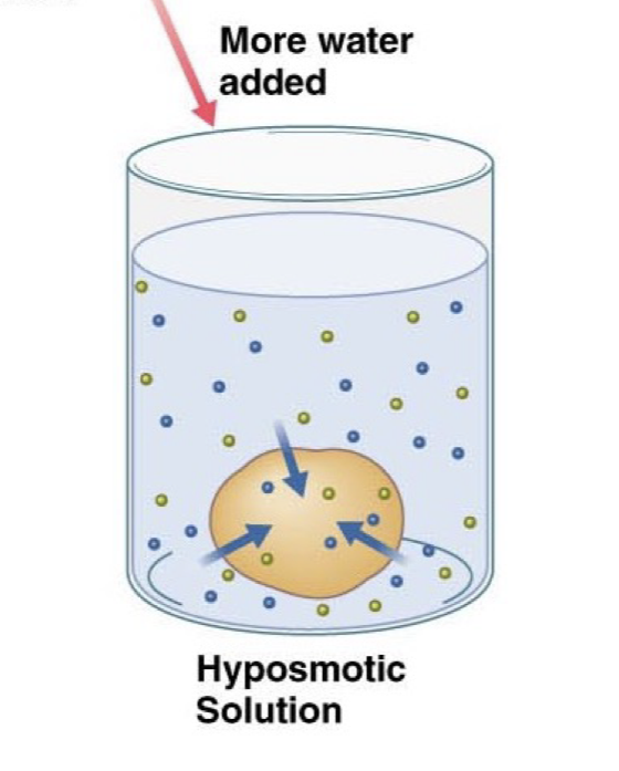
Hyposmotic Solution
The solution has much lower osmolarity than the cell, causing water to rush into the cell to try and balance these concentrations, eventually would lead to the cell bursting open.
Marine Environments
Lots of solute in the extracellular environment, so the osmolarity is higher in the external environment
Animals tend to lose water and gain salts, against their homeostatic osmolarities.
Freshwater environments
Typically, there is a low osmolarity, low solute concentration in freshwater environments compared to the animals who live in these waters.
As a result, animals tend to gain water and lose salts, against their homeostatic osmolarities.
Terrestial Environments
Animals simply tend to lose water, constant battle for being hydrated.
Animal Movement
Many animals tend to move and change environments during their lifetime and so they must be able to alter their homeostatic mechanism.
Osmotic Regulation
In order to meet and handle osmotic challenges, animals tend to practice two forms of regulation, either be osmoconformers or osmoregulators.
Animals need to have different mechanisms for handling the different solute concentrations in the environments they navigate.
Osmolarity is the total solute concentration in an environment or animal.
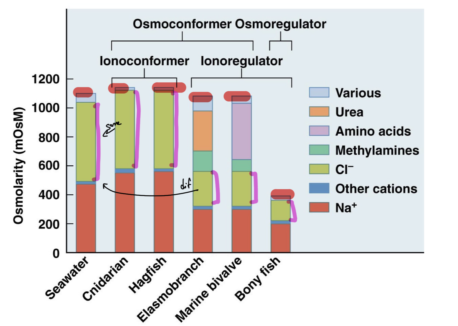
Osmoconformer
Conform to their external environment osmolarity, which is the total solute concentration.
Internal and external osmolarity is similar.
Match the solute content in their bodies to the content in the environment surrounding them
Marine invertebrates tend to be osmoconformers.
Osmoregulator
These animals maintain a constant osmolarity rather than changing to match their environment.
Osmolarity remains constant regardless of their external environment.
Most vertebrates are osmoregulators
Ionic Regulation
Animals also need to manage their individual ion concentration, how much of each ion they maintain in their bodies.
Animals have two mechanism to control their ionic concentrations, either be ionoconformers or ionoregulators.
Ionic regulation is the regulation of how much of each ion is kept in the organism as compared to the external environment and other animals.
Ionoconformer
Exert little control over their own ion profile and end up matching ion concentrations with the extracellular space
EXCLUSIVELY found in marine animals, such as many marine invertebrates
Ionoregulator
Have individual control over their ion profile of extracellular space, do not leave it up to environment.
Most vertebrates are ionoregulators, have different amounts of Na+ compared to the environment for example
Can control the individual ion concentrations in blood or extracellular space of body.
Osmotic and Ionic Regulation Together
Some animals can be both ionoconformers and osmoconformers, matching their total solute concentration to the environment as well as matching specific ion profile to the ion profile of the environment, same concentration of Na+, Cl-, etc.
However, some animals can also be ionoregulators but can have the same total solute concentration (osmolarity) as environment. So they would have different amounts of specific ions but end up with the same total solute concentration as the environment.
On the other hand, some animals are both ionoregulators and osmoregulators, controlling their own total solute concentrations and maintaining their own ion profiles as compared to their environment.
Perturbing Solutes
Solutes which cause damage and dysfunction in organisms, not good environments for organisms to live in
Marine invertebrates and ancient fish, such as lamprey and cartilaginous fish, which are osmoconformers would not fare well in perturbing solutes
Compatible Solutes
Solutes that are good and stable environments for osmoconformers to match with and live in.
Maintain relatively stable and are not extremely harmful for osmoconformers to live in.
Counteracting Solutes
Solutes which chemically oppose one another in an environment and can be regulated together to provide a stable environment for an osmoconformer. Like a chemical tug of war.
Protect chemical and cellular structure by actively synthesizing and regulating these counteracting solutes which provide an overall safe and neutral environment for the osmoconformers. One solute without its counteracting solute would also be harmful, but the cells maintain the proper concentrations of each to counteract one another.
Matching the external osmolarity could mean accumulating a high amount of a pertubring solute but also accumulating a high amount of a more stabilizing solute, which provides a net neutral effect on the osmoconformer’s physiological processes.
Through conforming, animals may accumulate large amounts of a harmful solute (urea) but to counteract this solute, they also accumulate large amounts of a counteracting solute, such as TMAO (methylines), which provide a net neutral environment in the end.
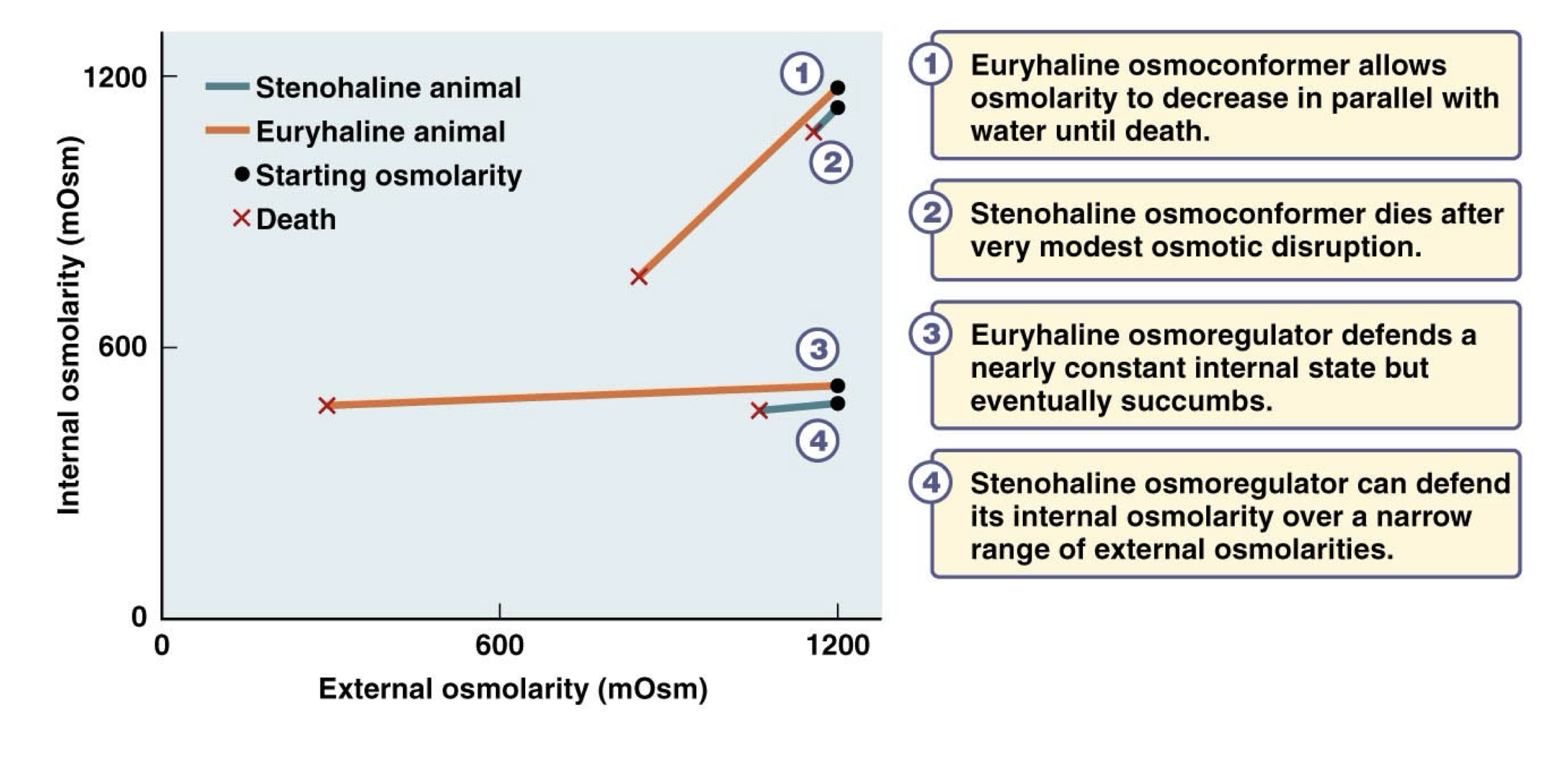
Stenohaline
Animals which can tolerate only a narrow range of external osmolarities before succumbing to the osmolarity differences and dying.
Think steno=stern=strict, tight range
Eurylhaline
Animals which can tolerate a wide range, can live in many different environments and tolerate a wide range of osmolarity concentrations.
Euryhaline and Stenohaline Osmoconformers and Osmoregulators
Euryhaline osmoconformers allow the osmolarity to decrease in parallel with decreasing water osmolarity until a certain point, at which they die.
Stenohaline osmoconformers die after a very modest/slight osmotic disruption
Euryhaline osmoregulators defend a nearly constant internal state across a wide range of water osmolarities but eventually submits and dies
Stenohaline osmoregulators also maintain a constant internal osmolarity, but only over a very narrow range of external osmolarities, eventually cannot regulate and die.
Epithelial Tissue Properties which Allow Ion Movement to Osmoconform/Osmoregulate
Four Features
Asymmetrical distribution of membrane transporters, so that solutes are selectively transported across the membrane. Cells have polarity, apical membrane and basolateral membrane which have different distribution of transporters for different solutes.
Cells are interconnected with tight junction to form an impermeable sheet of tissue. Little leakage of solutes between cells.
High cell diversity within the tissues
Abundant mitochondria because it takes a lot of energy to power all of the necessary transporters. Large Energy (ATP) supply
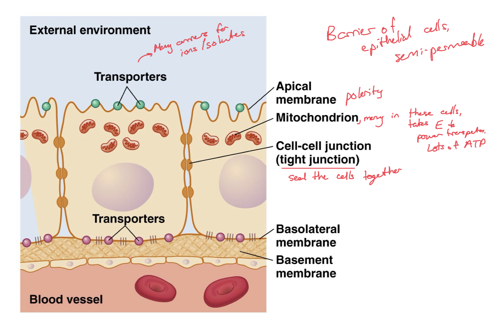
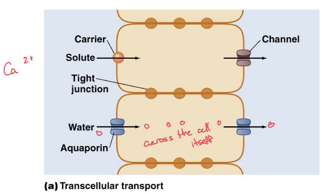
Transcellular Transport
Transport of a solute, like water through aquaporins, across the cell itself, right down the middle of a cell.
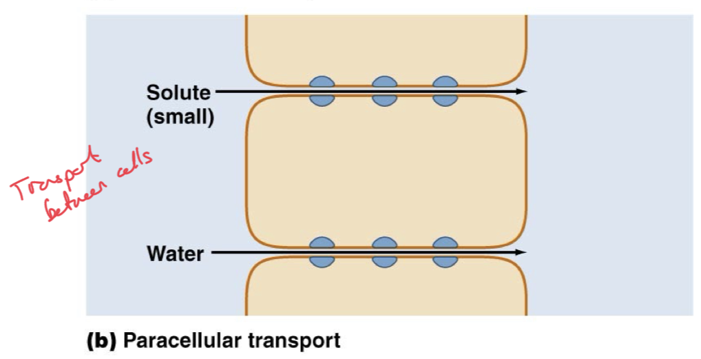
Paracellular Transport
Transport of a solute right down the crack between two cells. Pass/leak through the tight junctions.
Movement between cells, usually through more “leaky” epithelia rather than the “tight” epithelia
Only possible for small solutes which can leak through. Water can pass through paracellular transport.
More likely to occur when solute concentrations are higher, stronger push/gradient force.
Types of transporters for solute movement
Na+/K+ ATPase
Ion Channels (Cl-, K+, Na+)
Electroneutral cotransporters
Electroneutral Exchangers
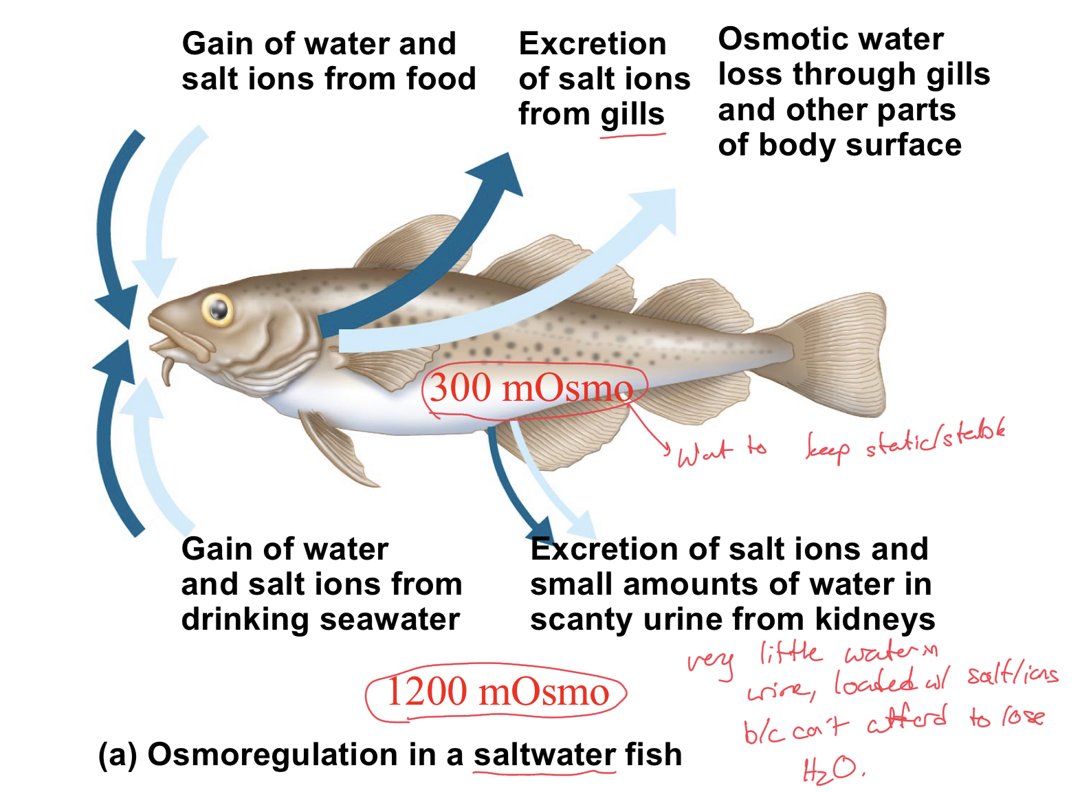
Osmoregulation in Saltwater Fish
These fish will inherently gain water and salt ions from their food and from drinking seawater. Their homeostatic osmolarity is much lower than that of the seawater environment.
As a result, these fish have to lose lots of salt ions and try to conserve as much water as possible.
Simply due to osmolarity differences, they experience osmotic water loss through their gills and other parts of body surface
Fish work to excrete salt ions from gills, and excrete mainly salt ions, only small amounts of water in scanty urine from kidneys.
Very little water in their urine, loaded with salts/ions because they cannot afford to lose H2O and want to get rid of as many salt ions as possible, as they want to maintain the lower osmolarity.
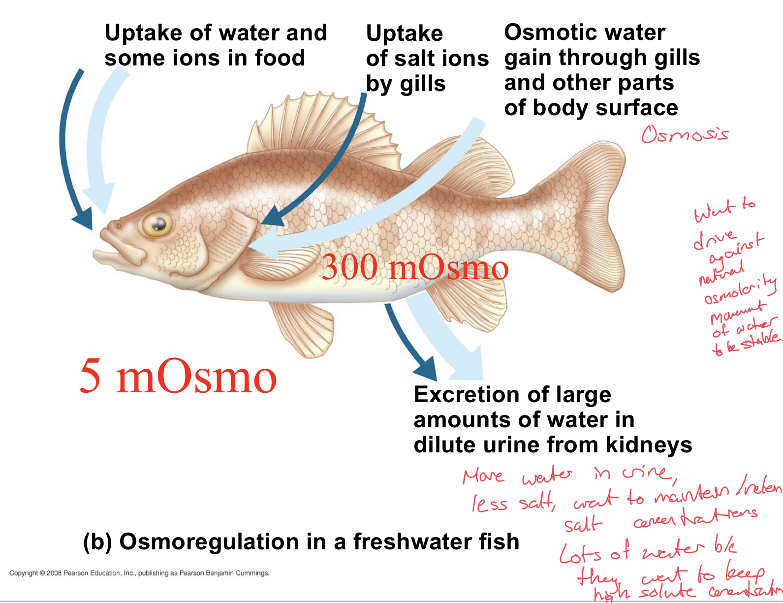
Osmoregulation in a freshwater fish
Fish naturally has a much higher osmolarity than the external aquatic environment.
As a result, there is naturally some osmotic water gain through the gills and other parts of body surface. Higher solute inside, so water moves in naturally.
Uptake of water and some ions in food.
Uptake of salt ions by gills
End up excreting large amounts of water, very dilute urine from kidneys
More water in urine, less salts in urine. Want to maintain/retain their salt/ion concentrations.
Want to keep the higher osmolarity so try to uptake salt ions and lose lots of water through urine, drive against the natural osmolarity movement of water.
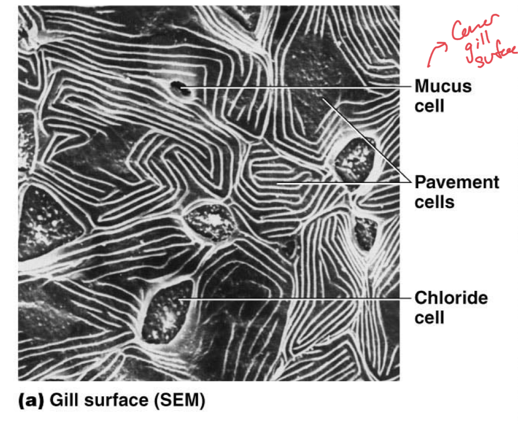
Epithelial Cells in Fish Gills
Fish gill lamellae are the major organ that controls osmolarity.
Lamellae are composed of mitochondria-rich chloride cells, pavement cells which can be mitochondria rich or poor
Pavement cells of lamellae carry out other tasks to maintain the water-ion balance.
Transport of ions is likely carried out by the mitochondria rich cells because they have lots of ATP production to power the transporters.

Saltwater-Freshwater Transitions
Some fish migrate between saltwater and freshwater and are known as diadromous fish
Ion functions of epithelia must change during migration to acomodate for the different ion concentrations and osmolarity of the new environments.
These changing ion functions are controlled by hormones, may need to conserve water or lose more water depending on where you are moving from/to.
Catadromous Fish
Live in freshwater and migrate to saltwater to spawn
Eels are a good example
Ion transport would change because in freshwater, try to conserve ions and excrete lots of water due to low osmolarity environment but in saltwater, will need to conserve water and have very concentrated urine, excreting lots of ions.
Anadromous Fish
Live in saltwater for majority of their lives and migrate to freshwater to spawn
Salmon are a good example of this
In this case, these animals would be used to excreting very little water and producing very concentrated urine, excreting lots of excess ions. However, in freshwater, this would need to change to excrete lots of water and conserve ions to maintain the higher osmolarity than freshwater environments.
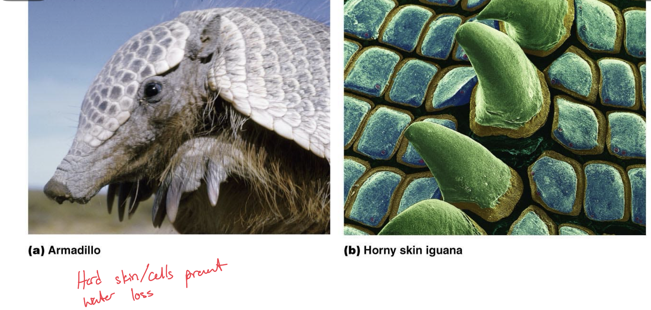
Land Animals and Water Conservation
Water conservation is a primary priority
Typically animals will need to reduce their water flux/loss
Do this by covering external surfaces with layer of hydrophobic molecules such as mucus
Cornified stratum corneum with keratin, which is a lipid modified protein, provides a protective outermost layer to epidermis to prevent dehydration
common in terrestrial amniotes (mammals, birds, reptiles)
Cuticles with chitin, such as in arthropods (insects, crustaceans, arachnids) also prevent the evaporation of water easily.
Integumentary System
Composed of basement membrane and epidermis.
Epidermis has keratinocytes and corneocytes
Corneocytes are specialized, dead keratinocytes which form the outermost layer of the skin, a dry cell layer known as the stratum corneum.
Corneocytes prevent water evaporation.
Sources of Water for Terrestial Animals
Dietary Water
Metabolic Water
Drinking
Dietary Water
Water found in the food sources we ingest.
Water that is preformed in plant and animal tissue
Metabolic Water
Water generated as a result of oxidative phosphorylation
Breakdown of glucose and lipids for ATP also results in water as a byproduct. Last step of aerobic respiration.
Drinking
Water that we drink that goes into our body this way
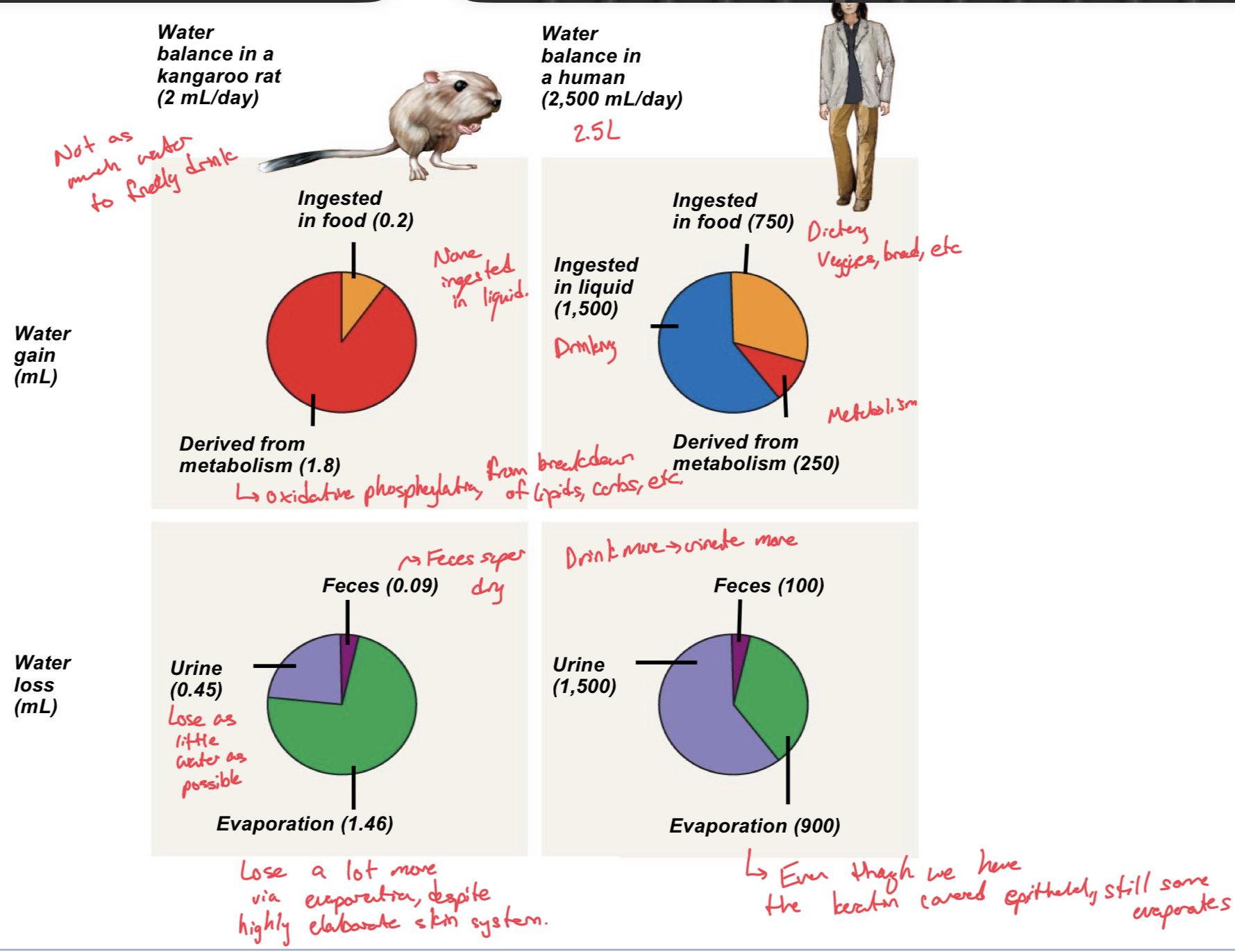
Different Water-Conservation Adaptations
Desert animals have very little external water sources available to them, almost no water to just drink.
Instead, desert animals, like the kangaroo rat gain water from ingested food and MOSTLY from their metabolic processes which produce water as a byproduct
In contrast, humans get most of their water from ingesting it in liquids, as well as getting water that is preformed in foods. Only a smaller portion of our water comes from our metabolic processes.
As far as water loss, desert animals try to conserve as much water as possible when it is being filtrated in the kidneys, so they only lose about a quarter of it to urine, very little is lost in feces, but most is lost through evaporation due to heat and conserving it otherwise. Have highly elaborate integumentary systems, but the greatest loss is still in evaporation.
As for humans, most of our water loss is through urine, then our evaporation, and the least exits through feces. Despite us having a keratin covered epithelium, some water still escapes through evaporation.
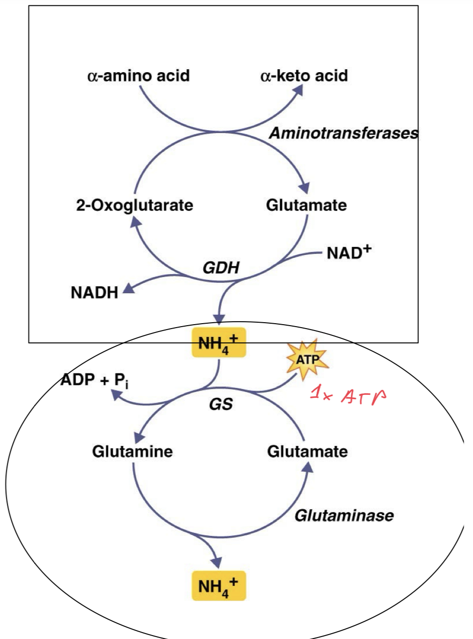
Nitrogen Excretion
Ammonia is a byproduct produced from the breakdown of amino acids, because they obviously have amine chains.
Ammonia is toxic and must be excreted
It can be excreted in three forms: the unprocessed ammonia, uric acid, and urea, depending on the organism.

Type of Nitrogen Compound Excreted
The type of nitrogen compound excreted depends on the animal and the animal’s environment
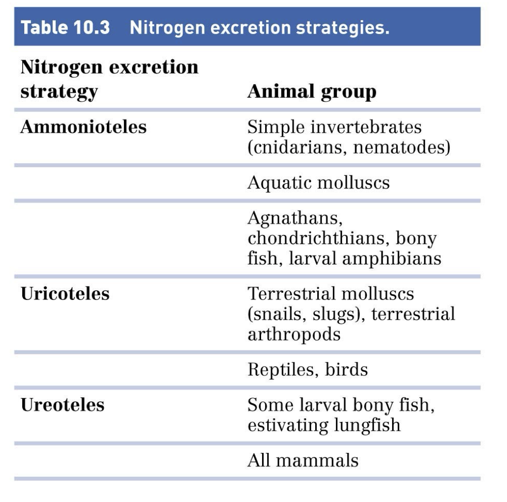
Ammonioteles Nitrogen Secretion
Ammonioteles are animals that excrete ammonia as is, because they are in a water-rich environment.
Aquatic animals usually excrete ammonia.
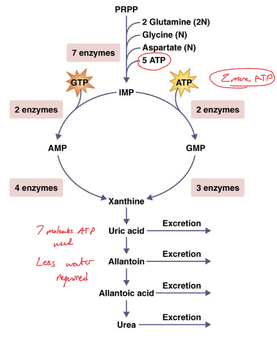
Uricoteles
Animals that secrete nitrogen in the form of uric acid.
Birds are a common animal which secrete nitrogen in this form.
Cannot carry as much nitrogen.
Ureoteles
Terrestial animals that secrete nitrogen in the form of urea
Cannot carry as much nitrogen
Humans are a form of animal which secrete nitrogen in this form.
Ammonia Secretion
Advantages: Ammonia released by the deamination of amino acids, requires very little energy to produce ammonia, 1 ATP
Disadvantages: Highly toxic, requires large volumes of water to store and excrete safely/properly, hence why mainly aquatic animals do this. Has to be very dilute urine.
Uric Acid Excretion
Advantages: Few toxic effects and can be excreted in small volumes of water, urine does not need to be very dilute.
Disadvantages: Expensive to produce, takes the most energy to produce, uses up 7 molecules of ATP total.
Urea Excretion
Advantages: Only slightly toxic, not as toxic as ammonia. Relatively inexpensive to produce, energetically cheaper to produce than uric acid. Uses up 5 ATP for 1 molecule of Urea.
Disadvantages: Urea is a perturbing solute, so it naturally will disrupt macromolecular processes/functions at any normal concentration in the animal.
Six Roles of Kidney
Ion balance
Osmotic balance
Blood Pressure
pH balance
excretion of metabolic wastes and toxins
hormone production
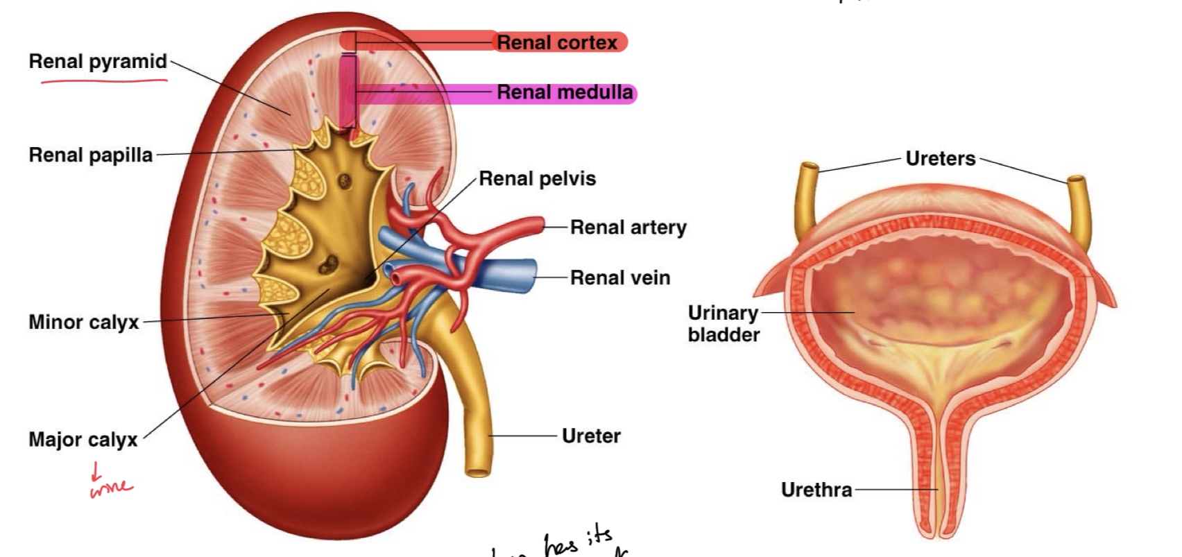
Kidney Structure
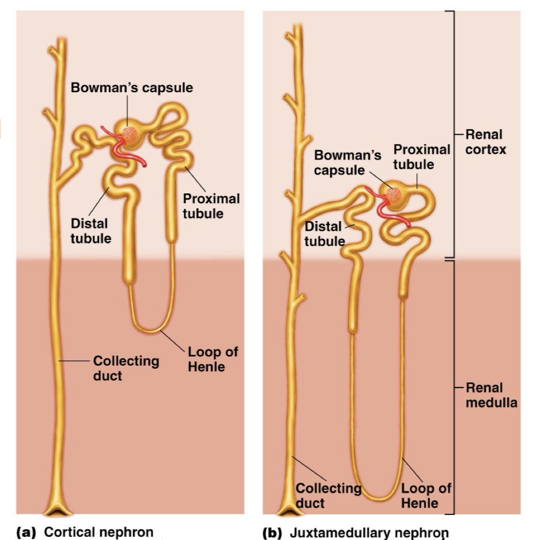
Kidney has many many nephrons which span the renal cortex and the deeper, renal medulla.
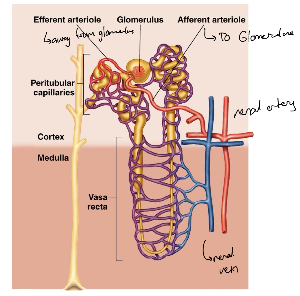
Each nephron has its own blood supply to filter
Main blood network is the renal artery and the renal vein
Minor and major calyx collect urine from the renal pyramids and direct it to the renal pelvis to be excreted.
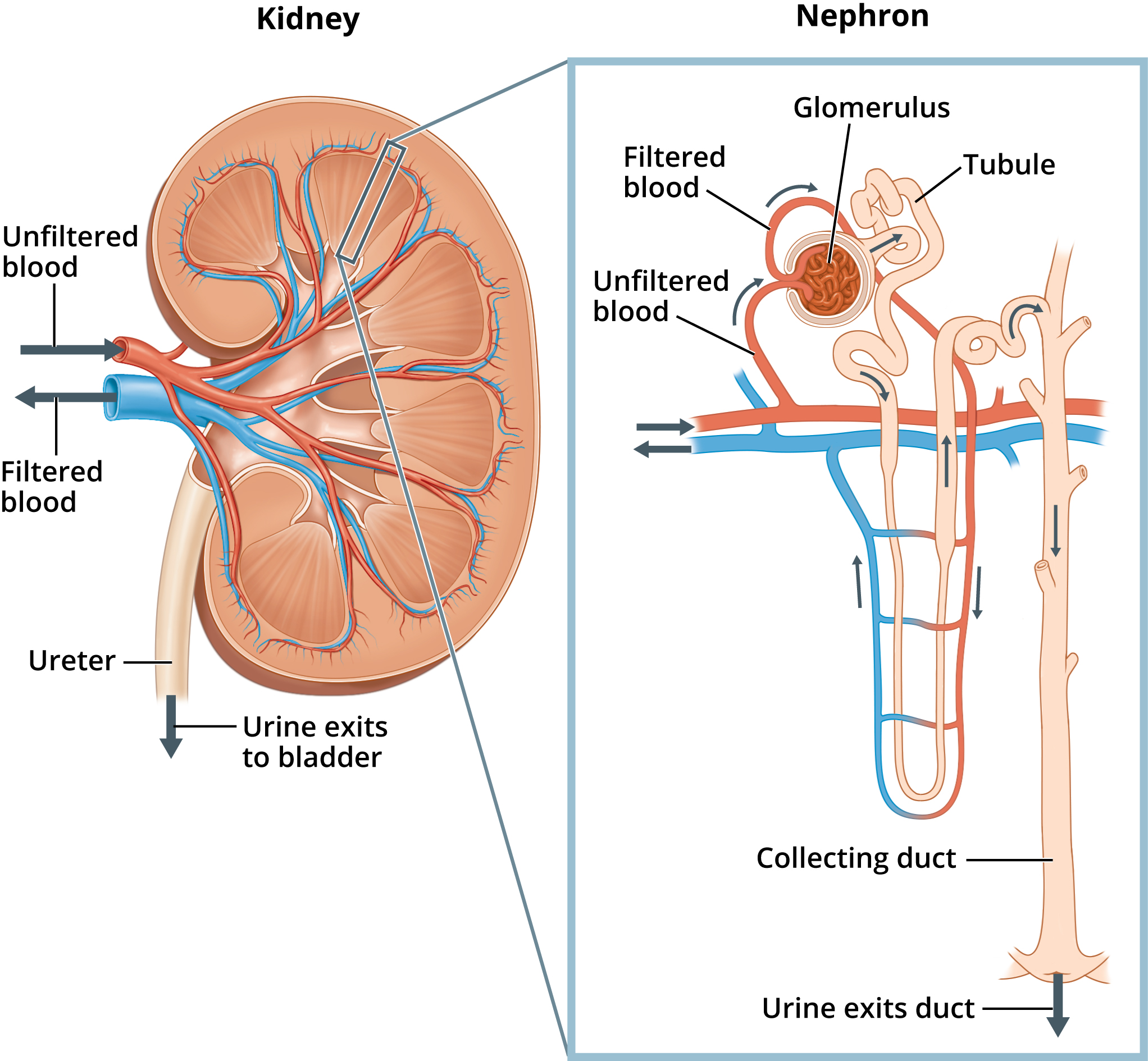
Nephron
The functional unit of the kidney
There are cortical nephrons as well as juxtamedullary nephrons
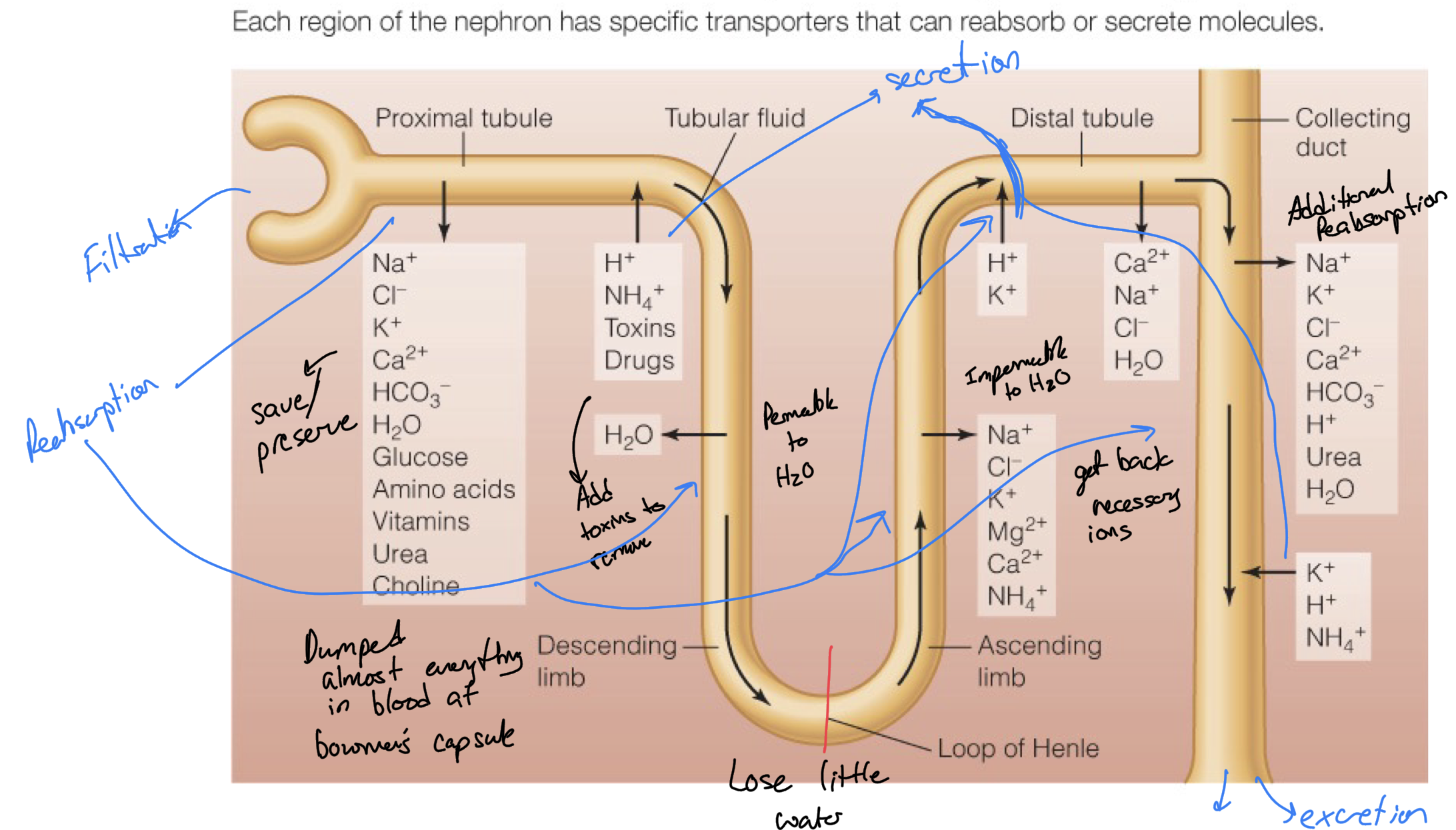
Composed of the renal tubule which is broken up into proximal tubule, descending limb, loop of Henle, ascending limb, distal tubule, and it connects to the collecting duct
In the proximal tubule of the nephron, all of the contents are removed from blood, except for blood cells and larger molecules such as proteins. Some waste products are also secreted into the proximal tubule to be carried away
In the Descending Loop, the tubule is impermeable to solutes but is highly permeable to water with many supporting aquaporins, so lots of water is removed from the filtrate, making it more concentrating, and returning water to blood and cells.
In the Ascending Loop, the tubule is now impermeable to water but it is permeable to solutes such as ions Na, Cl which can get reabsorbed by the blood as necessary.
The distal tubule is an area where more secretion occurs, body dumps more waste into the filtrate and also does more filtration, pulling ions like Ca2+, Na+, Cl-, and more water
The collecting duct also does some more secretion as well as filtration, making urine more dilute in the end.
Collecting duct of kidneys
Collects metabolic waste from many nephrons, not part of the structure of each nephron, instead shared among nephrons, links them all together technically.
Vasculature of the Nephron
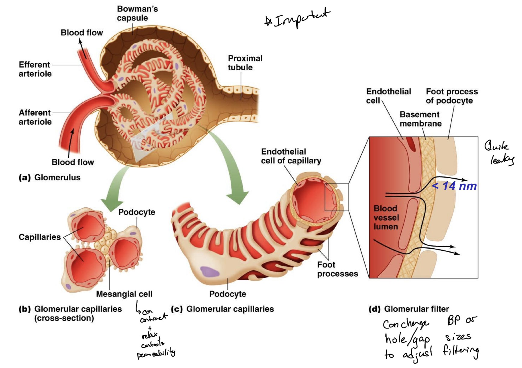
Afferent arteriole from the renal vein goes to the glomerulus which is covered by bowman capsule, which is part of the renal tubule covering a knot of blood vessels.
Then the efferent arteriole leaves from the glomerulus and surrounds the renal tubule to be present for reabsorption of nutrients and further secretion of waste.
One arterioles around one nephron, so that blood gets filtered through all of those nephrons specifically, to get the best filtration, more focused filtration.
Material exchange occurs at glomerulus and things get absorbed back into the blood or waste is secreted from blood into renal tubule, through proximal and distal tubule.
Glomerulus
Ball of capillaries in the nephron
Surrounded by Bowman’s capsule, part of the renal tubule
Capillary beds surround the renal tubule.
Majority of material exchange occurs here, take all of the things in your room and throw them into the hall, to later be sorted through by different parts.
Takes all possible solutes and water out of blood to be filtered/sorted through, reabsorbed, or excreted after collecting duct.
Nephron Composition
Functional unit of the kidney
Composed of the renal tubule
Renal Tubule
Main portion of the nephrons
Lined with transport epithelium
Various segments with specific transport functions
Four Processes of Urine Production/Kidney Filtration
Filtration
Reabsorption
Secretion
Excretion
Filtration
Liquid components of the blood are filtered into Bowman’s capsule
Water and smaller solutes can cross this glomerular wall
Blood cells and large macromolecules, like proteins, are too large to leak through and thus are not filtered
Glomerular capillaries are very leaky because they have podocytes with foot processes which help to form the filtration structure. The foot processes engulf the arteriole, providing small gaps for solutes and water to leak through, do not fully seal up capillary, allowing things to pass through endothelial cells, basement membrane of capillaries, and the gaps created by podocytes.
Mesengial cells surrounding the capillaries in Bowman’s capsule are smooth muscle-like cells which control blood pressure and filtration within the glomerulus, regulating filtration rate.
Filtrate taken from the blood then flows from Bowman’s capsule into the proximal tubule.
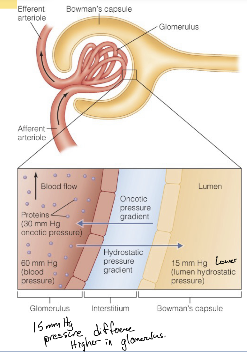
Glomerular Filtration Rate
GFR
The amount of filtrate produced per minute
Measures how efficient your kidneys are filtering blood
Determined by the pressure across the glomerular wall
Three main forces driving this rate:
Glomerular capillary hydrostatic pressure
Bowman’s Capsule Hydrostatic Pressure
Oncotic Pressure, osmotic pressure due to protein concentration in blood.
Pressure Gradiants in the Glomerulus
Different pressure gradients between the glomerulus and bowman’s capsule are what drive the movement of filtrate (solutes+H2O) to be filtered or stay in the blood.
There is a net 15mm Hg of pressure pushing solutes out from blood, through podocytes into bowman’s capsule
There is 60mm Hg in the glomerulus which is just blood pressure, pushing water and small solutes outward, which is a form of hydrostatic pressure, generated by the presence of water and its force exerted by being present.
There is also an inward pressure generated by proteins of 30 mm Hg, known as oncotic pressure, pulling the filtrate inward due to the higher concentration of proteins in blood. Proteins cannot be filtered through to Bowman’s capsule, so end up with higher protein concentration in arterioles. To balance this, an osmotic gradient/pressure is generated, pulling water back.
In Bowman’s capsule there is also an outward pressure, attempting to keep filtrate out and push back against the flow of solutes and water, but it is only 15 mm Hg, known as lumen hydrostatic pressure
The net is 60 mm Hg outward (out of blood, to Bowman) -45 mm Hg inward (towards blood)= +15mm Hg towards Bowman’s capsule, describing that flow is not just uncontested, but net pressure pushes the filtrate outwards from the arterioles of each nephron.
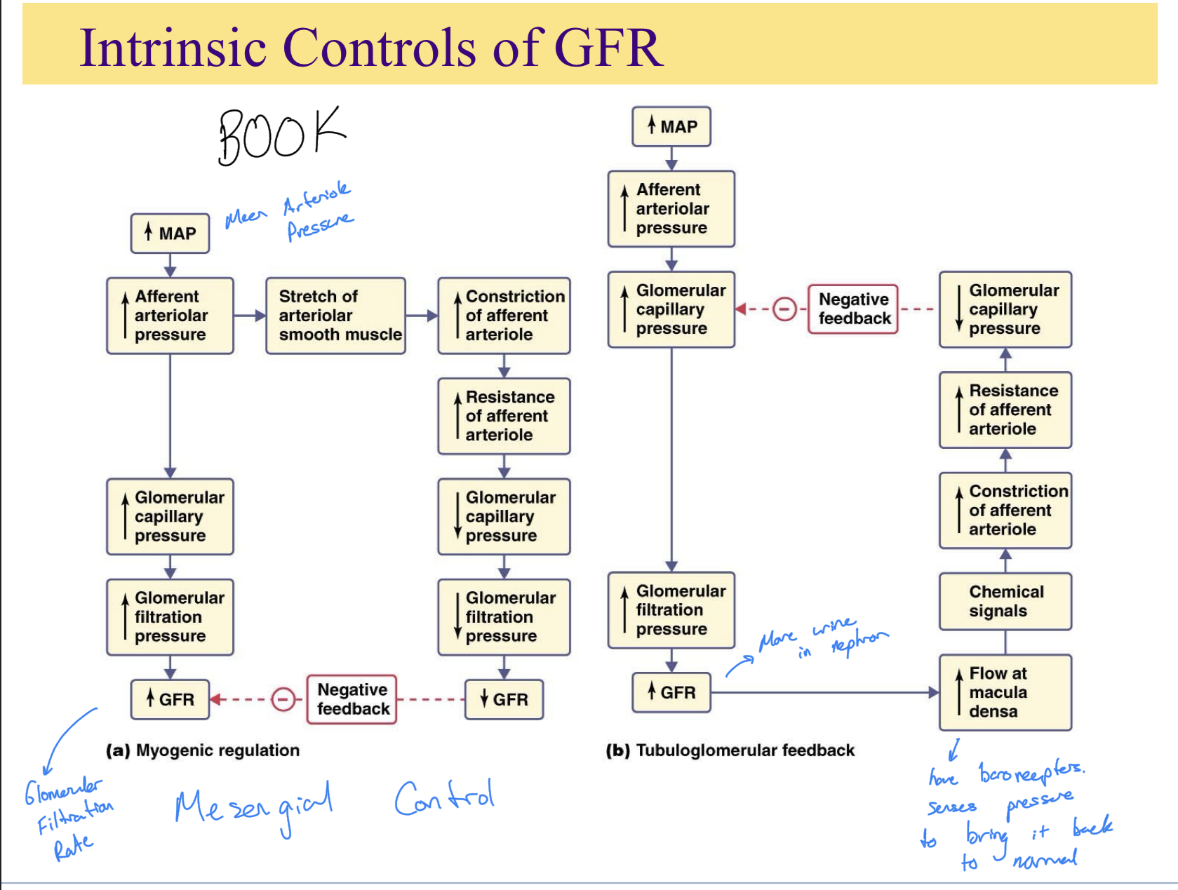
Intrinsic Regulators of GFR
Mesengial Control
Myogenic Regulation
Tubuloglomerular Feedback
Mesengial Control
Altered Permeability of Glomerulus
Myogenic Regulation
Constriction/Dilation of afferent arteriole
Tubuloglomerular Feedback
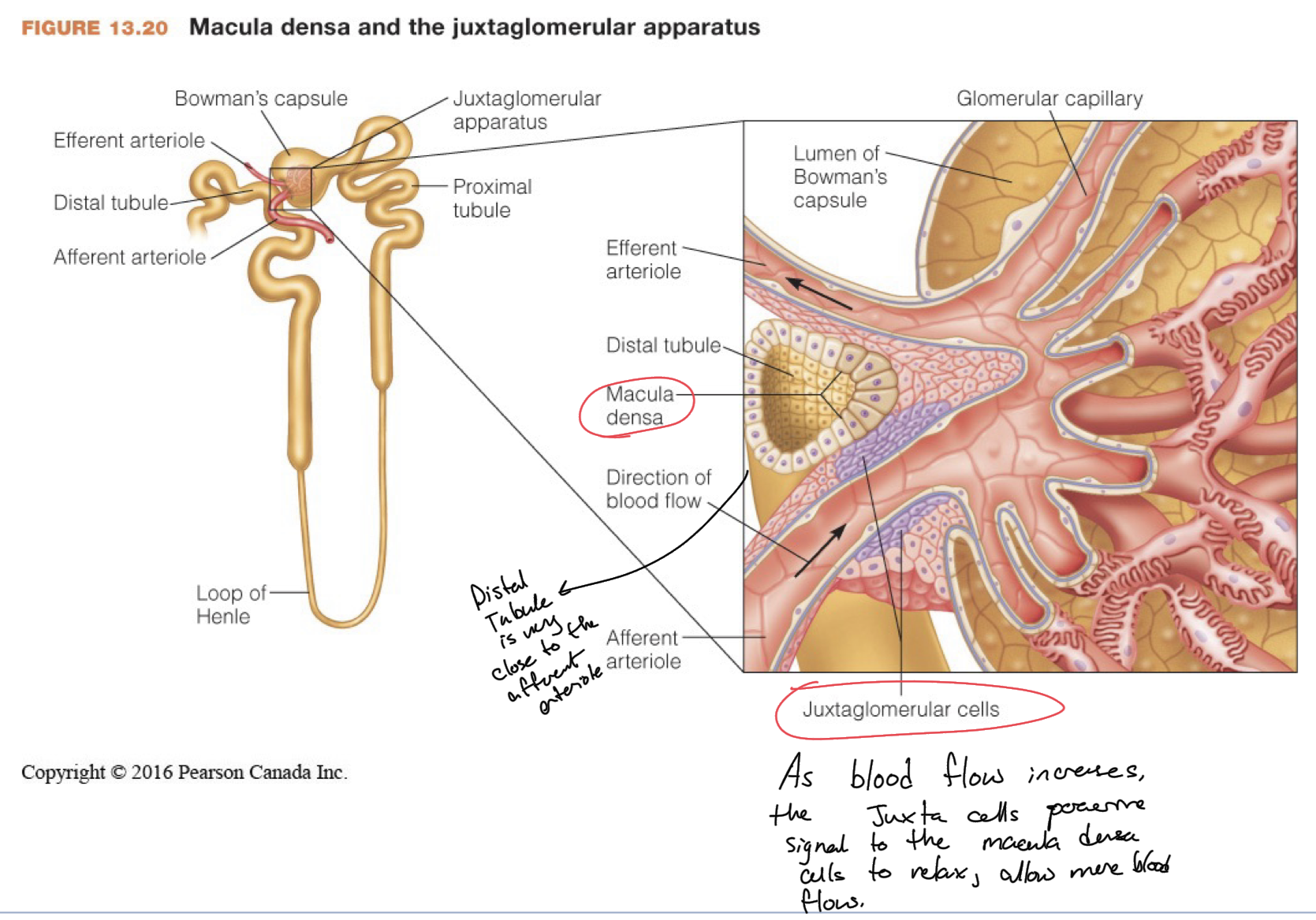
Works via the Juxtaglomerular Apparatus
Macula Densa Cells in the distal tubule, which is anatomically close to glomerulus, paracrine signal to juxtaglomerular cells of the afferent arteriole to alter the diameter of afferent arteriole.
Intrinsic Controls of GFR
(KNOW/UNDERSTAND PATHWAYS)
Tubule Regions + Functions