Week 1a: Imaging in Rehabilitation X-Ray
1/39
Earn XP
Description and Tags
COMPLETE
Name | Mastery | Learn | Test | Matching | Spaced | Call with Kai |
|---|
No analytics yet
Send a link to your students to track their progress
40 Terms
Why should a PT study study imaging?
1.) A more comprehensive evaluation is obtained
2.) The information the physical therapy clinician seeks is often of a different nature from the information the physician seeks and even of a different nature from what may be described in the radiologist's report
*Professional Collaboration requires an understanding of what each party has to offer
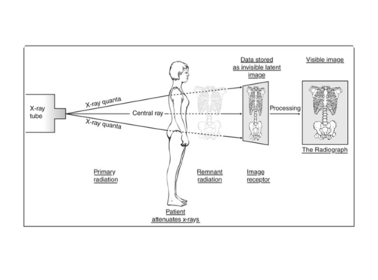
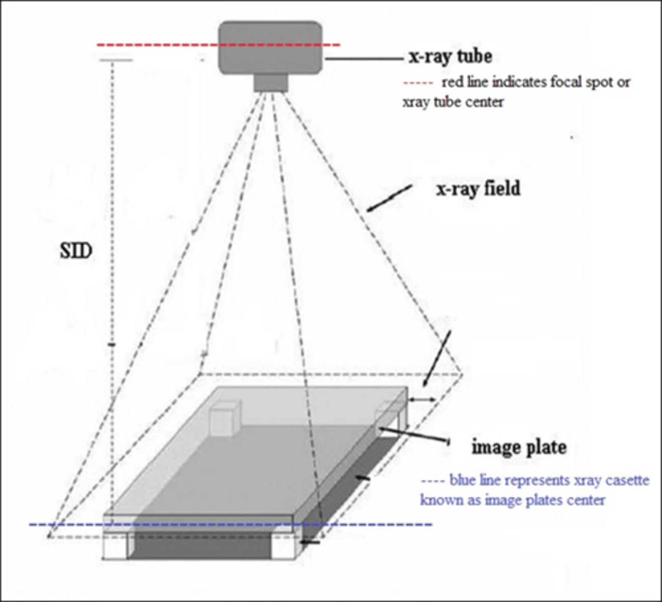
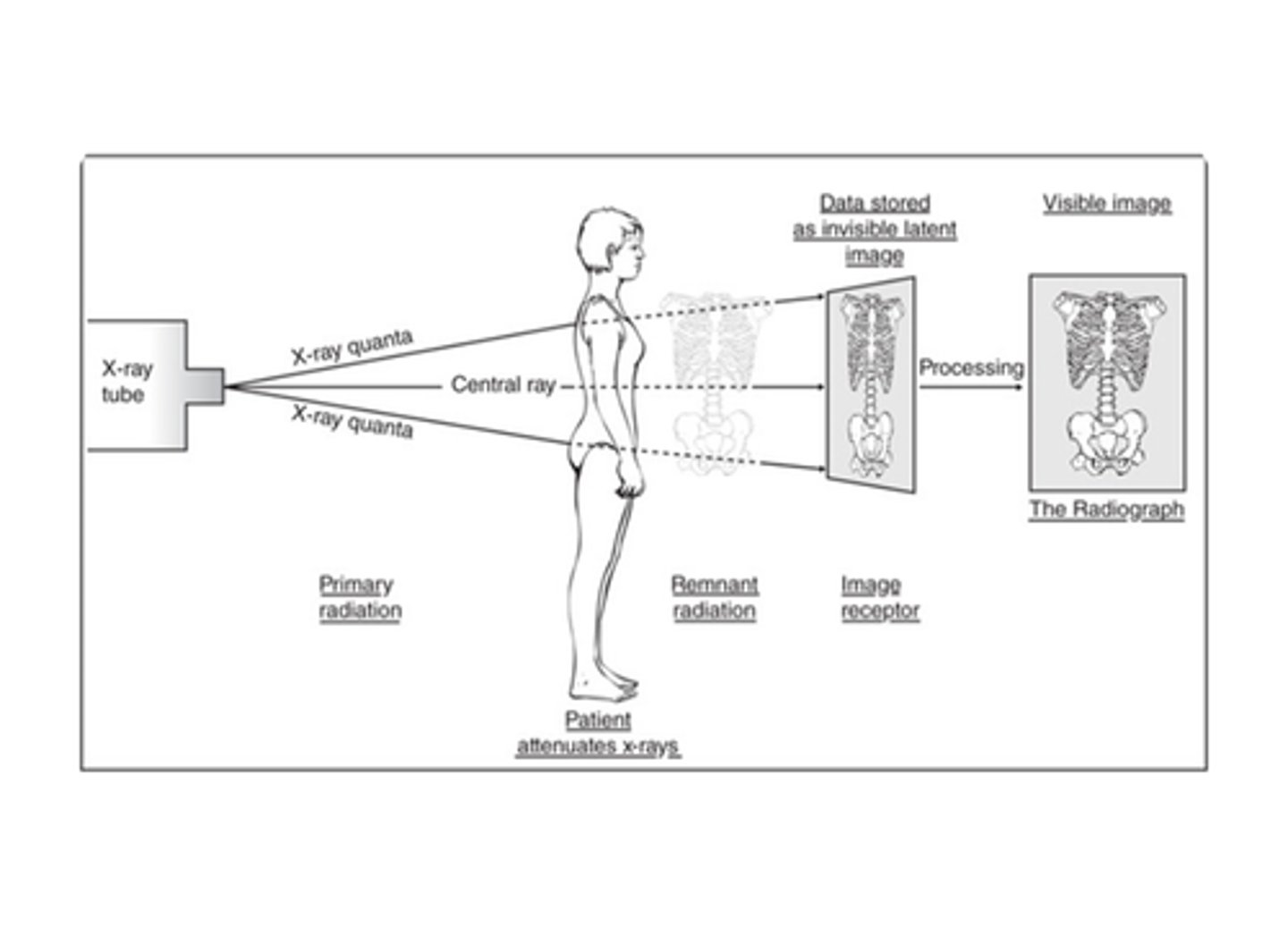
Basic X-Ray Imaging System
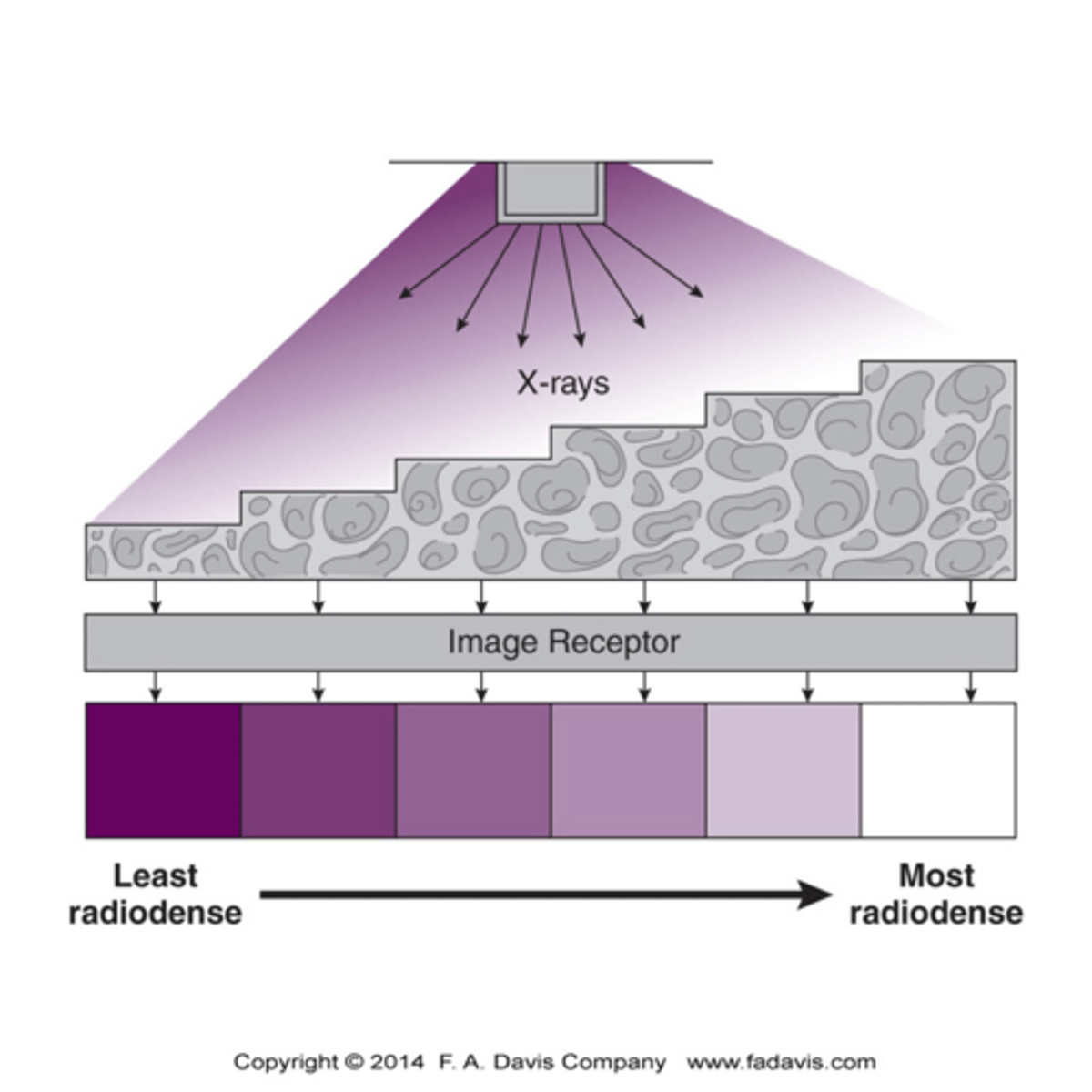
The X-ray beam is attenuated in different amount, depending on the density of the tissue it has passed through or not passed through. The remnant beam is intercepted by an interpretation device called the image receptor (latent image) and converted into a visible image based on shades of gray (4 main shades).
The receptors can be:
1) film or screen radiograph,
2) Fluoroscopic video,
3) Computer or digital image.
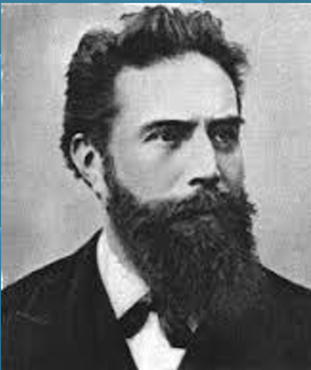
Who discovered X-Rays and was awarded the Nobel Prize in Physics in 1901?
Discovery of x-rays by Willhelm Conrad Rontgen in 1895
- Electromagnetic radiation in the wavelength range of X-rays
- Awarded the 1st Nobel Prize in Physics in 1901
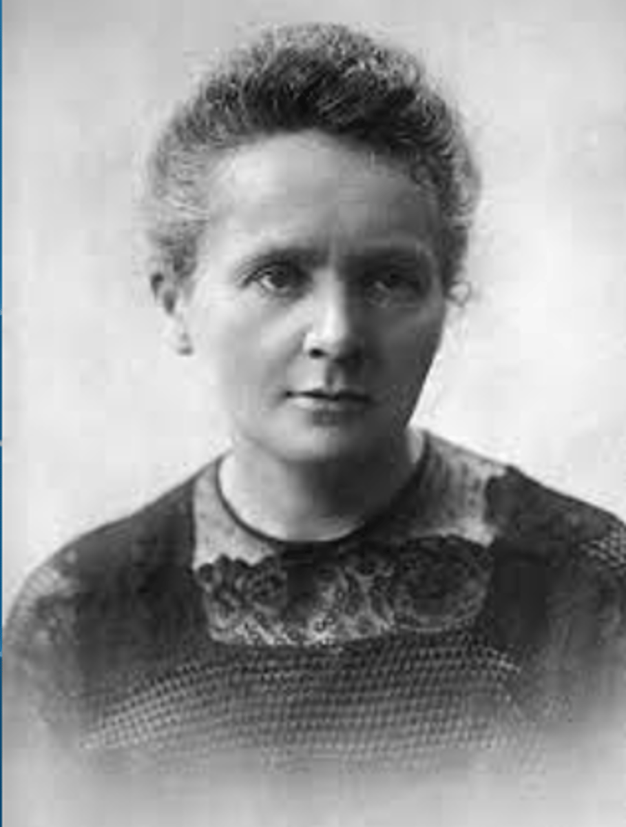
Who discovered radioactive elements in 1898?
Marie Curie:
- The theory of radioactivity-isolated radioactive isotopes using polonium and radium
Definition of Radiograph?
A recorded image of an anatomic part acquired by the passage of x-ray through the body
What is the first imaging study done following a clinical examination?
Conventional Radiograph (X-Ray)
What is the most efficient image to look at bone and joint abnormalities?
Conventional Radiograph (X-ray)
What are the three things required to produce an X-Ray image?
- Source of electrons
- Force to move them rapidly
- Something to stop them rapidly
What are the shades of gray on a radiograph determined by?
Tissue Radiodensity
- More dense = More radiopaque (white/light)
- Less dense = More radiolucent (Gray/Black)
What are the two determinants of radiodensity?
- Composition (atomic number and volume density)
- Thickness

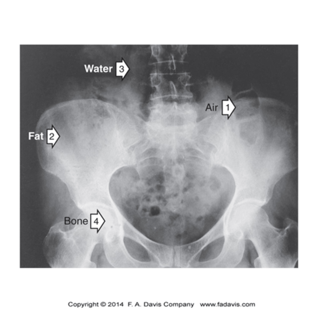
What are the four tissue radiodensities in the human body?
- Air (black)
- Fat (Gray-Black)
- Water: (Gray) All soft tissue and fluids (blood, muscles, nerves, cartilage, tendon, and ligaments)
- Bone (White)
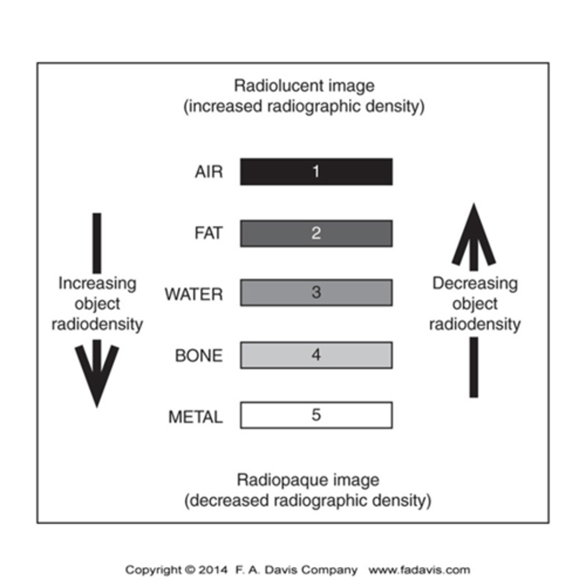
What is the relationship between absorption of x-rays (amount of the object radiodensity)?
Inverse Relationship
- Radiopaque = Bone
- Radiolucent = Air
What is the relationship between radiodensity based on thickness?
The thicker an object is the more radiodensity it possesses relative to thinner object made of the same substance
What does radiopaque mean?
- Not easily penetrated by x-rays, therefore has a greater radiodensity
- Structures that absorb x-rays block them from reaching the receptor
- Appears white on radiograph
Examples of Radiopaque structures in body
Bone (borders, process, ridge, spine, tubercles, tuberosity)
What does radiolucent mean?
- Easily penetrated by x-rays, therefore has less radiodensity
- Structures that don't absorb x-rays and allow them to pass through (greater radiation reach the receptor)
- Appears Black/Dark
Examples of Radiolucent structures
Air and Fat
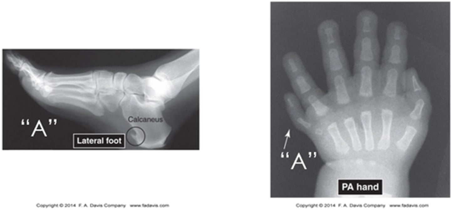
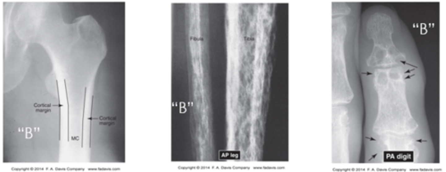
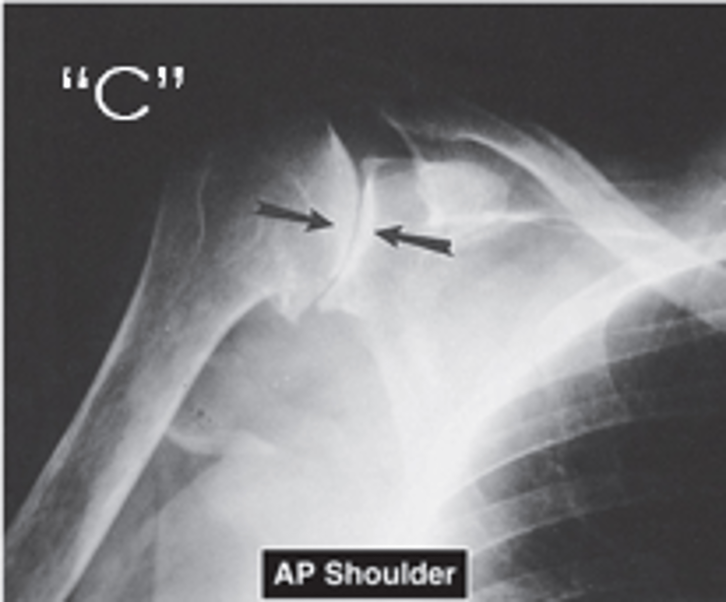
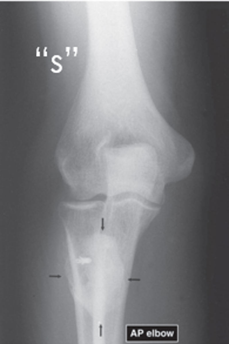
What is meant by the term, one view is no view?
At least TWO images, as close to 90 degrees are required to view all three dimensions of a structure
What are the 4 image quality factors that determine the quality of a radiograph and what property do they fall under?
Photographic Properties
- Density
- Contrast
Geometric Properties
- Details
- Distortion
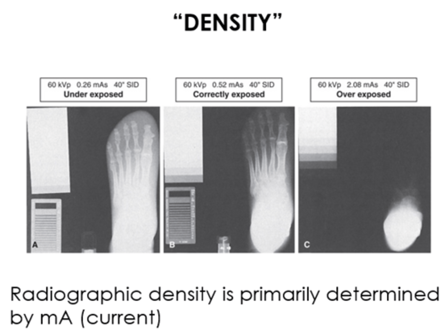
What is Density?
Defined as the amount of blackening on the radiograph, and it does this by varying the current and exposure time
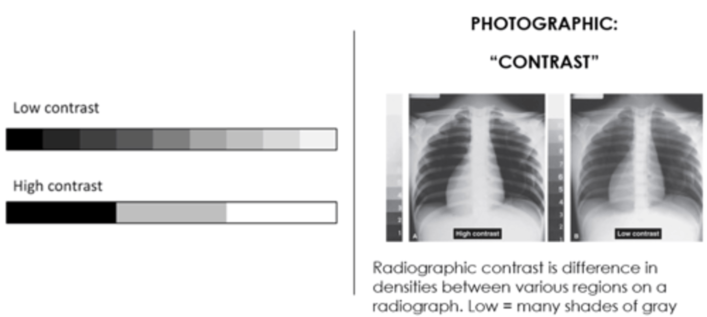
What is Radiographic contrast?
The difference among various adjacent radiographic densities
Greater variation = high contrast
Less variation = low contrast
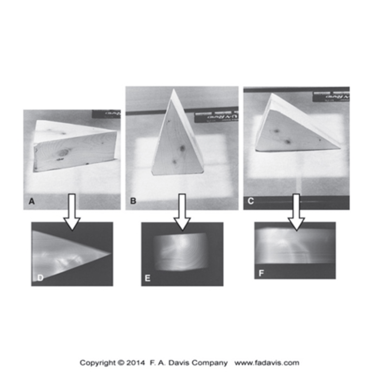
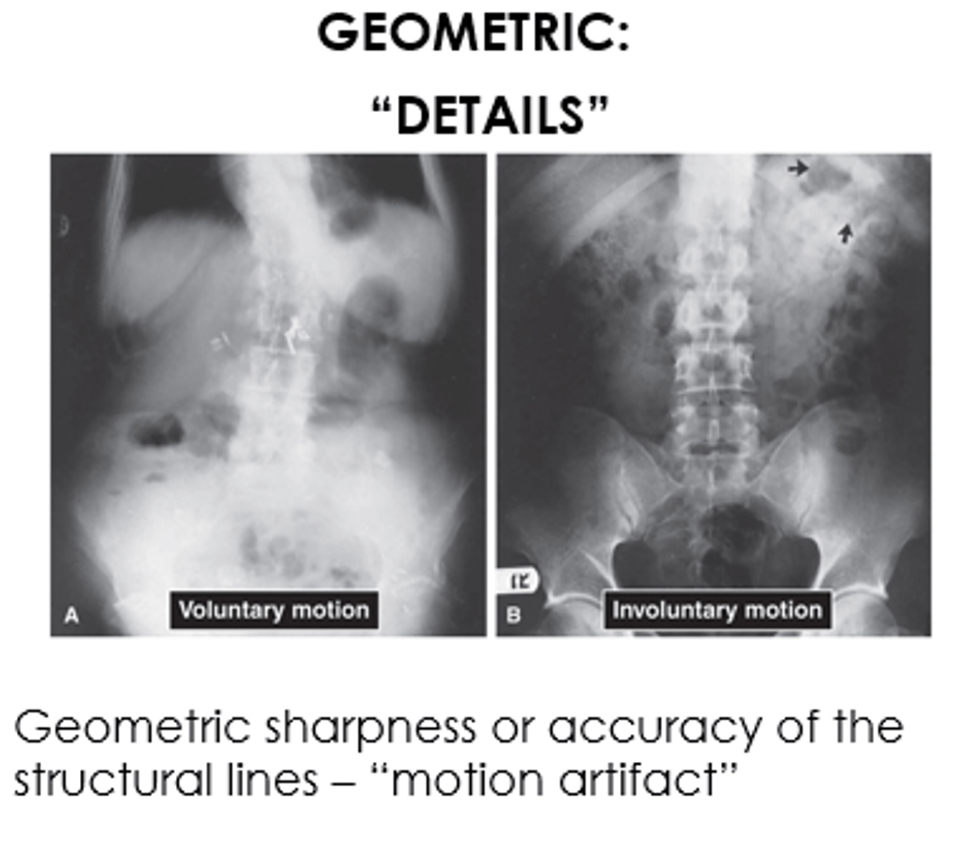
What is Detail?
- Geometric sharpness or accuracy of the structural lines recorded on the radiograph
- Usually because of motion artifact
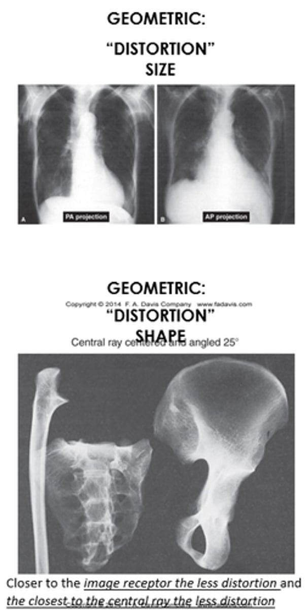
What is Distortion?
- Difference between actual object and the recorded object. Usually either size or shape distortion
- Closer to the image receptor the less distortion and the closest to the central ray the less distortion
What is Magnification?
Beam source - image receptor distance / beam source - patient distance.
Human vision can only see 32 shades of gray
Radiographic image interpretation requires what four things?
- Foundations in imaging technology
- Dimensional perception and knowledge of anatomy
- Organized search pattern
- Characteristic patterns of pathology
What does the ABCs stand for?
- Alignment
- Bone density
- Cartilage Spaces
- Soft Tissues
What are the factors involved in Alignment?
General Skeletal Architecture
- Gross size, appearance and number of bones
General Contour of Bone
- Normal shape and contour
Alignment of Bones to adjacent Bones
- Normal joint articulation and spatial relationship
What are the factors involved in Bone Density?
General Bone Density
- Contrast of soft tissue and bone (shades of grey)
- Contrast between cortical and cancellous bone
Textural Abnormalities
- Normal trabecular architecture
Local Bone Density Changes
- Sclerosis at areas of increased stress such as weight bearing surfaces or site of ligamentous, muscular, or tendon attachment
What are the factors involved in Cartilage Spaces?
Joint Space Width
- Well preserved joint spaces imply normal cartilage or disk thickness
Subchondral Bone
- Smooth surface
Epiphyseal Plates
- Normal size relative to epiphysis and skeletal age
What structures are Soft Tissue?
Muscles
- Look for wasting, swelling, edema, hemorrhage, or tumor
Fat pads and Fat lines
- Displacement is usually due to swelling
Joint Capsule
- Distended due to swelling or effusion
Periosteum
- Normally rather indistinct can have periosteal reaction
Miscellaneous
- Gas, calcification
What are the advantages of X-Rays?
- Fast
- Inexpensive
- Provides a relatively low radiation dose
- Provides an excellent definition of bone
- Screens for significant portion of pathologies
- Is valuable in directing which imaging study to use next if more information is needed to define a condition
What are the disadvantages of X-Rays?
- Radiographs can show only significant changes in bone density, Therefore, if a disease is slow to alter density, the evidence of the disease may not become visible until in its advanced stages
- Radiographs are 2-dimensional, and the 3rd dimension is limited by superimposition of tissues
- Soft tissue not well-defined
What are 3 diseases that may not become visible until in its advanced stages?
- Osteoporosis
- Avascular Necrosis
- Stress Fractures
What are examples of Non-Conventional X-Rays?
Fluoroscopy
Arthrography
Myelography
Arteriography & Angiography
Contrast-enhanced (inject or ingest a contrast medium)
What is Fluoroscopy?
A real time dynamic, continuous, or continuous radiographic exam used to guide interventional procedures
- Radiograph = Photograph
- Fluoroscopy = Movie
What is an Arthrography?
- An image made of a joint after it has been injected with a contrast
- There are conventional arthrography and MRI and CT arthrography
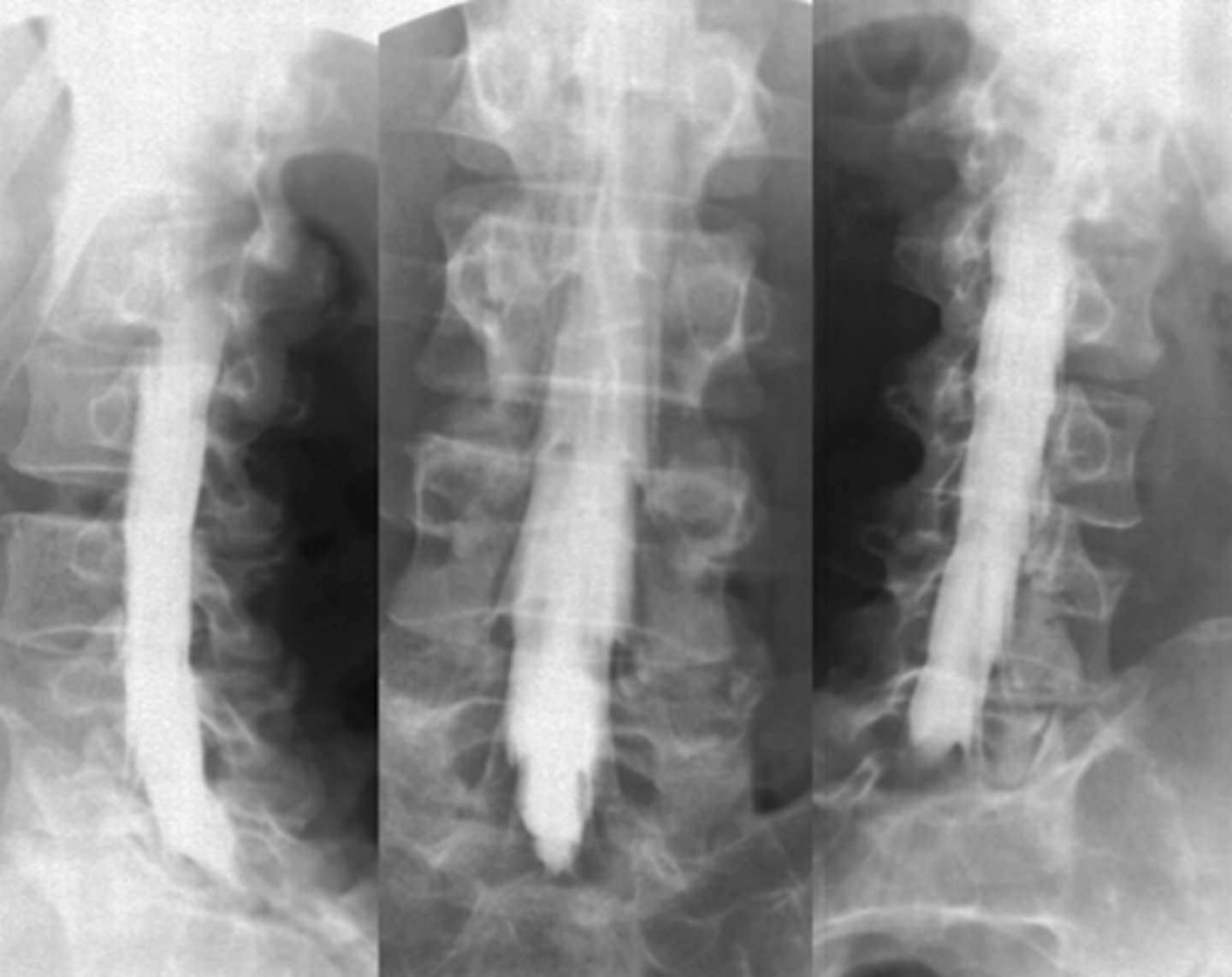
What is a Myelography?
- An image made with an injection of contrast medium into the subarachnoid space to examine the spinal cord and nerve roots
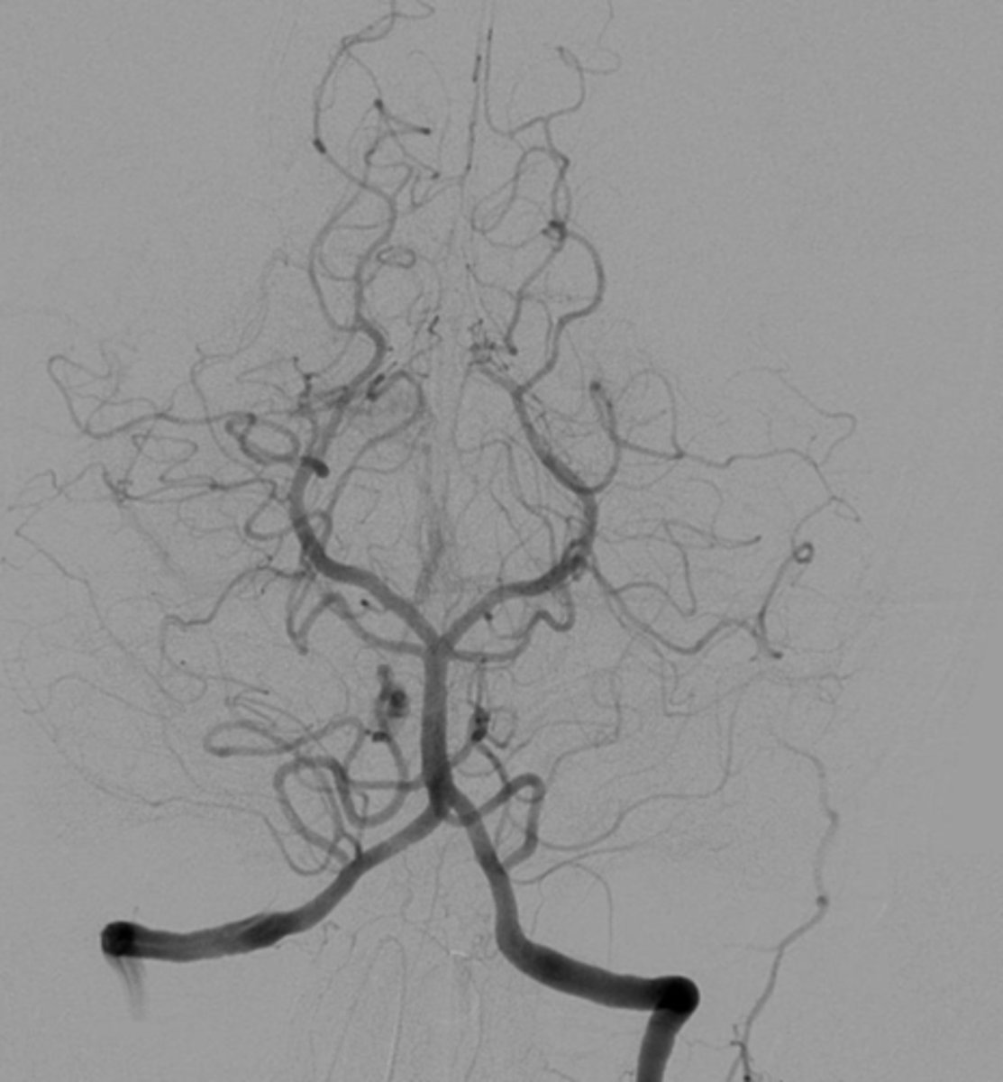
What is Arteriography & Angiography?
- Is an X-Ray examination of the arteries, or blood vessels. To make the arteries visible on x-ray, a type of dye called "contrast" is injected
- Is a medical imaging technique used to visualize the inside, or lumen, of blood vessels and organs of the body, with particular interest in the arteries, veins, and the heart chambers
What is Contrast-enhanced (inject or ingest a contrast medium)?
- Radiolucent = "Air", called a negative contrast
- Radiopaque = "Barium sulfate or iodine", called a positive contrast
What is the imaging chain?
- Humans play a critical role in the imaging system by their interpretation of the image and by their correlation of clinical findings with imaging information.
- The clinicians responsibility is to recognize always that if the results of any imaging study do not fit the physical findings, further clinical evaluation and diagnostic investigation are warranted.