Leg, Ankle, Foot, Vertebral Column, Back; Joints and Ligaments of the Lower Limbs
1/179
There's no tags or description
Looks like no tags are added yet.
Name | Mastery | Learn | Test | Matching | Spaced | Call with Kai |
|---|
No analytics yet
Send a link to your students to track their progress
180 Terms
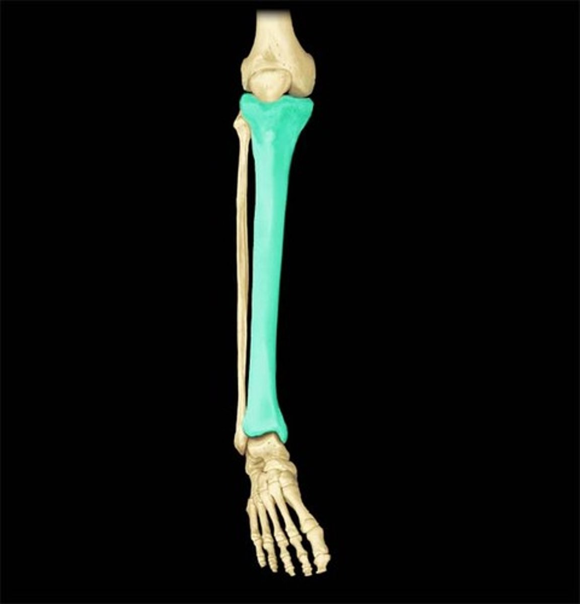
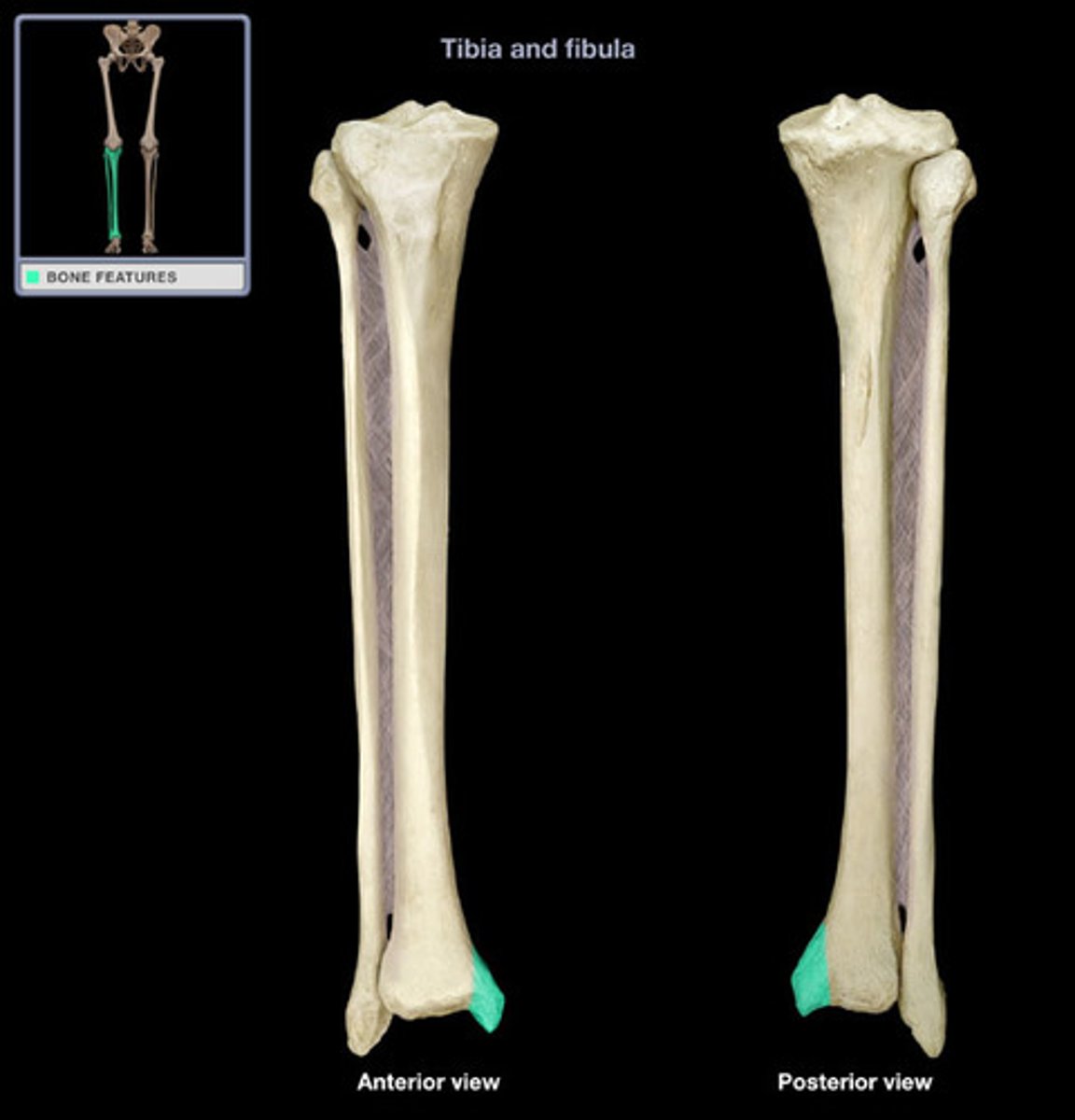
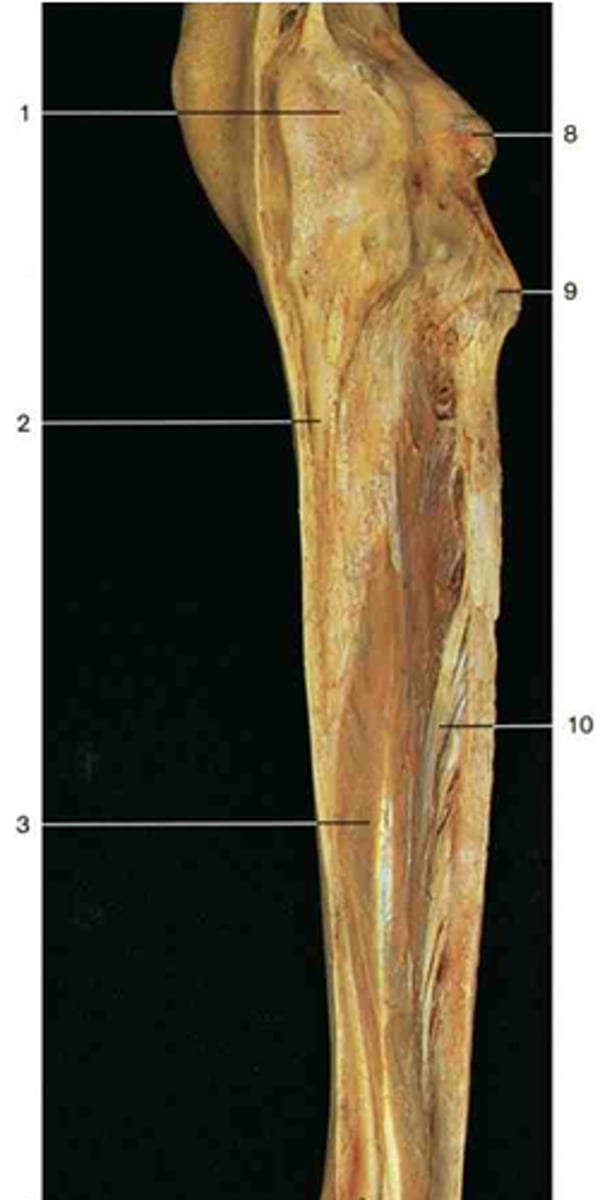
Tibia
The medial and larger bone of the lower leg.
Interosseous border
The sharp edge on the tibia and fibula where the interosseous membrane attaches. Number 4 and 6.
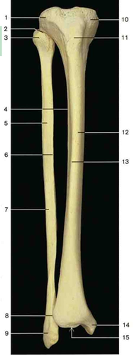
Tibial plateau
The top, flat portion of the tibia articulating with femoral condyles.
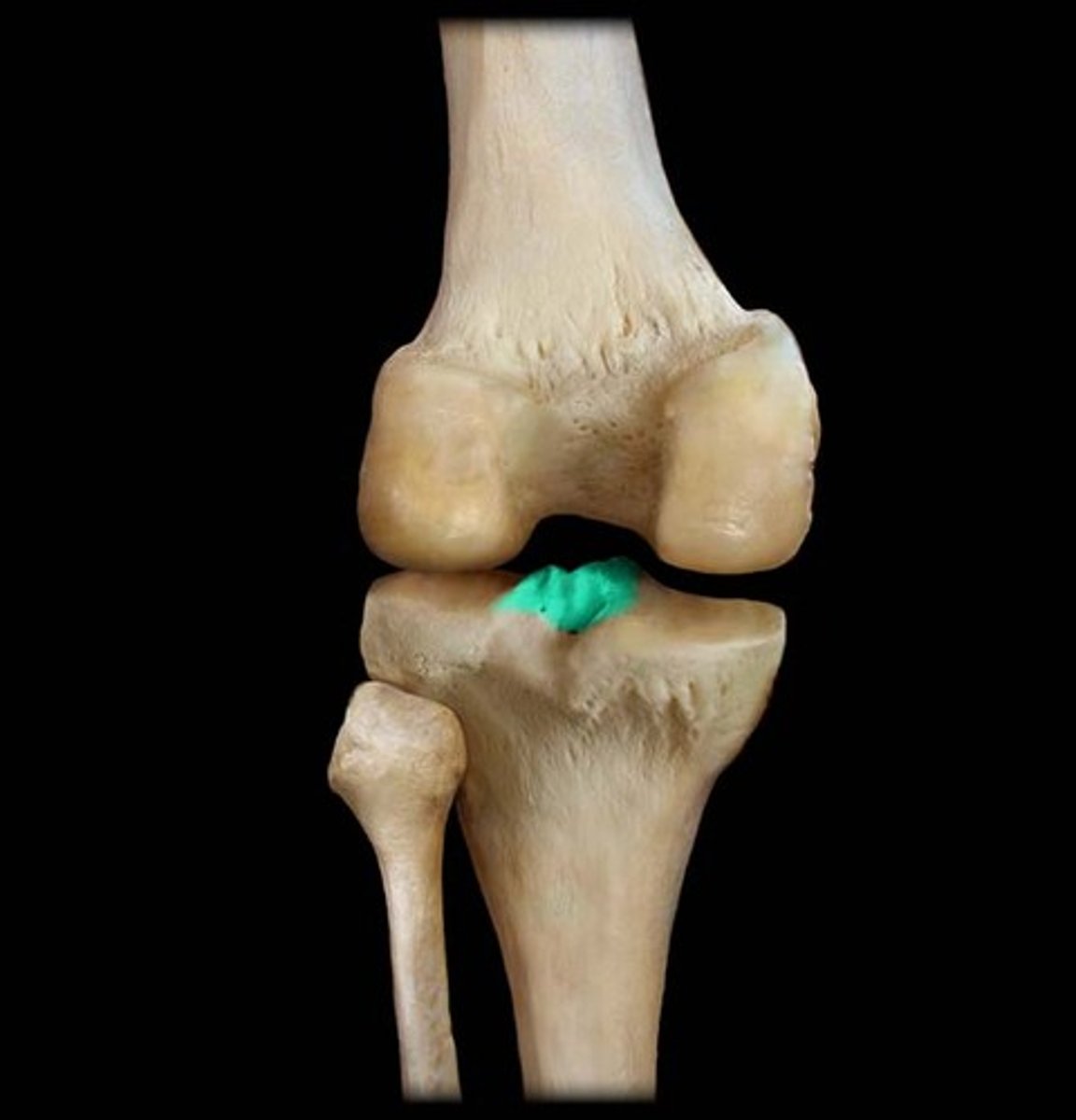
Intercondylar eminence
Irregular projection located between the two condyles.
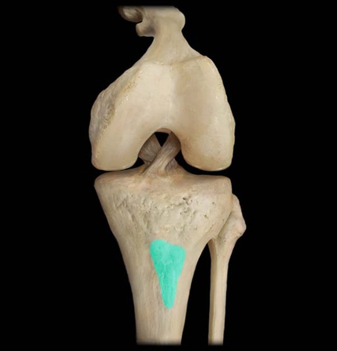
Tibial tuberosity
Point where the patellar ligament and quadriceps tendon attaches.
Medial malleolus of tibia
Medial process on distal end, forms medial bump of ankle.
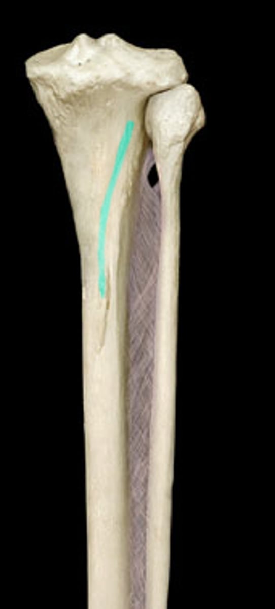
Soleal line of tibia
A raised line on the proximal, posterior aspect of the tibial body.
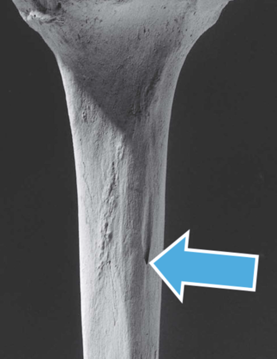
Nutrient foramen of tibia
Located on the posterior surface of the tibia, near the soleal line or slightly below it.
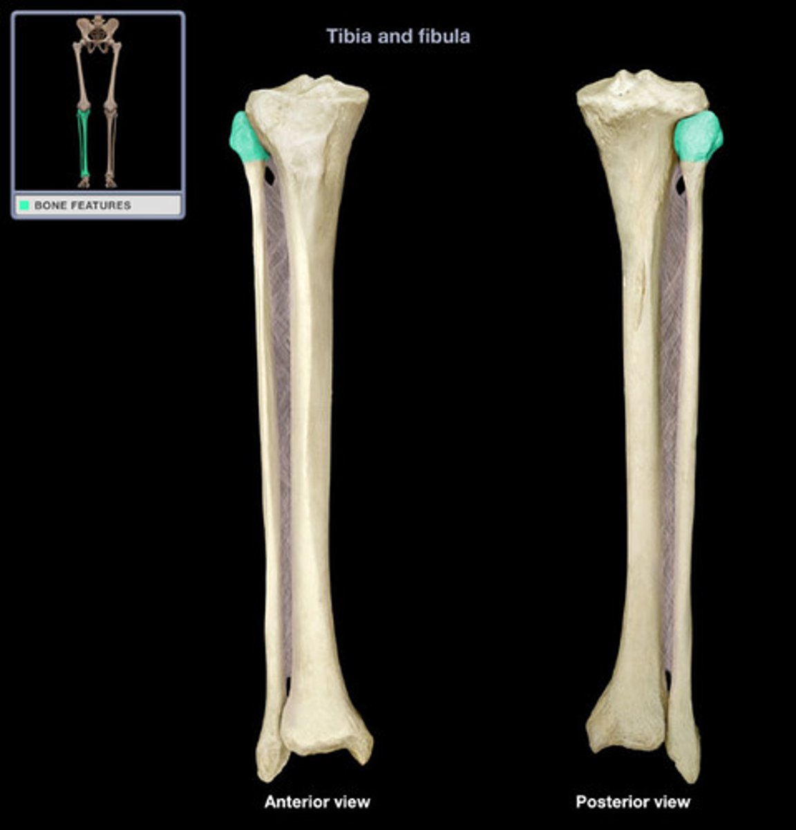
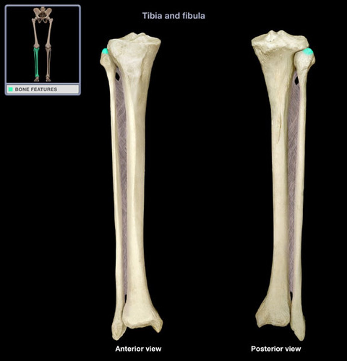
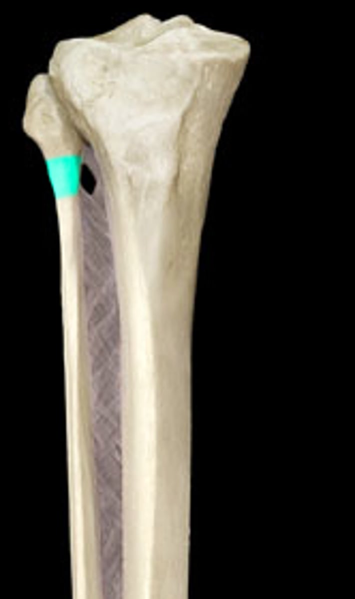
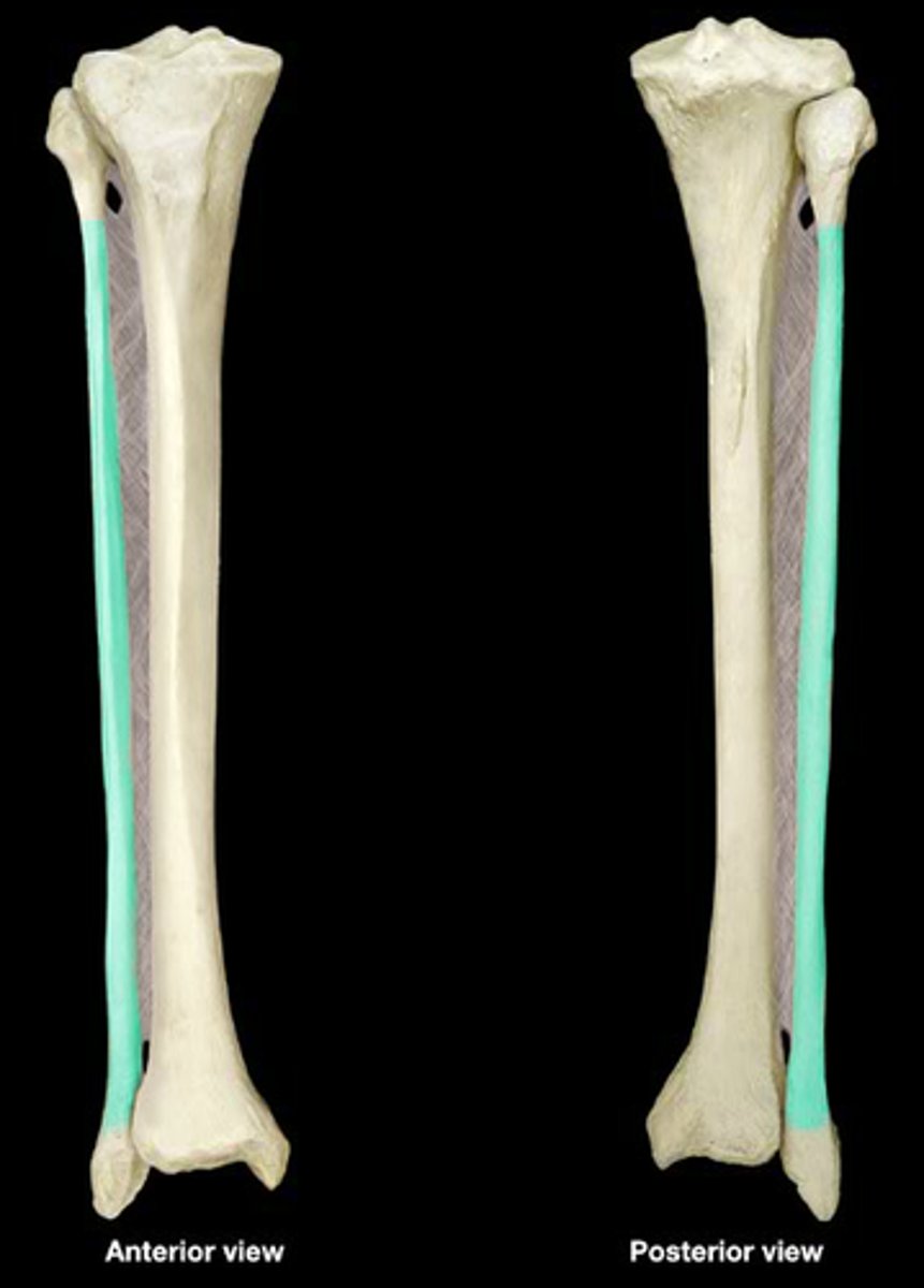
Fibula
The lateral and smaller bone of the lower leg.
Head of fibula
Proximal end of fibula.
Apex of fibula
The point of the head of the fibula.
Neck of fibula
Tapered area just below the head of the fibula.
Shaft of fibula
Elongated, slender portion located between the expanded ends of the fibula.
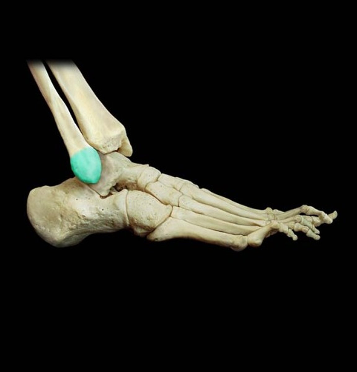
Lateral malleolus of fibula
Forms the lateral bulge of the ankle and articulates with the talus.
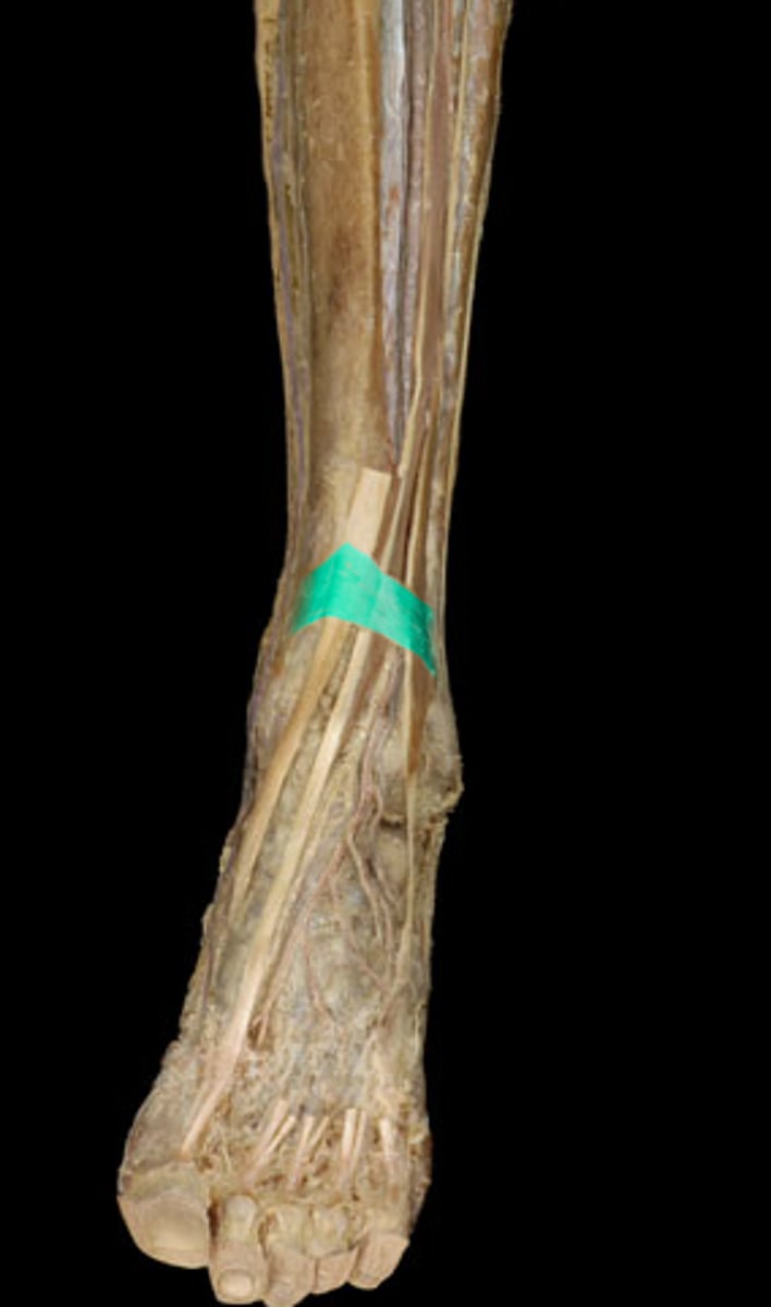
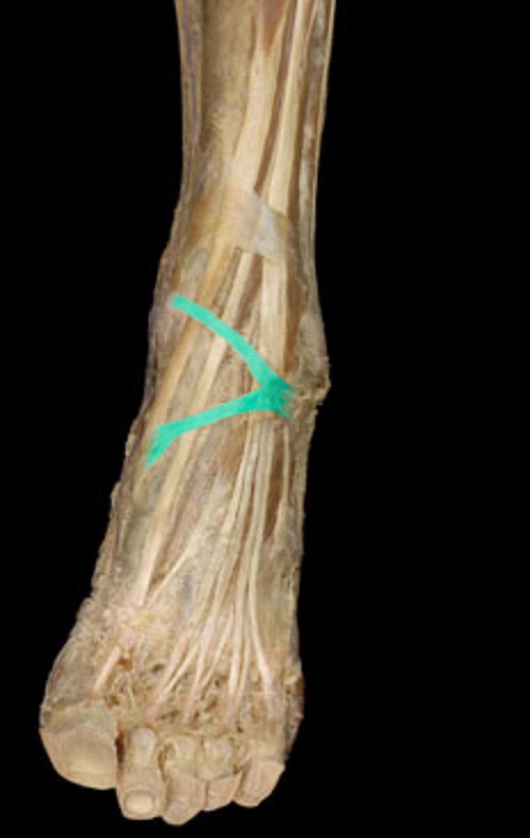
Superior extensor retinaculum
Transverse ligament of the ankle.
Tibialis anterior
The most medial and superficial dorsiflexor; also inverts foot.
Extensor hallucis longus
Lying between the tibialis anterior and extensor digitorum longus, it extends the big toe.
Extensor digitorum longus
Found superficially, just lateral to the tibialis anterior; extends toes.
Fibularis tertius
While not always present, it lies at the level of the extensor digitorum longus, just lateral to it.
Deep fibular nerve
Rising between the fibularis longus and neck of fibula, it innervates the extensor digitorum and hallucis brevis.
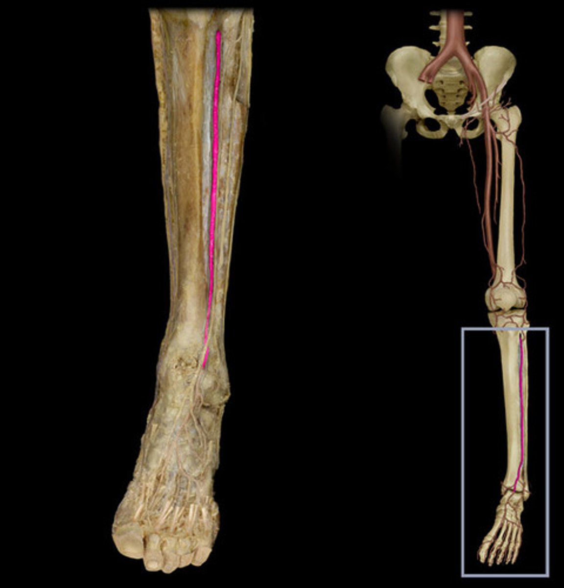
Anterior tibial artery
It starts at the inferior border of the popliteus, ending at the ankle joint. It supplies the anterior muscle compartment of the leg.
Fibularis longus
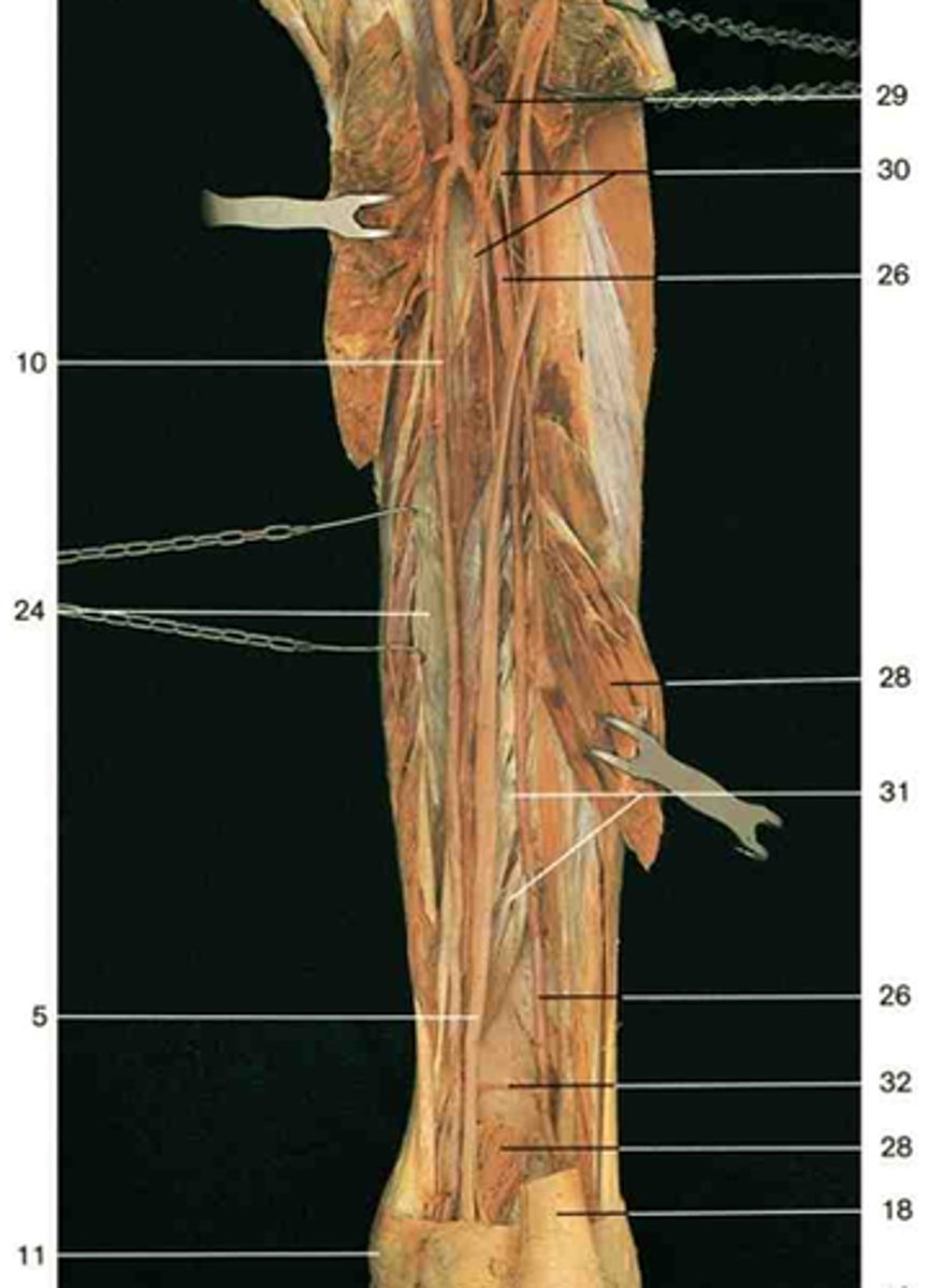
Partly covered by the soleus, above the fibularis brevis, and lateral to the extensor digitorum longus and fibularis tertius. Everts and weakly plantarflexes the foot. Number 5.
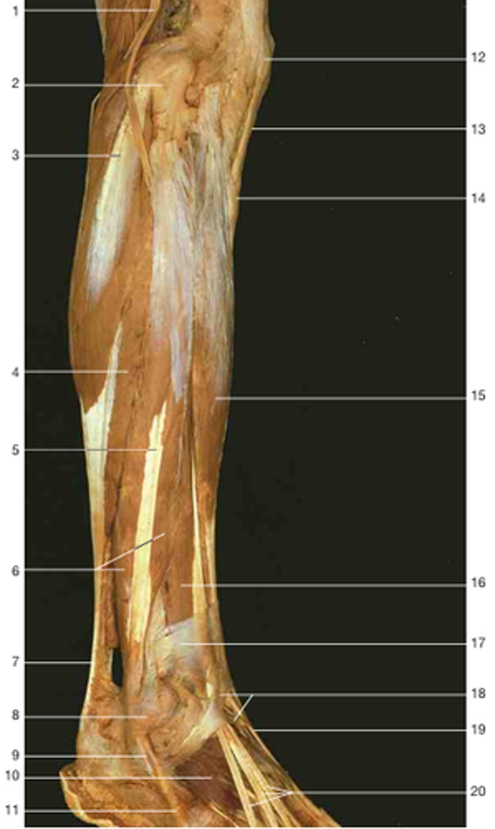
Fibularis brevis
This lateral muscle is directly under the fibularis longus. Everts foot, weakly plantarflexes. Number 6.

Superficial fibular nerve
Arising at the fibular head, it travels between the fibularis longus and brevis (innervating them) and emerging in the lower third of the leg, to innervate the skin there. Number 11.
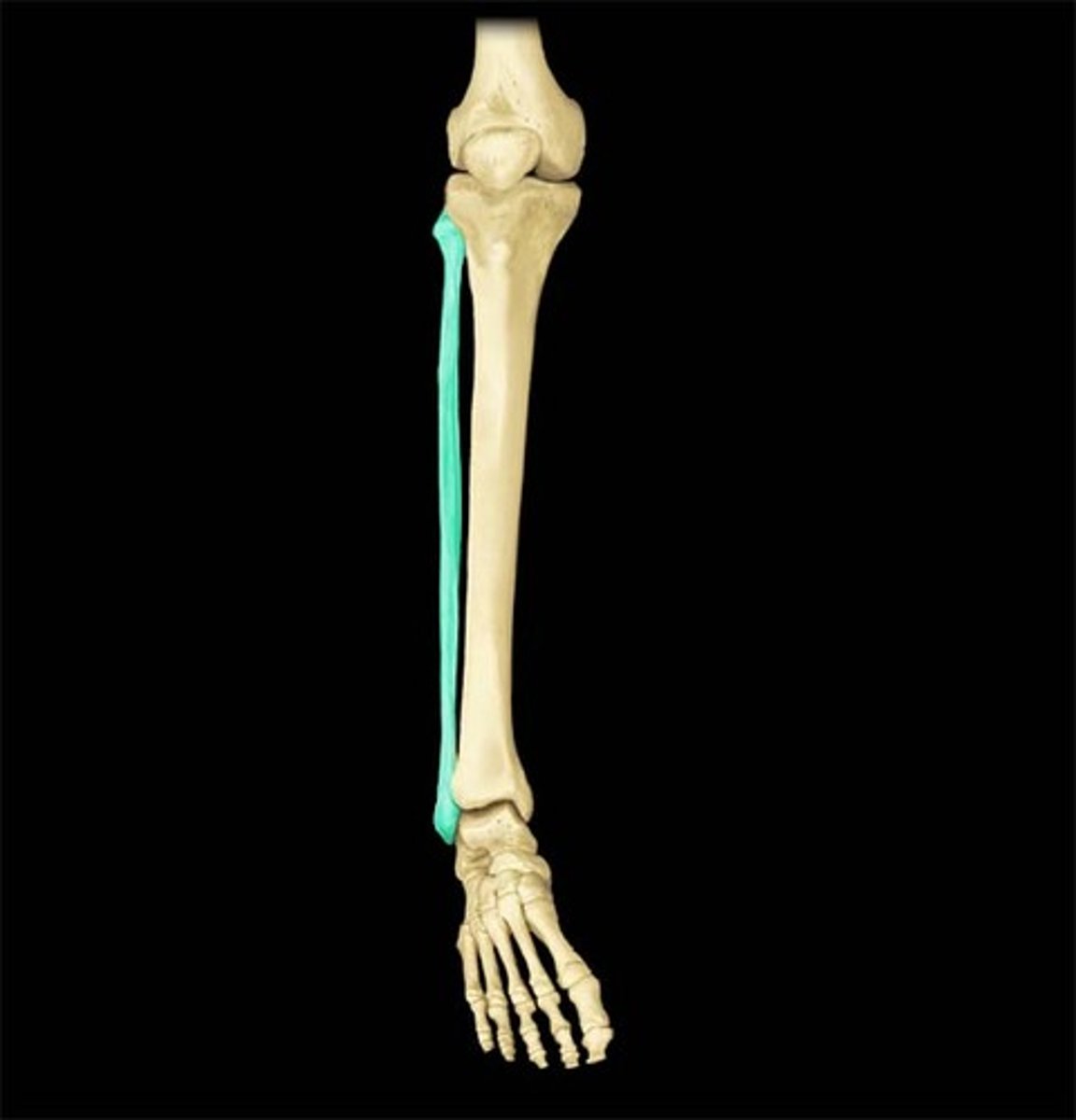
Triceps surae
Consists of the gastrocnemius and soleus, sharing the calcaneal tendon.

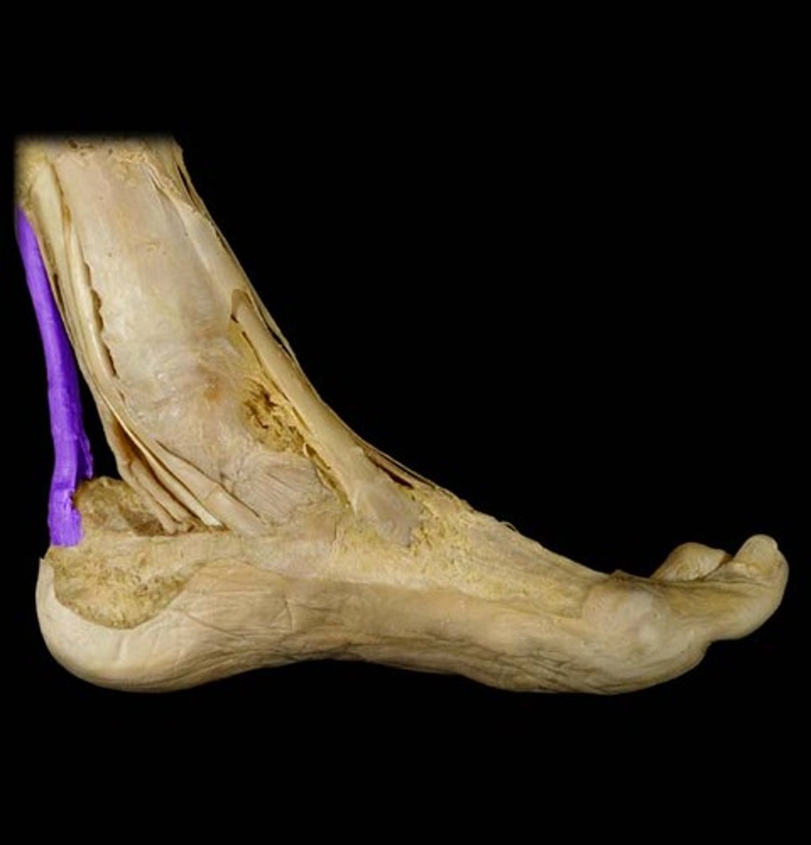
Calcaneal tendon
Also known as the Achilles tendon; the thickest and strongest tendon.
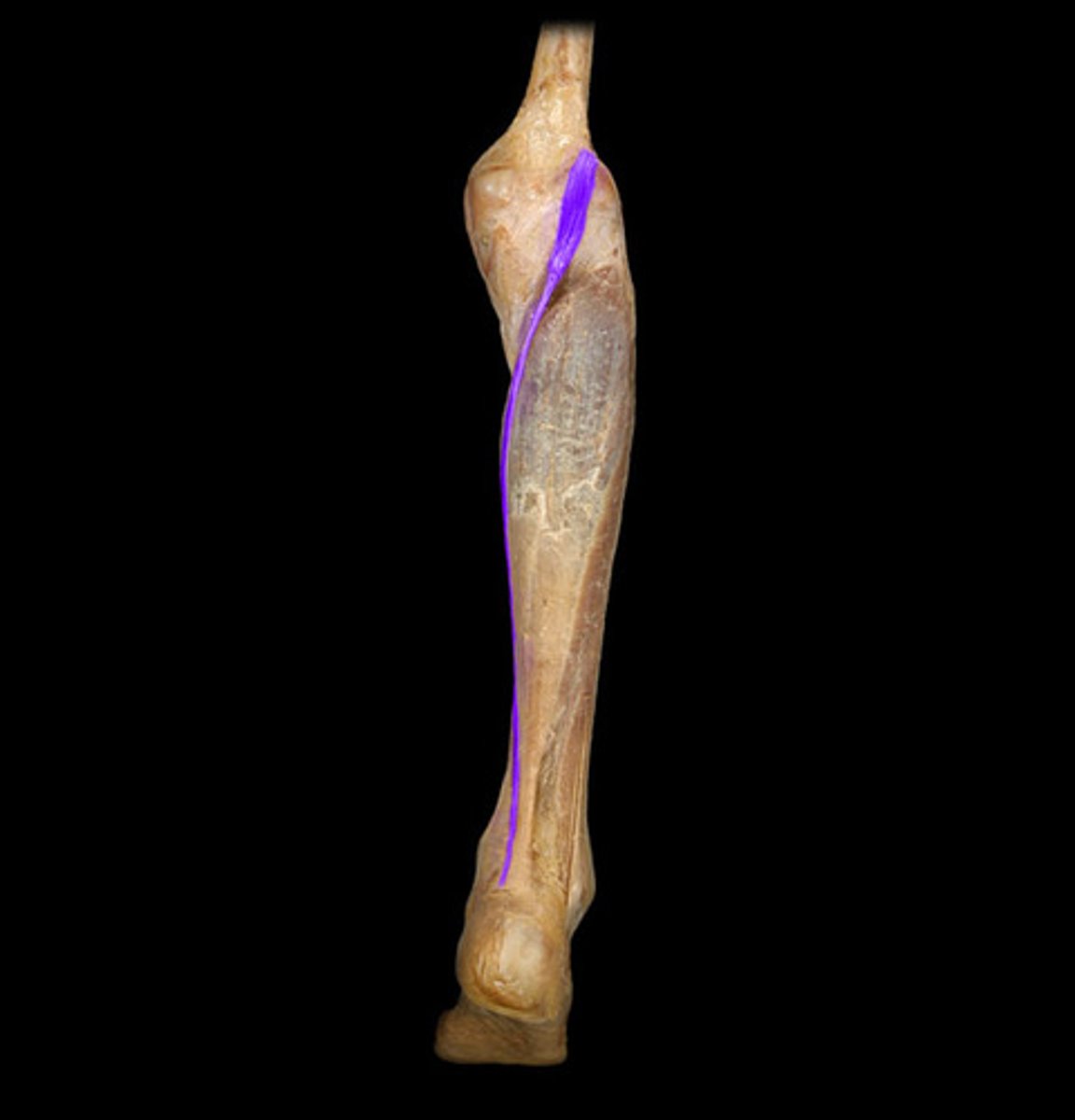
Plantaris
Has a long, thin, tendon running between the gastrocnemius and soleus, then inserts to the calcaneal tendon. Assists in ankle plantarflexion and knee flexion.
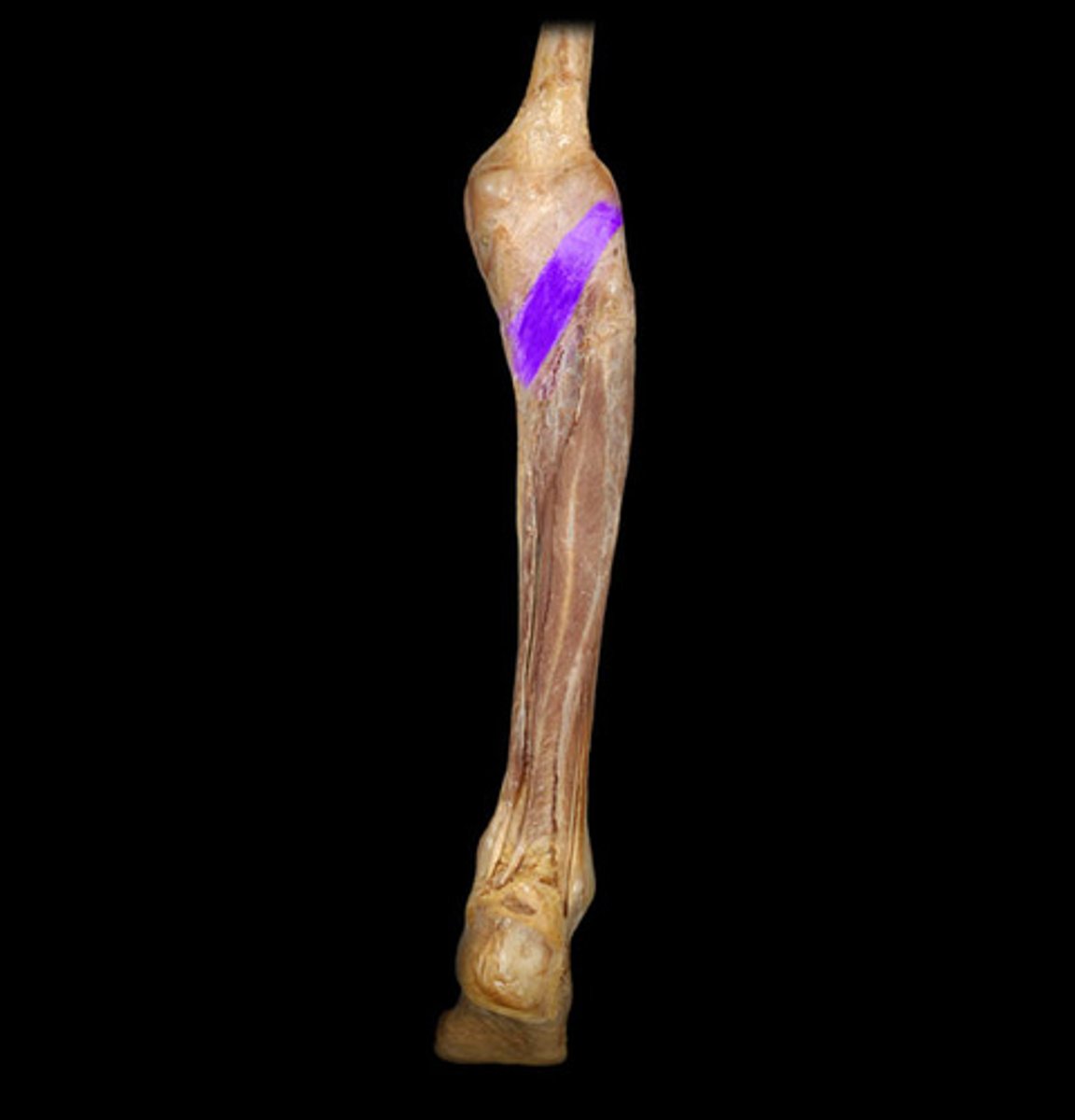
Popliteus
Found under the gastrocnemius and plantaris, it runs obliquely and weakly rotates the knee medially.
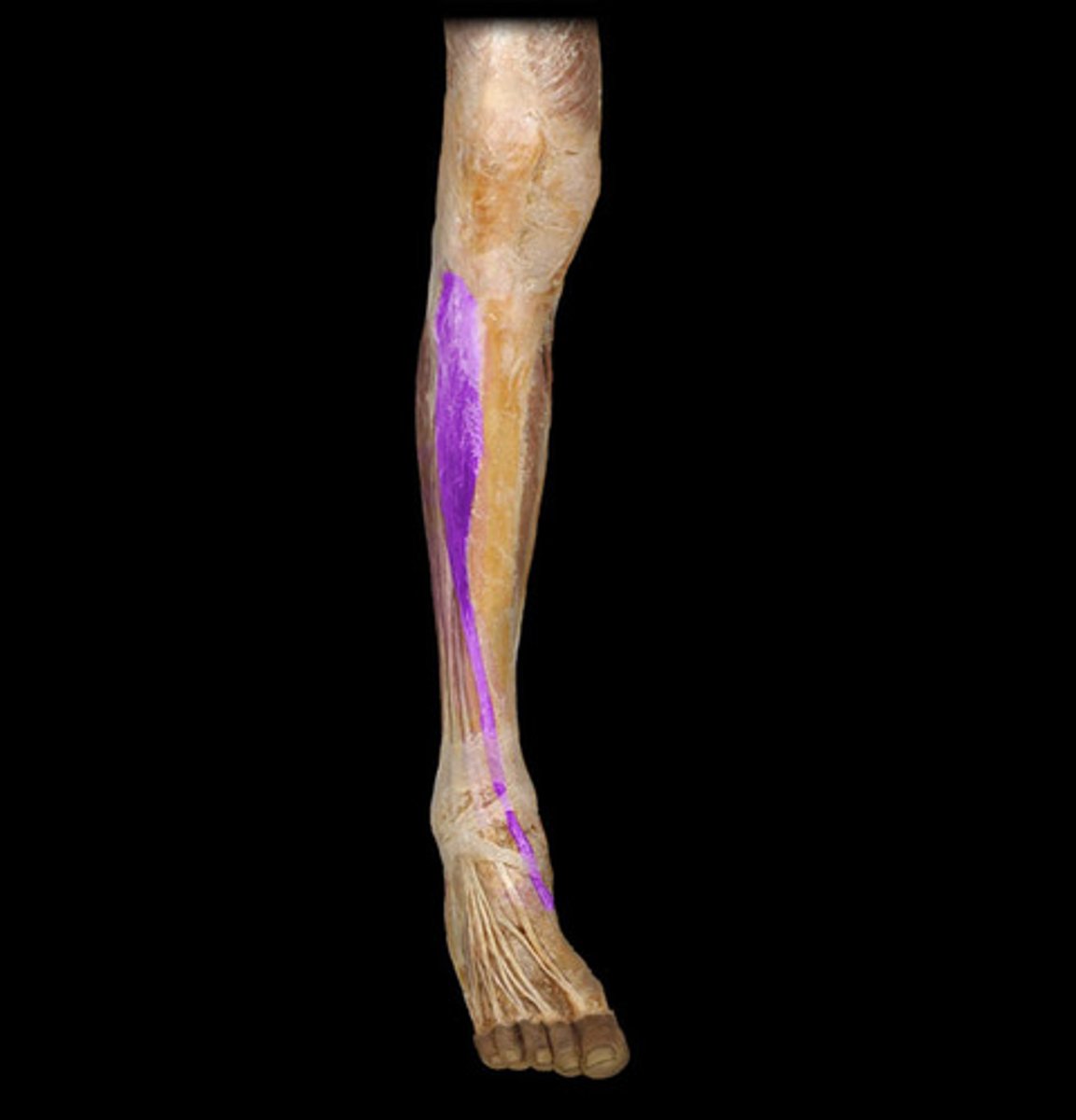
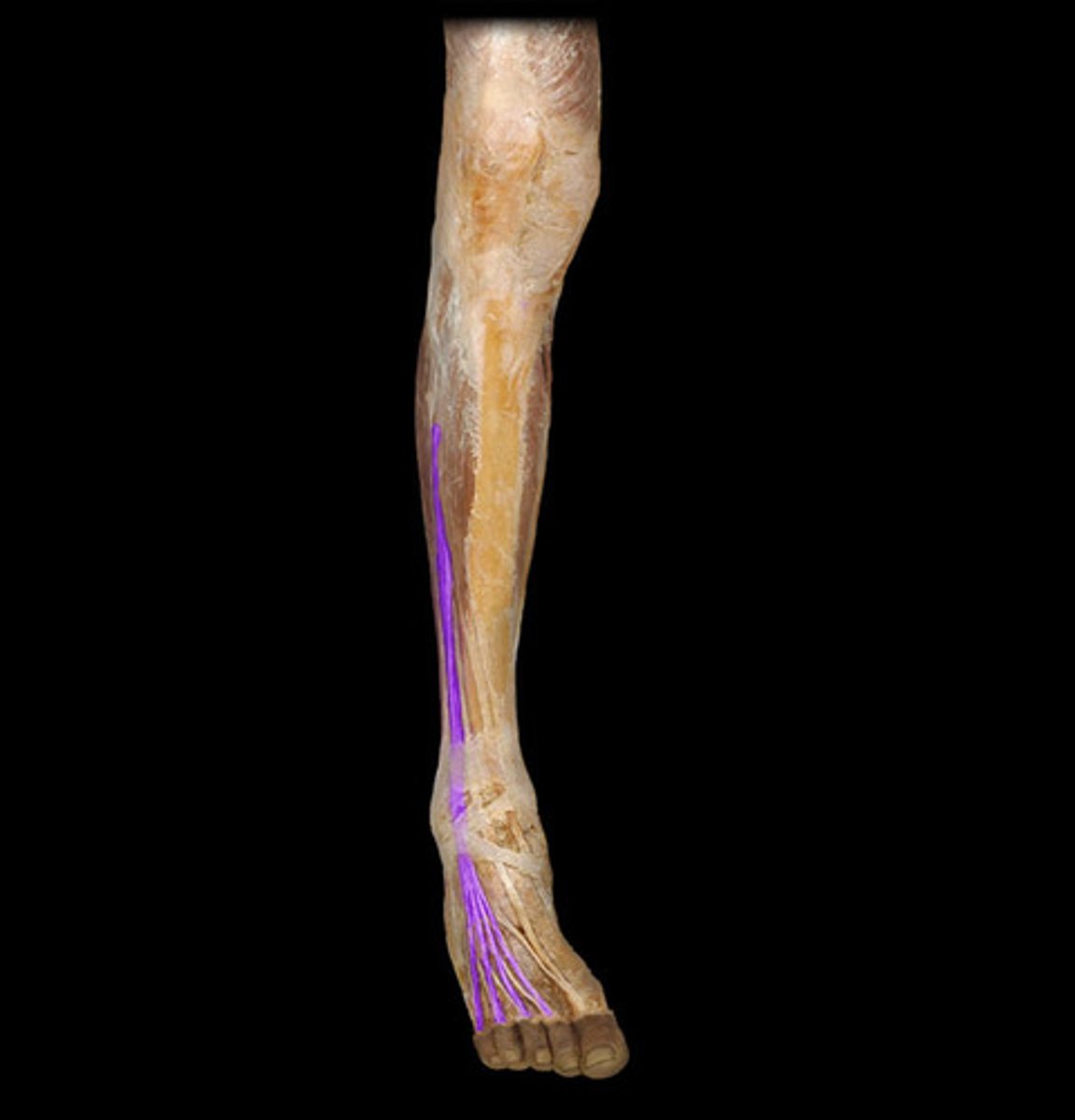
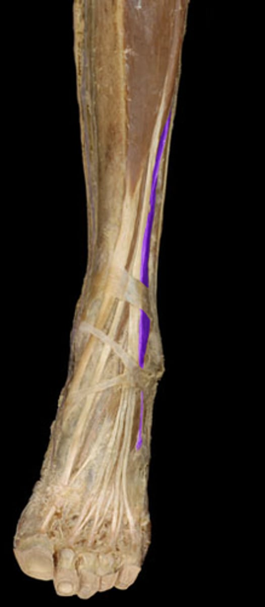
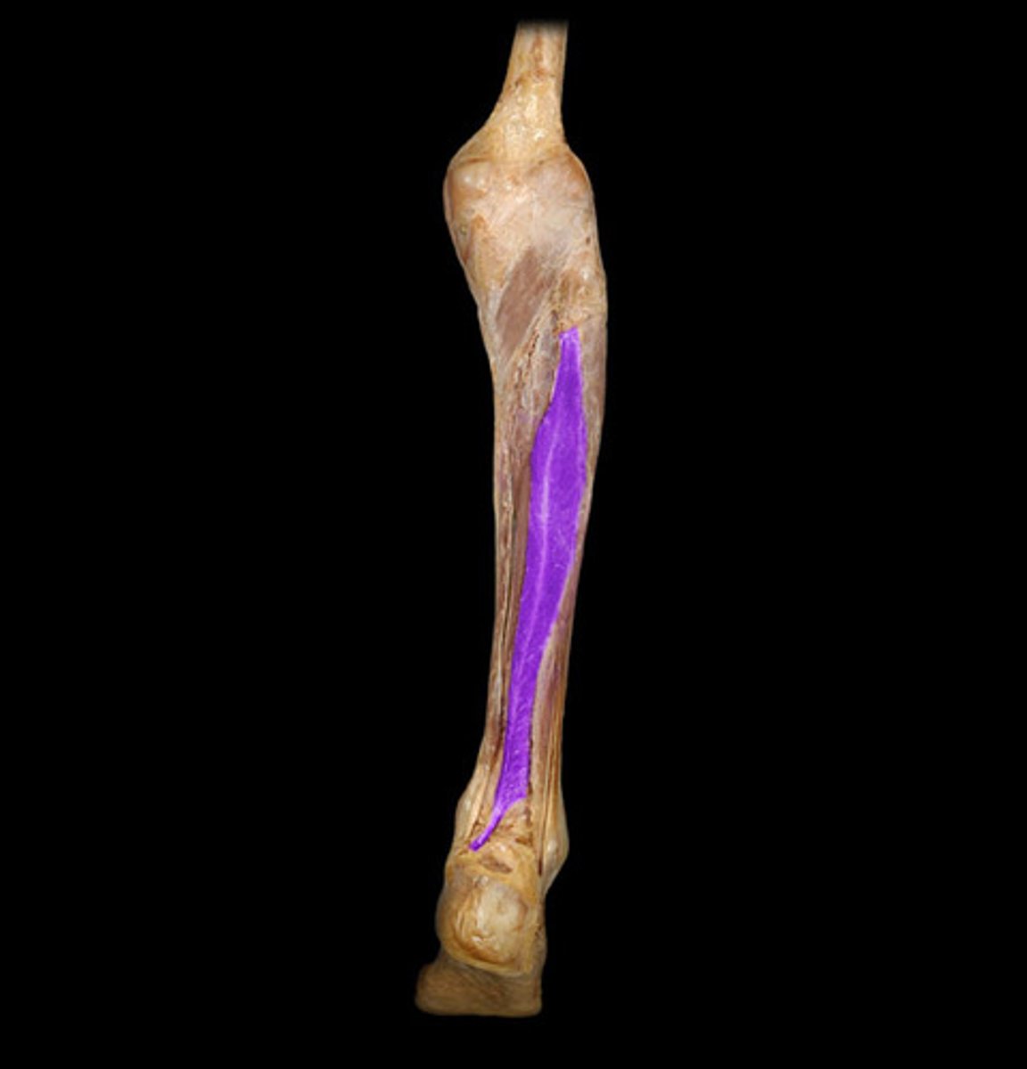
Flexor hallucis longus
Found under the triceps surae and lateral to the flexor digitorum longus, it flexes the big toe.
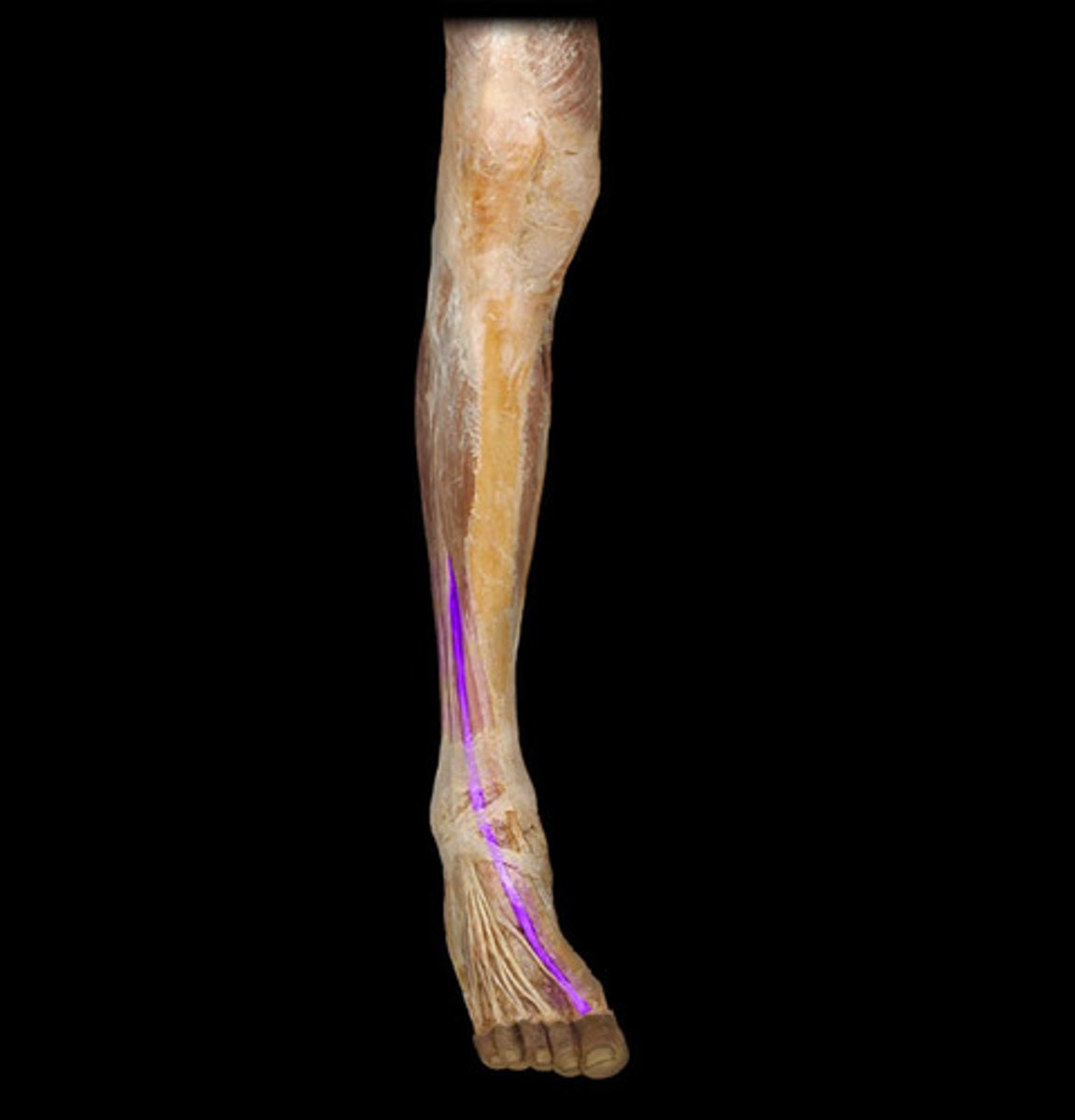
Flexor digitorum longus
Found under the triceps surae and posterior tibial artery, and medial to the flexor hallucis longus. It flexes the big toe. Number 24.
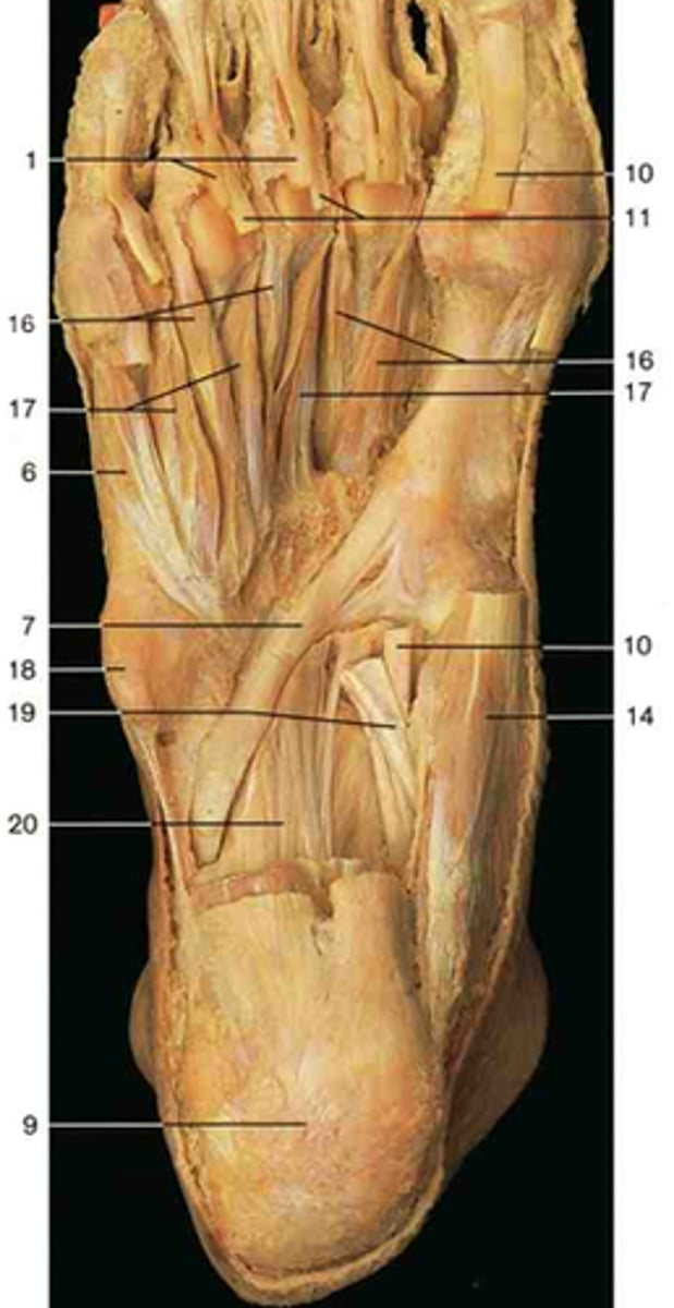
Tibialis posterior
The deepest posterior leg muscle, directly attached to the posterior tibio-fibula and interosseous membrane. For foot inversion and plantarflexion. Number 10.
Flexor retinaculum
A strong fibrous band located on the medial side of the ankle.
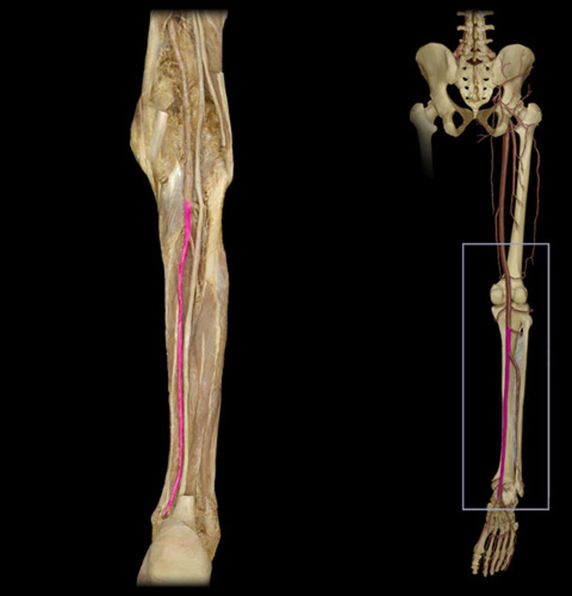
Posterior tibial artery
Larger branch of the popliteal artery that supplies the posterior leg and foot. Under the triceps surae, flexor digitorum longus, and tibialis posterior. Medial to fibular artery.

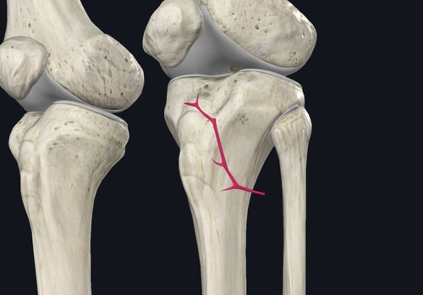
Nutrient artery of tibia
Arises from the posterior tibial artery on the medial side, and pierces the tibialis posterior to enter the tibial shaft's nutrient foramen. Hence, it is the posterior tibial artery along the entire length, only becoming the nutrient artery when entering the fossa.
Fibular artery
Found under the flexor hallucis longus, lies on the tibialis posterior, lateral to the posterior tibial artery, and medial to fibula. Supplies the posterolateral leg compartments.
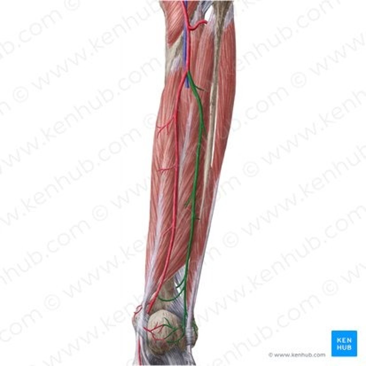
Circumflex fibular artery
A branch of the posterior tibial artery that courses around the neck of the fibula. Supplies the knee area.
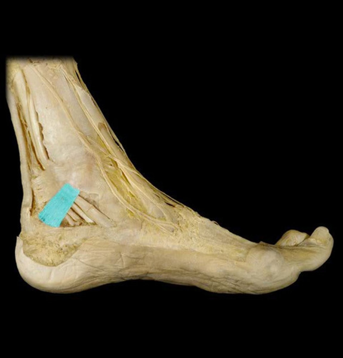
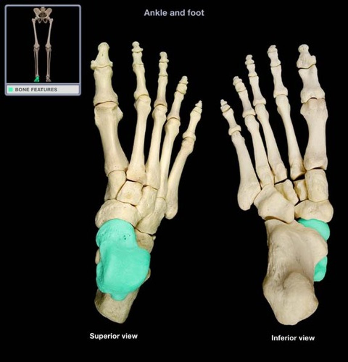
Talus
Bearing the weight transmitted by the tibia, it is the only foot bone articulating with the leg bones.
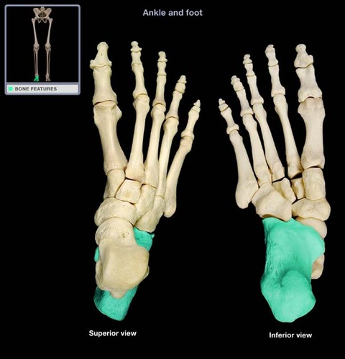
Calcaneus
The largest, strongest foot bone.
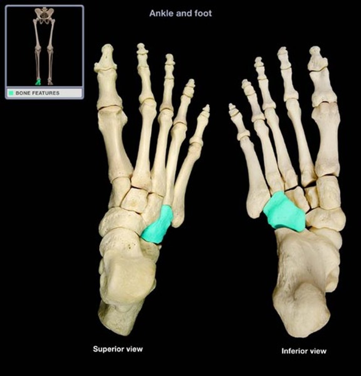
Navicular
A boat-shaped bone found between the talus and cuneiforms.
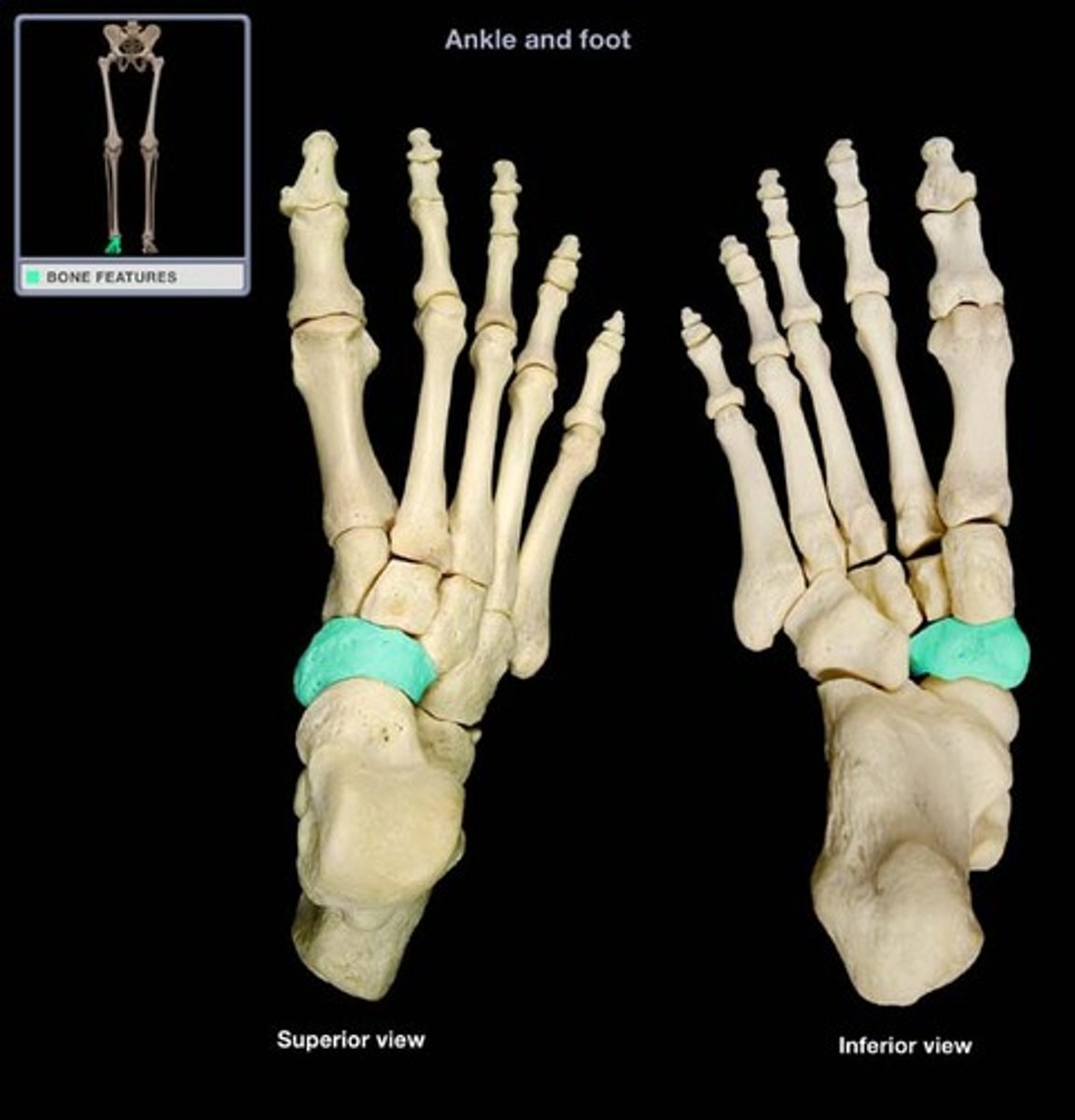
Cuboid
Most lateral bone in the distal row of the tarsus.
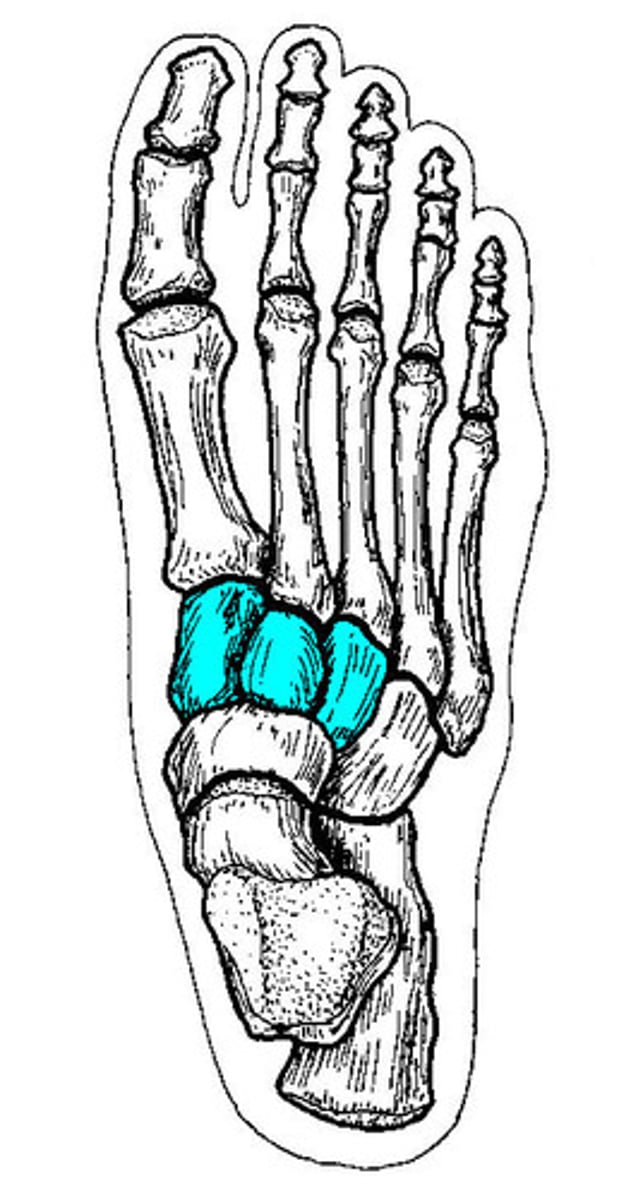
Cuneiform
A group of 3, wedge-shaped bones. With medial, intermediate, and lateral parts.
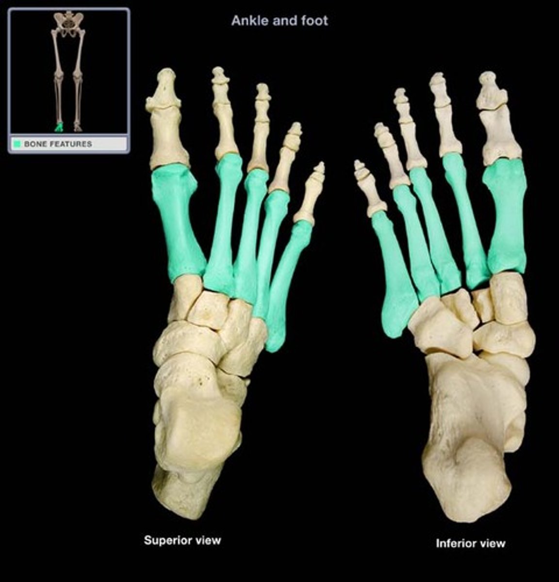
Metatarsals
The first set of bones in the forefoot.
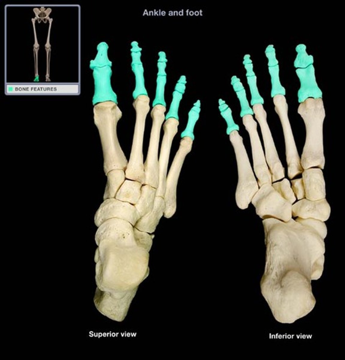
Phalanges
The second set of bones in the forefoot, consist of proximal and distal parts, except for the 1st digit.
Dorsal skin of foot
Thin, loose, low in fat.
Plantar skin of foot
Thick skin, many sweat glands, high in fat, very sensitive.
Deep fascia of foot
Also called the plantar fascia.
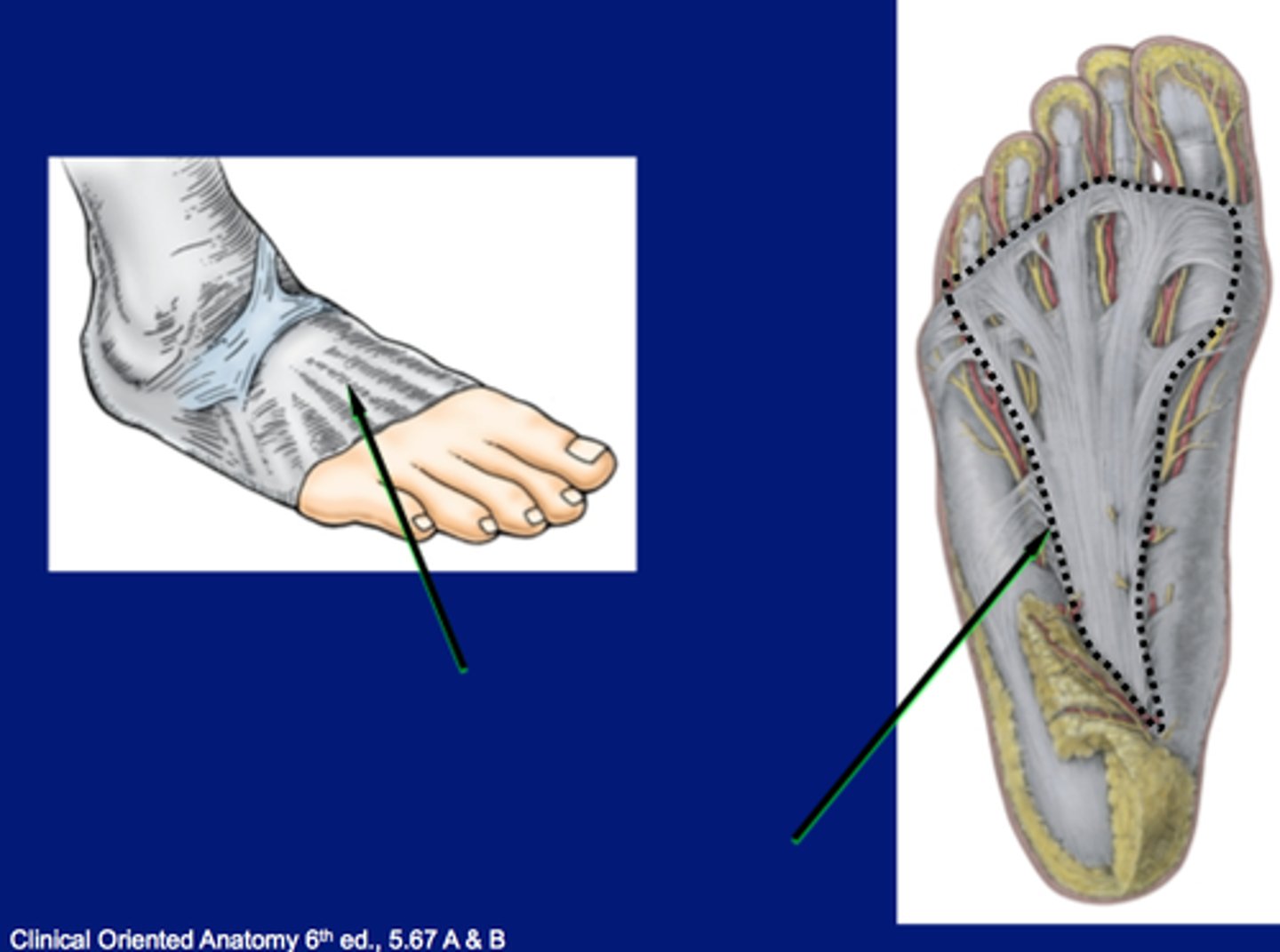
Inferior extensor retinaculum
Y-shaped band of deep fascia forming a strong loop around the tendons of fibularis tertius and extensor digitorum longus muscles.
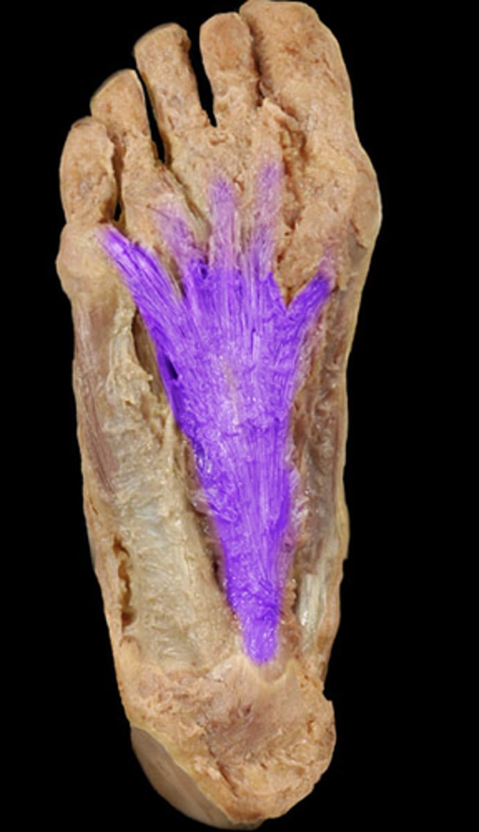
Plantar aponeurosis
Longitudinal bundles of dense fibrous connective tissues.
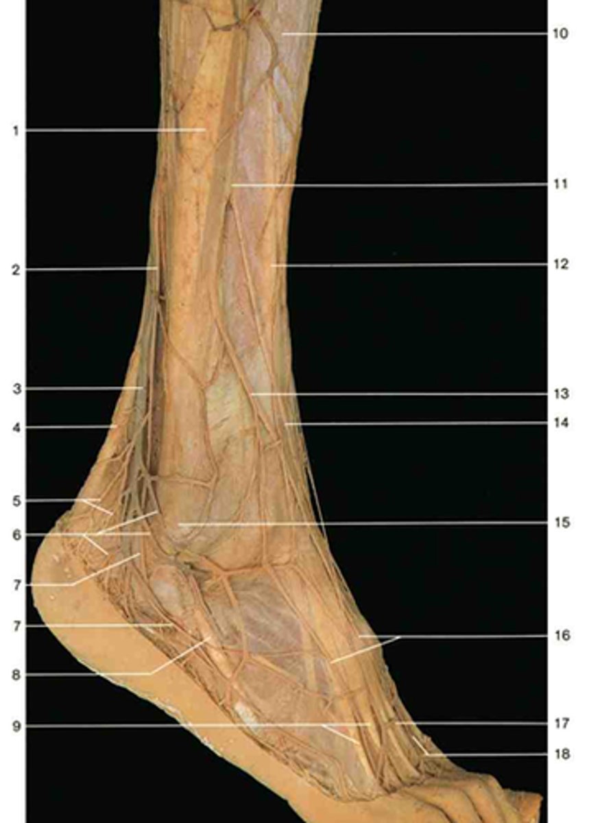
Extensor digitorum brevis
A superficial muscle on the dorsal side of the foot. Extends toes 2-4.
Extensor hallucis brevis
Part of the extensor digitorum brevis, but appears separate and moves the big toe.
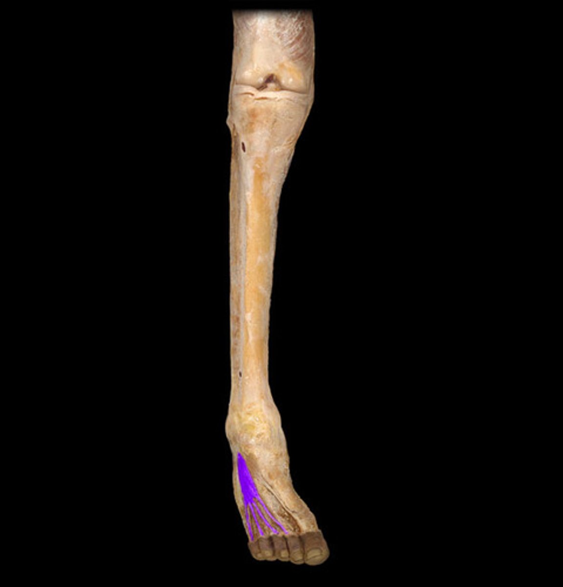
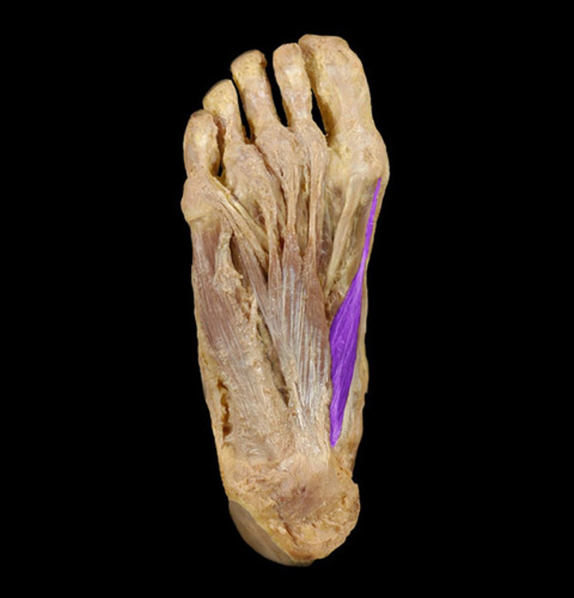
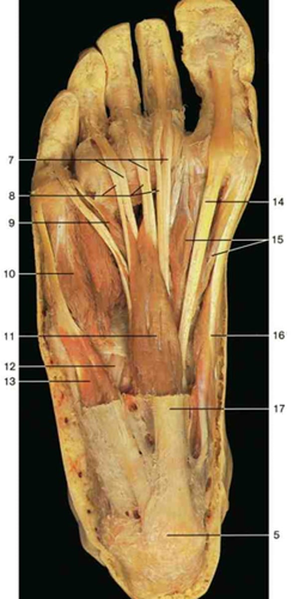
Abductor hallucis
Abducts and flexes big toe.
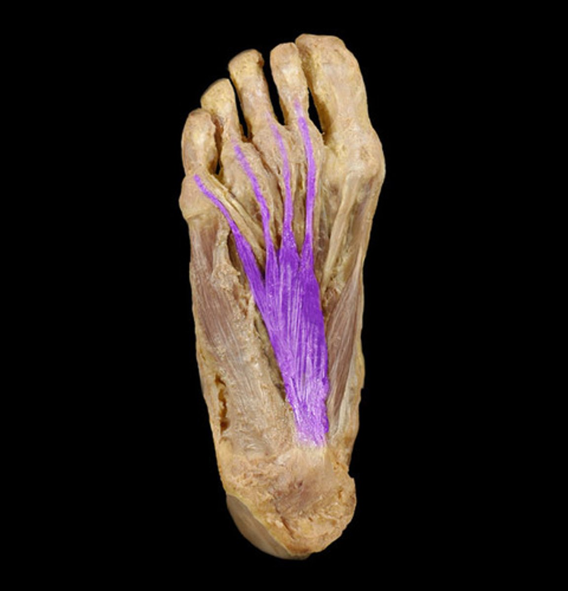
Flexor digitorum brevis
Muscle that flexes the toes and helps maintain balance while walking and standing.
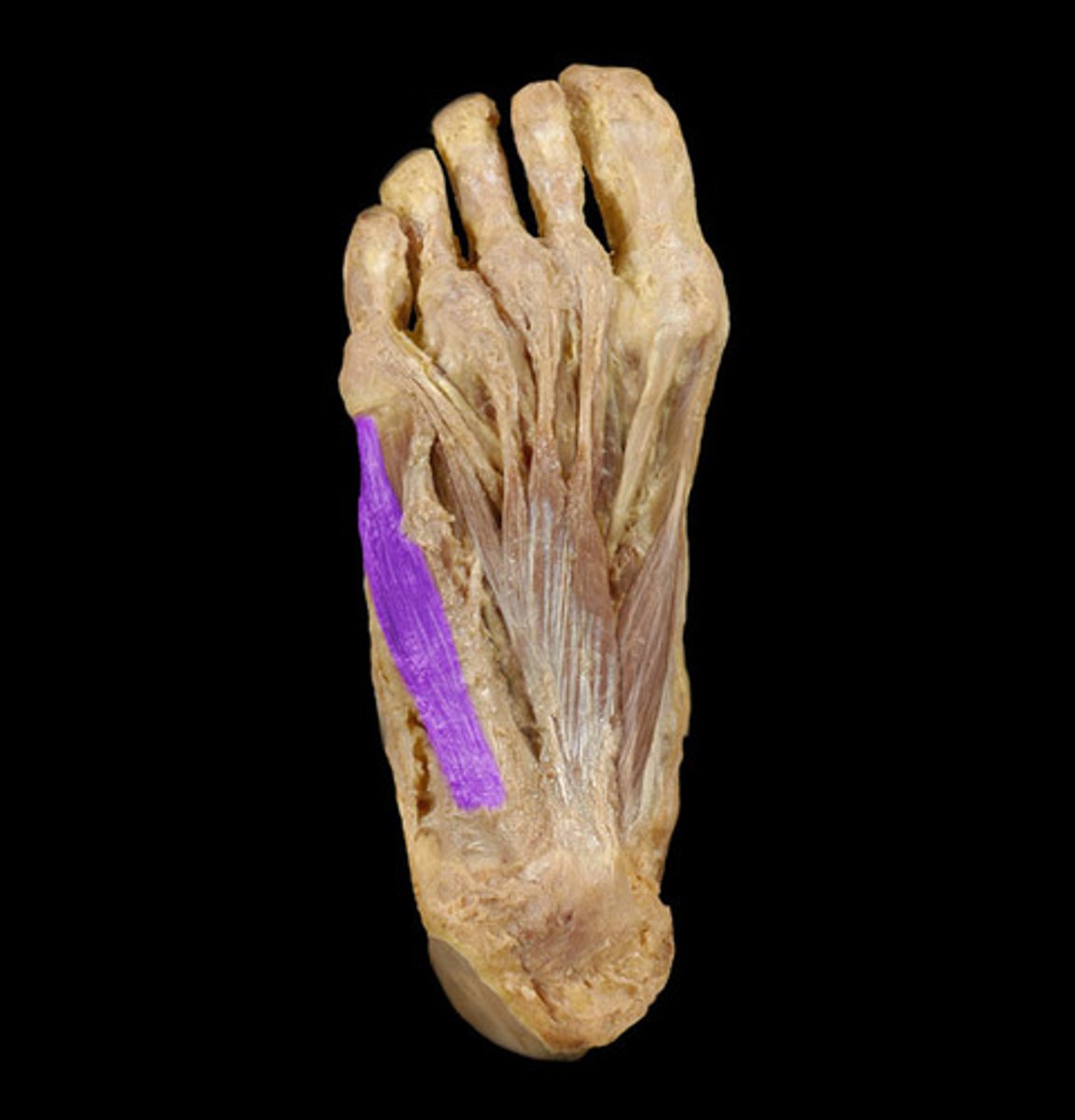
Abductor digiti minimi
Flexes and abducts 5th toe.
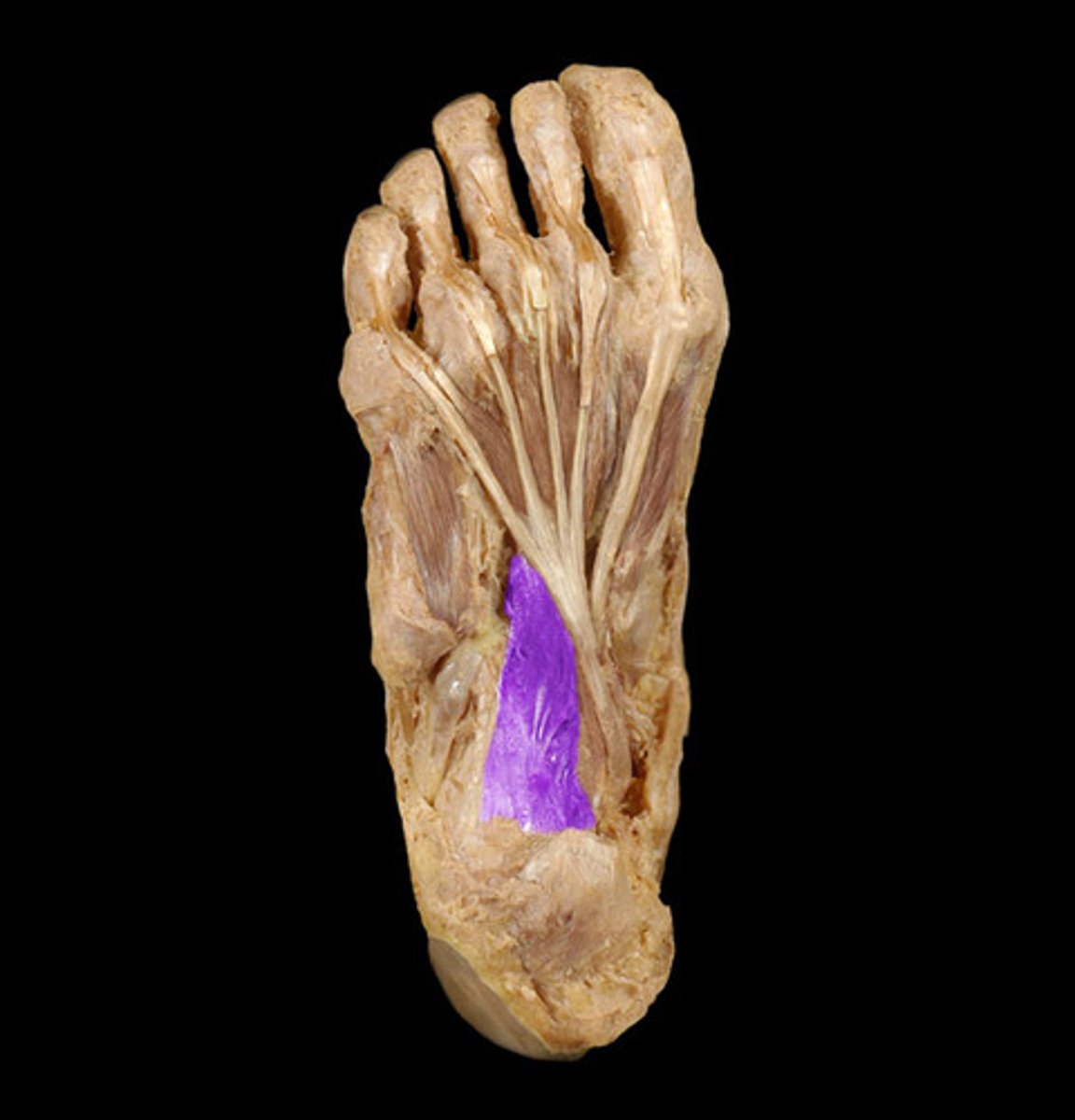
Quadratus plantaris
Assists the flexor digitorum longus in flexing 2nd to 5th toes.
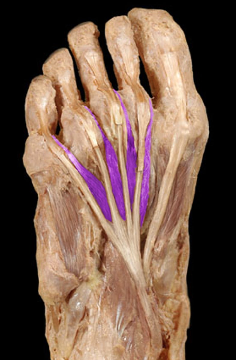
Lumbricals
Flexes metacarpophalangeal joints and extends interphalangeal joints of toes 2-5. Found under the flexor digitorum brevis, but above the plantar interossei.
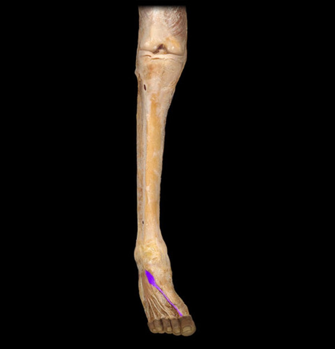
Flexor hallucis brevis
Flexes the big toe at the metatarsophalangeal (MTP) joint. Found under the abductor hallucis and extensor hallucis brevis, but above the flexor hallucis longus. Number 15.

Flexor digiti minimi brevis
Flexes the proximal phalanx of the 5th toe at the metatarsophalangeal (MTP) joint. Medial to the abductor digiti minimi. Number 10.
Plantar interossei
Adduct toes 3-5 toward the second toe. Found under the lumbricals, but above the metatarsals, and medial to the corresponding toe. Number 17.
Dorsal interossei
Abduct toes 2-4 away from the second toe. Found under the lumbricals, but above the metatarsals. Number 16.

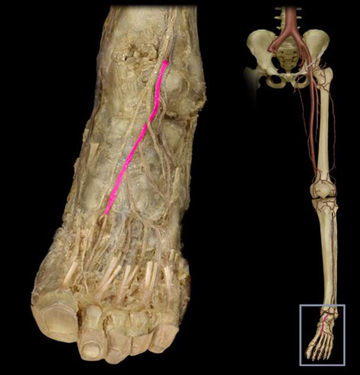
Dorsalis pedis artery
The continuation of anterior tibial artery, supplies the forefoot. Under the extensor hallucis longus.
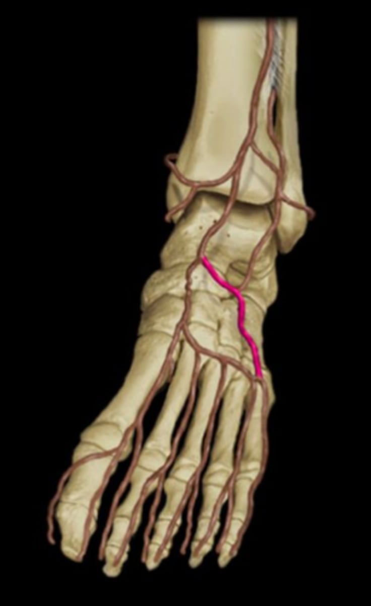
Lateral tarsal artery
Arises from dorsalis pedis near ankle joint. Found deep to muscles and meets with arcuate artery, to make an arch. Under the extensor digitorum brevis.
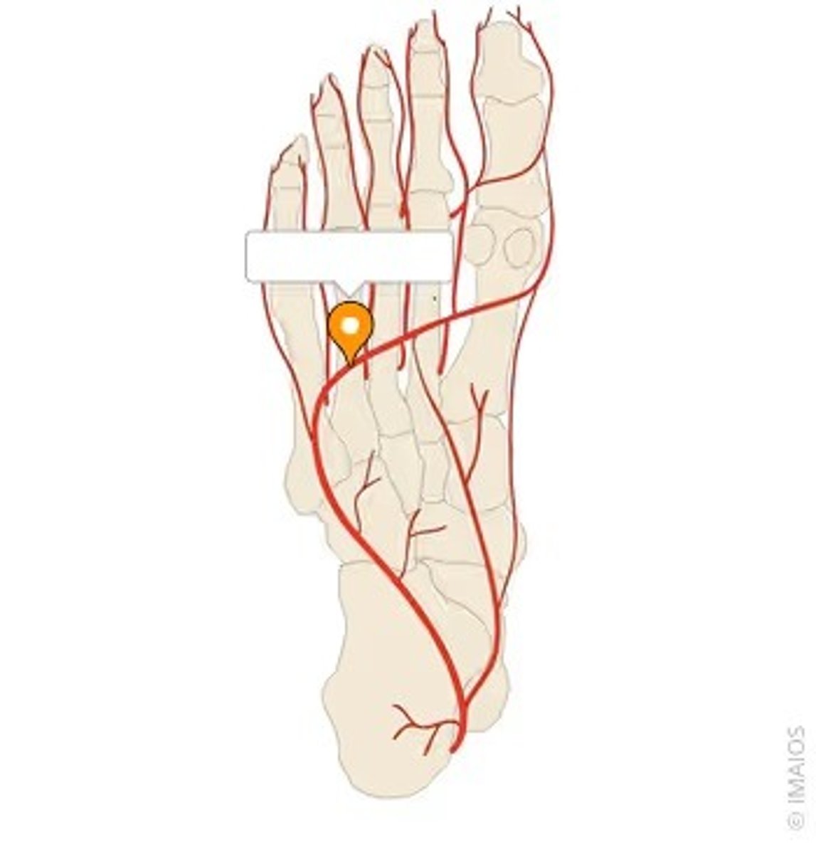
Deep plantar artery
Passes deeply between the interosseous of the first digit, joining the lateral tarsal artery to form the deep plantar arch. Pierces between 1st and 2nd metatarsals.
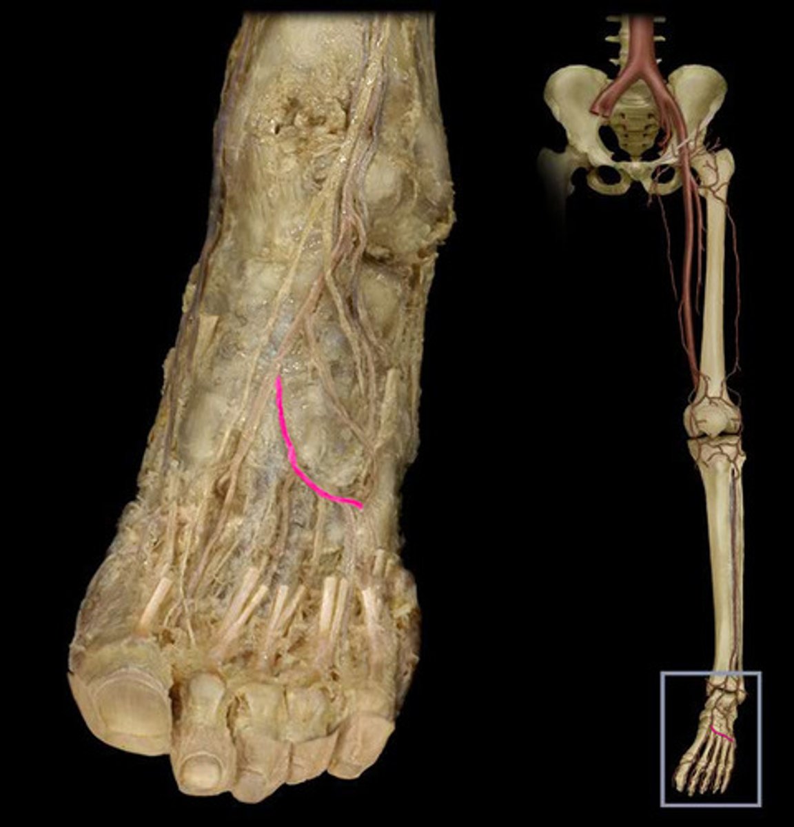
Arcuate artery
Anastomoses with the lateral tarsal artery to form an arterial loop. Gives rise to 2nd-4th dorsal metatarsal arteries. Under the extensor digitorum brevis.
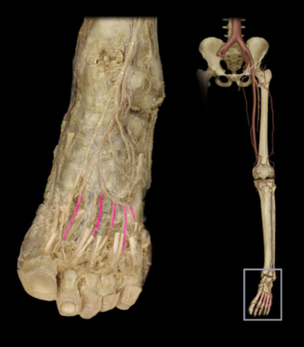
Dorsal metatarsal arteries
Branches from the arcuate arteries; supply blood to the dorsal digital arteries. Under the extensor digitorum longus.
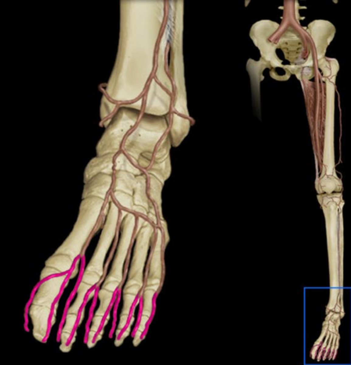
Dorsal digital arteries
Branches from the dorsal metatarsal arteries and supply blood to the digits. Under the extensor digitorum longus.
Deep plantar arch
Supplies the plantar surface of the foot. Formed by the lateral plantar and deep plantar branch of the dorsalis pedis artery. Under the adductor hallucis, but above the plantar interossei.

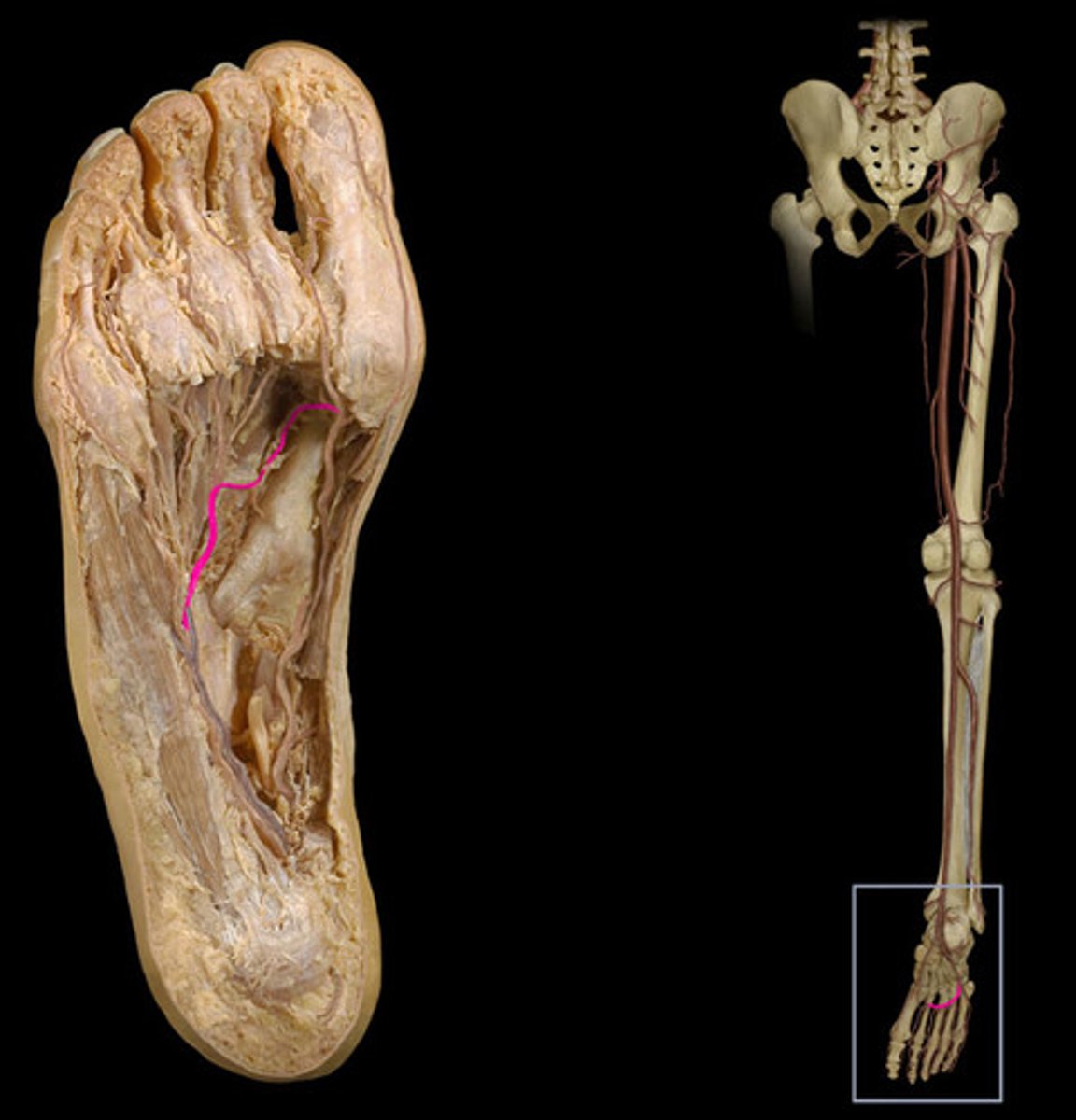
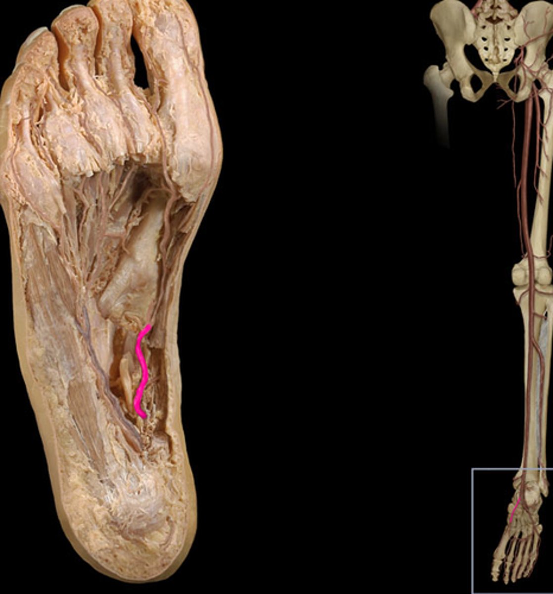
Medial plantar artery
Arises from the bifurcation of the posterior tibial arteries; supplies blood to the medial plantar surfaces of the foot. Found under the abductor hallucis.
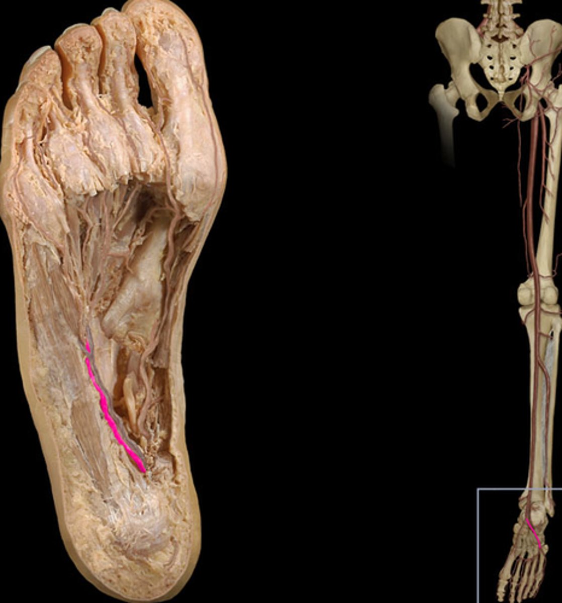
Lateral plantar artery
Arises from the bifurcation of the posterior tibial arteries; supplies blood to the lateral plantar surfaces of the foot. Found under the abductor hallucis.
Superficial plantar arch
Found above the flexor digitorum brevis, and under the skin. Supplies part of the foot and toes.
Dorsal venous network
Found superficially on the dorsum of foot, which it drains.
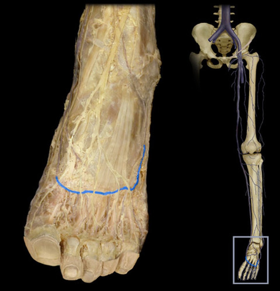
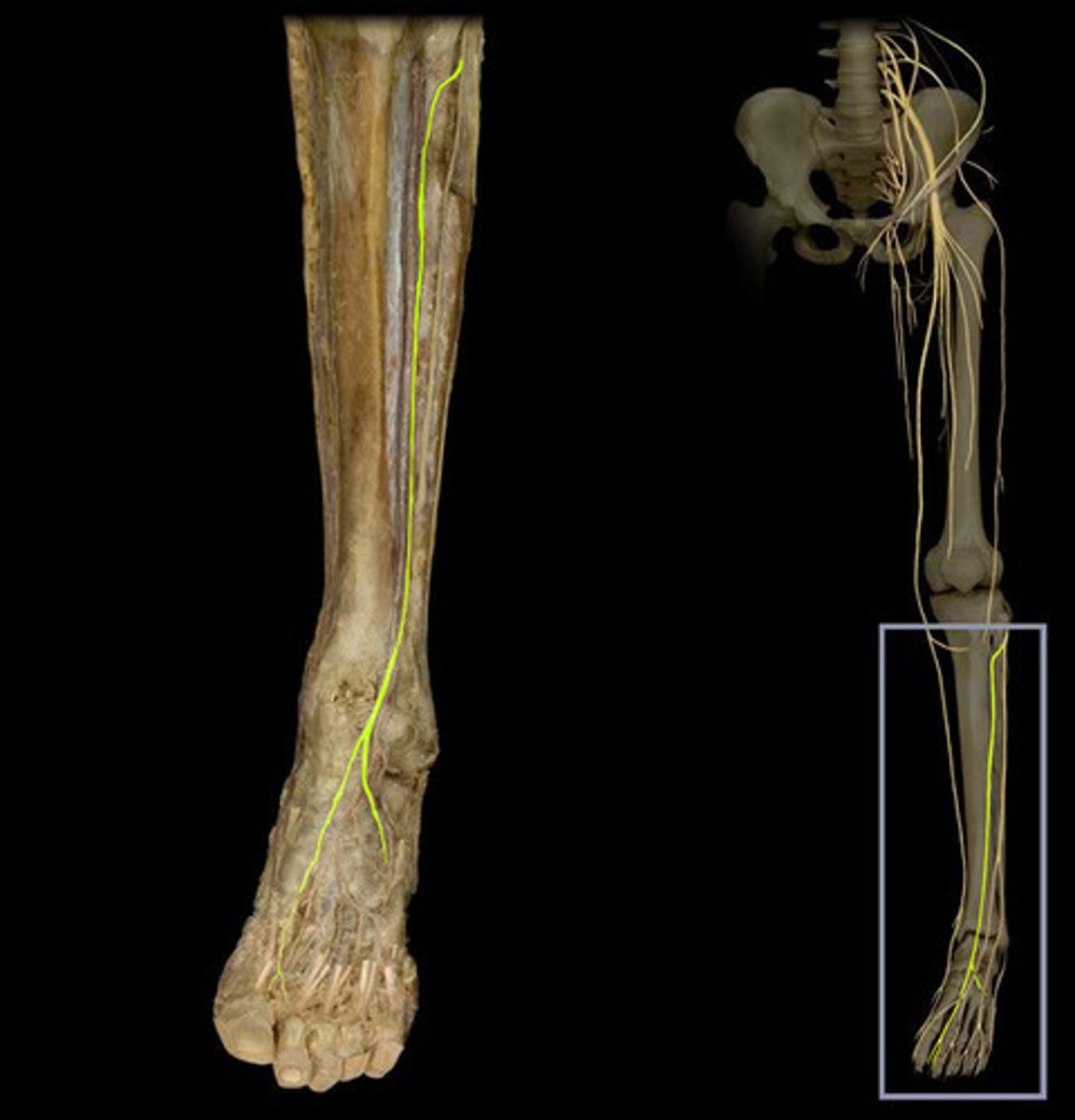
Saphenous nerve
Runs along the great saphenous vein, to innervate the medial leg and foot.
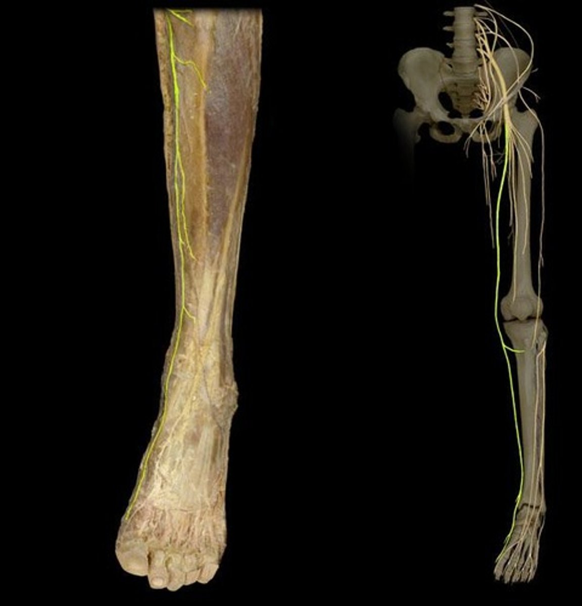
Sural nerve
Runs along the small saphenous vein, innervating the posterolateral foot and ankle.
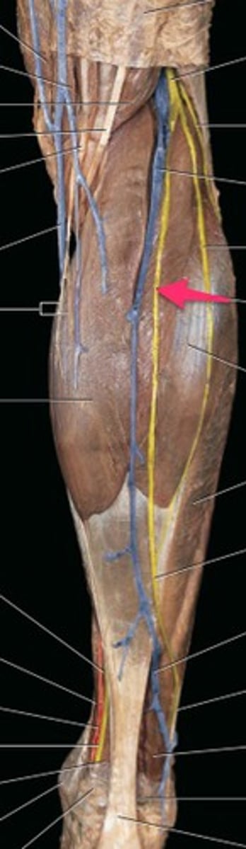
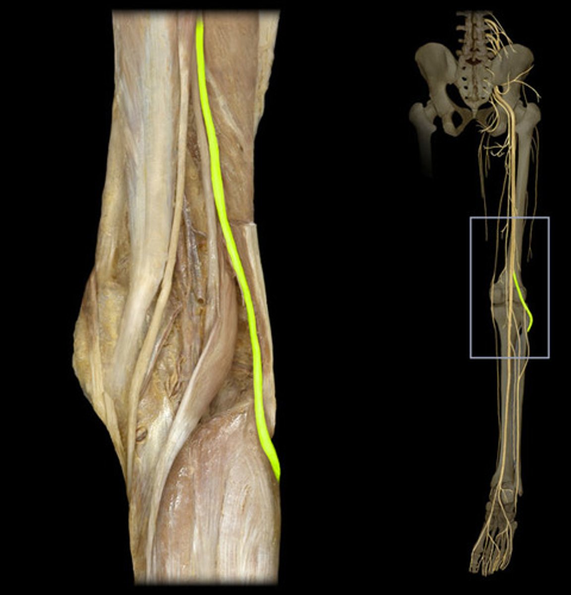
Common fibular nerve
Branch of the sciatic nerve wrapping around the fibular head. Innervates the anterolateral leg and foot.
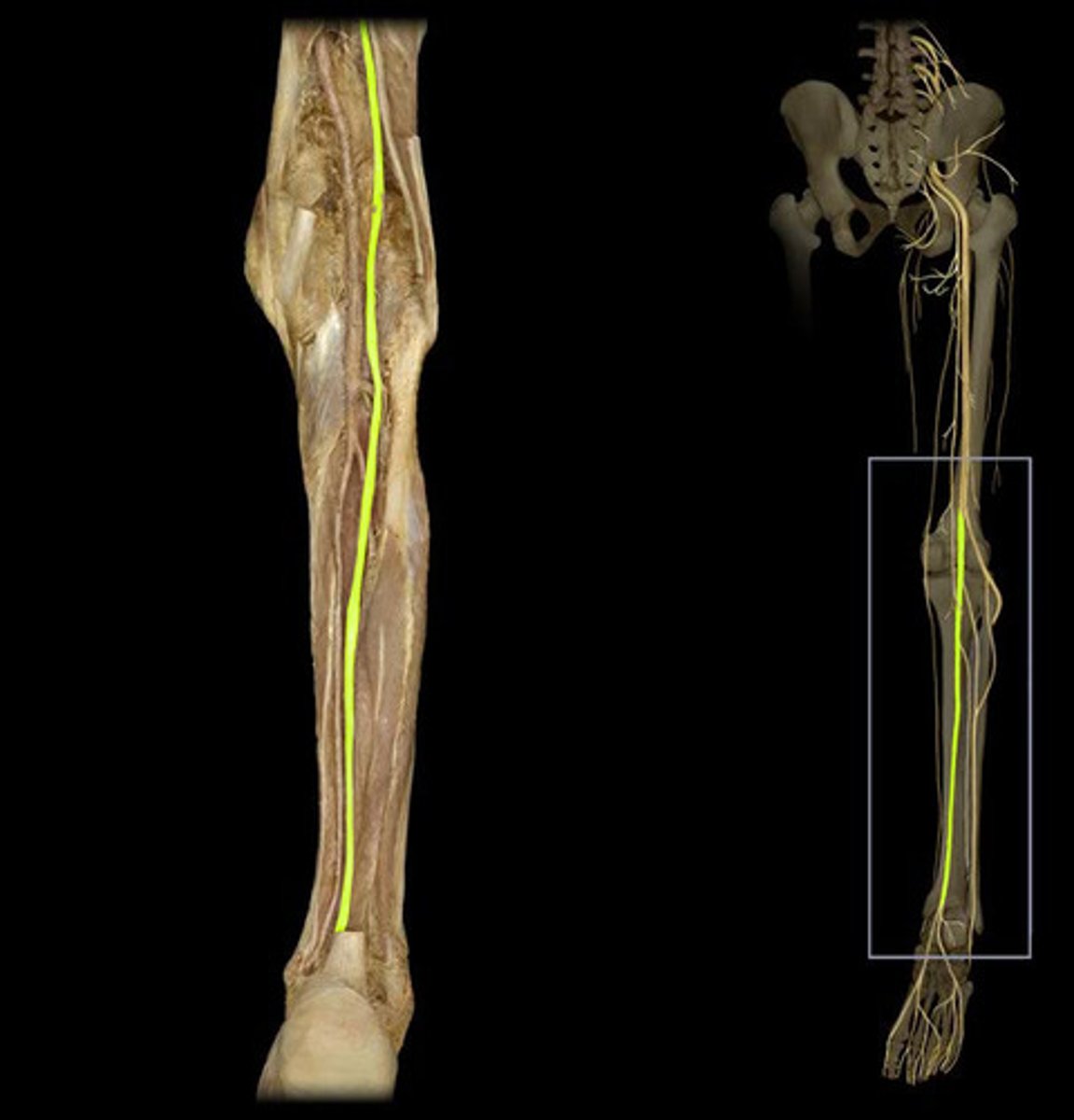
Tibial nerve
Posterior to the tibialis posterior and flexor digitorum longus, passing under the flexor retinaculum. Innervates the posterior leg and sole of foot.
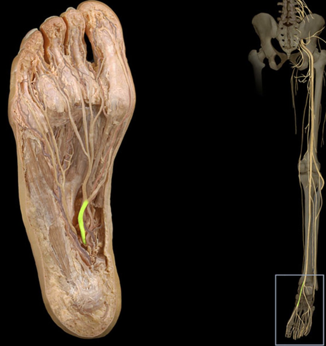
Medial plantar nerve
Found above the flexor digitorum brevis, but under the abductor hallucis, both of which it innervates.
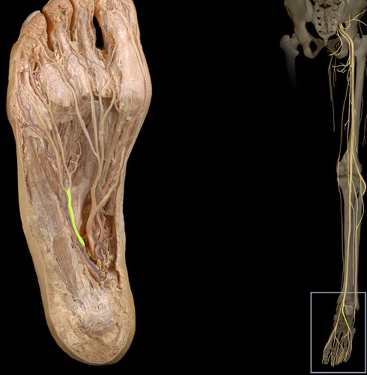
Lateral plantar nerve
Found under the abductor hallucis, then travels toward the 5th digit, which it innervates.
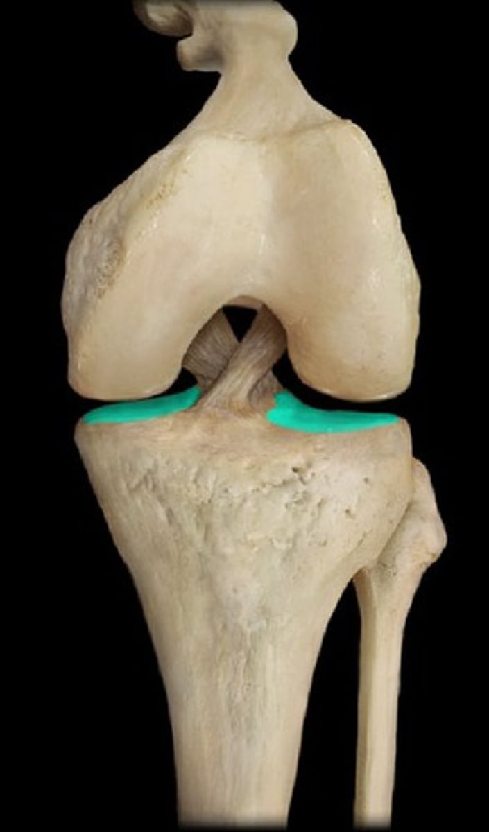
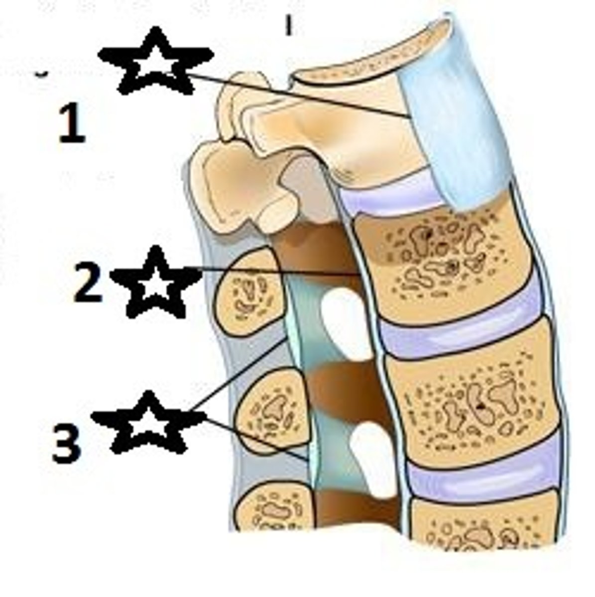
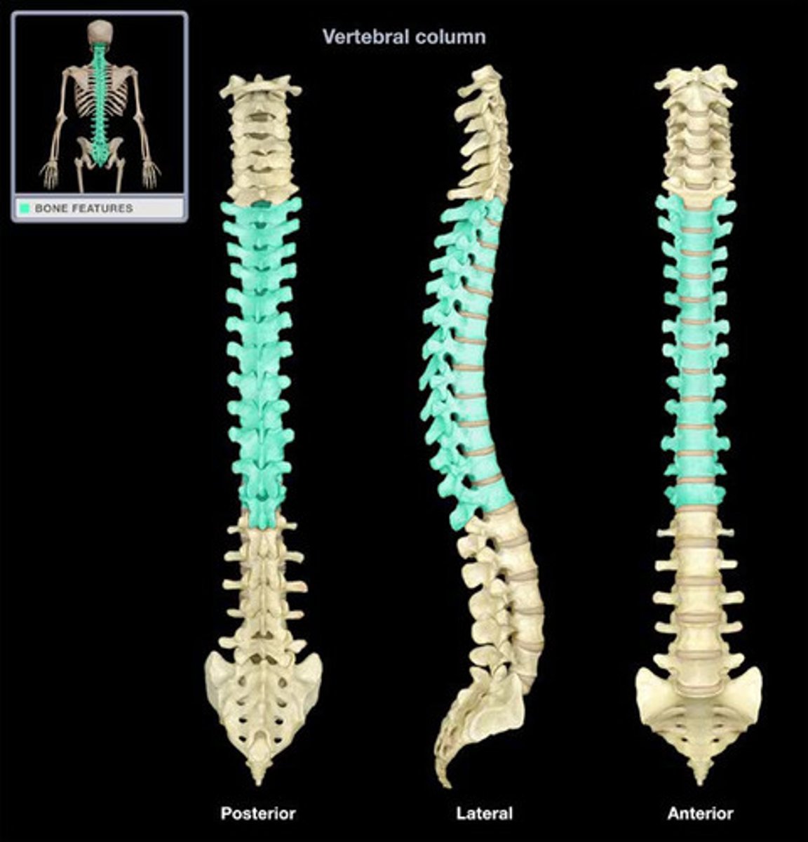
Anterior and posterior longitudinal ligament
Connects and supports vertebral bodies. Numbers 1 and 2.
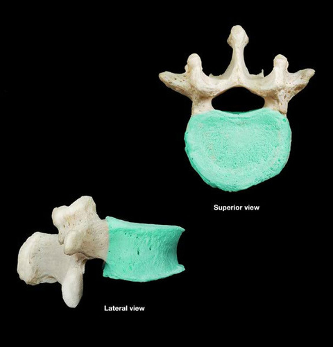
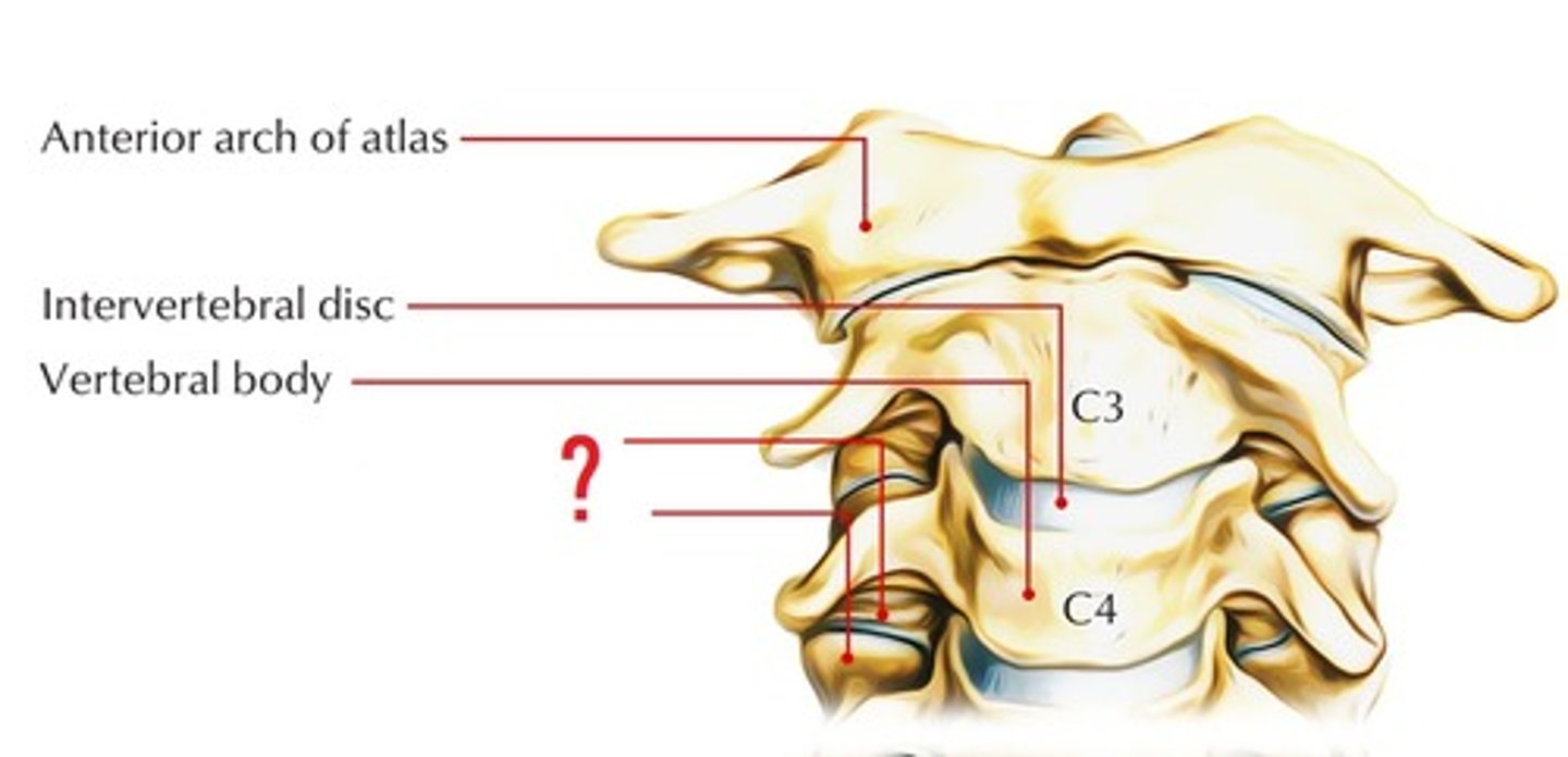
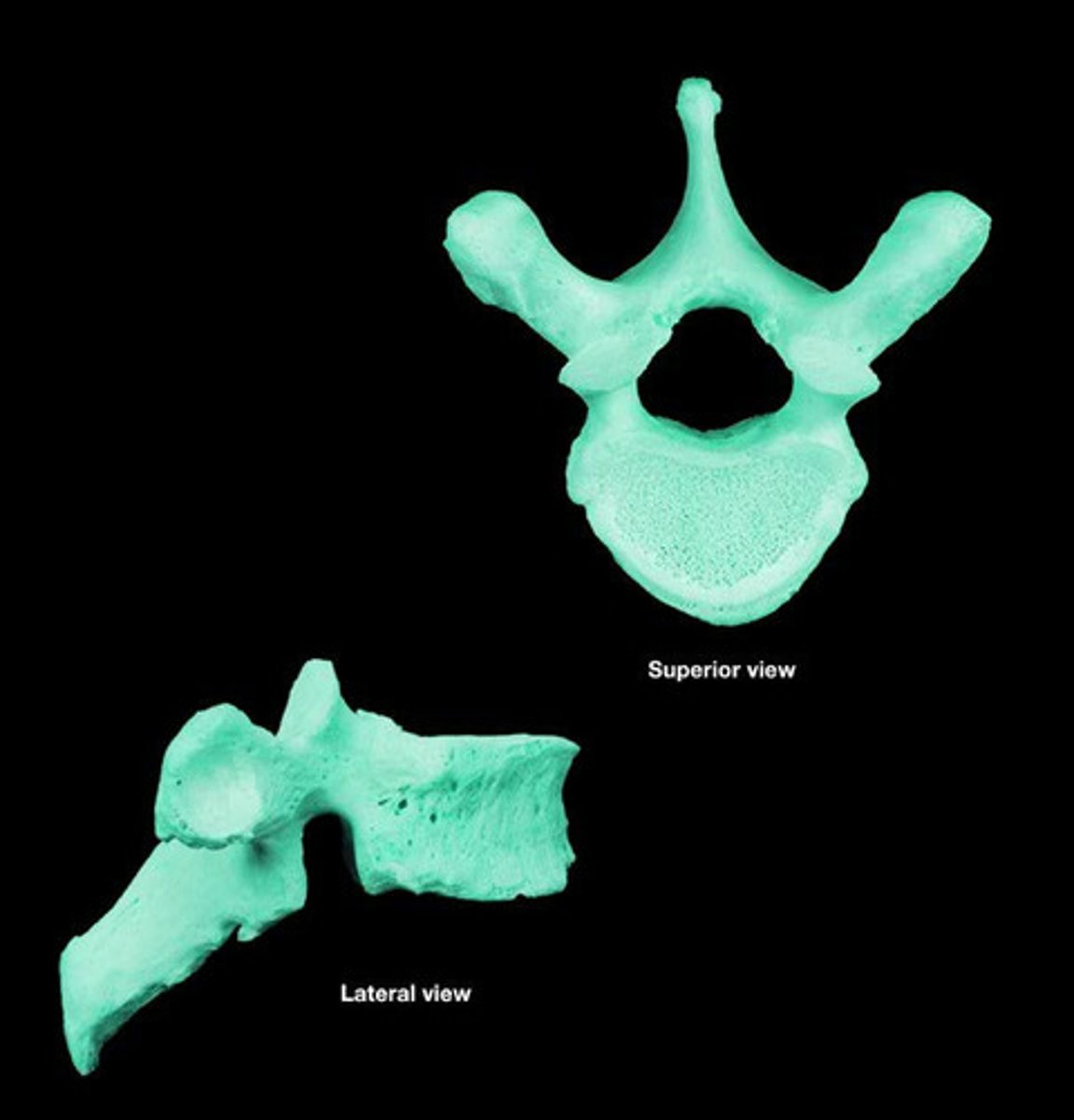
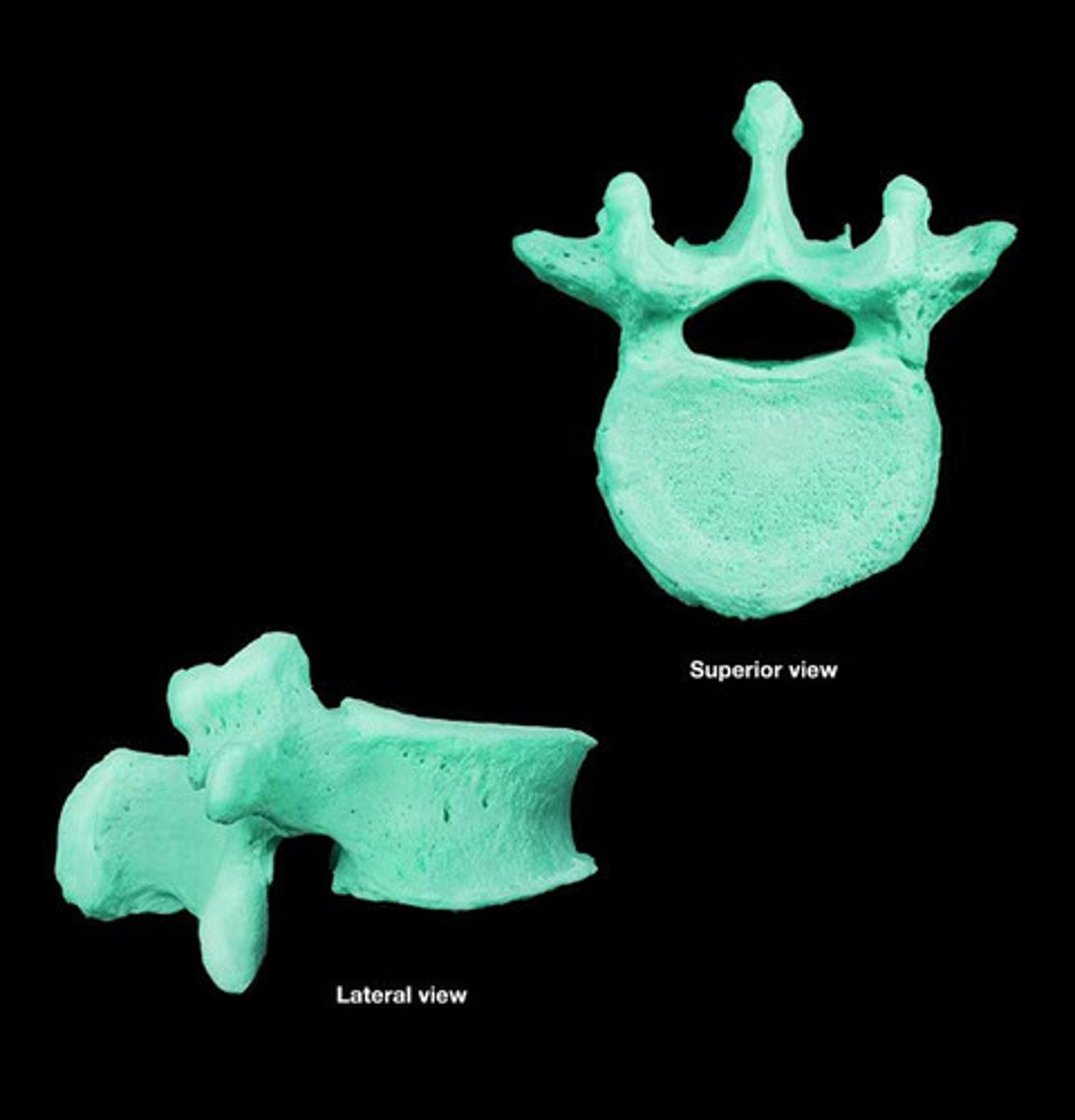
Vertebral body
Main portion of the vertebra, separate from the arches of the vertebra.
Vertebral arch
Structure that encloses the nerve cord.
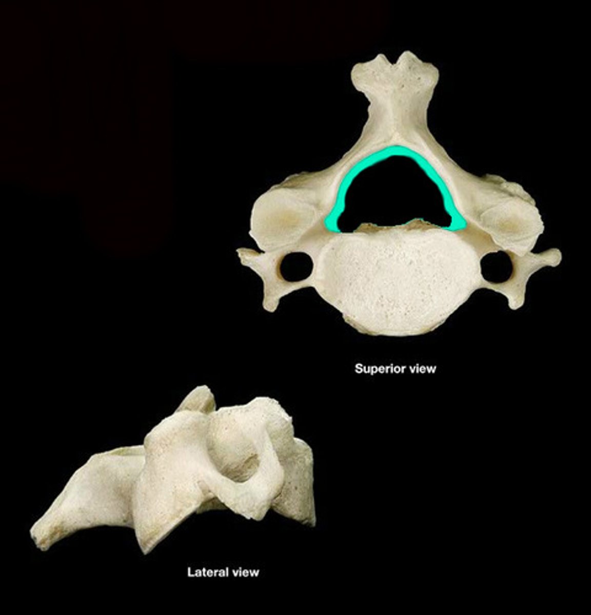
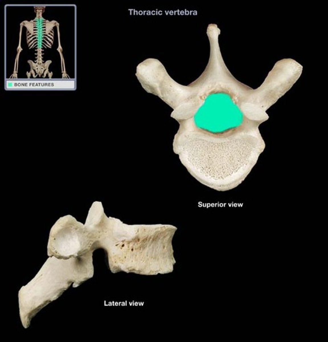
Vertebral canal
Contains the spinal cord.
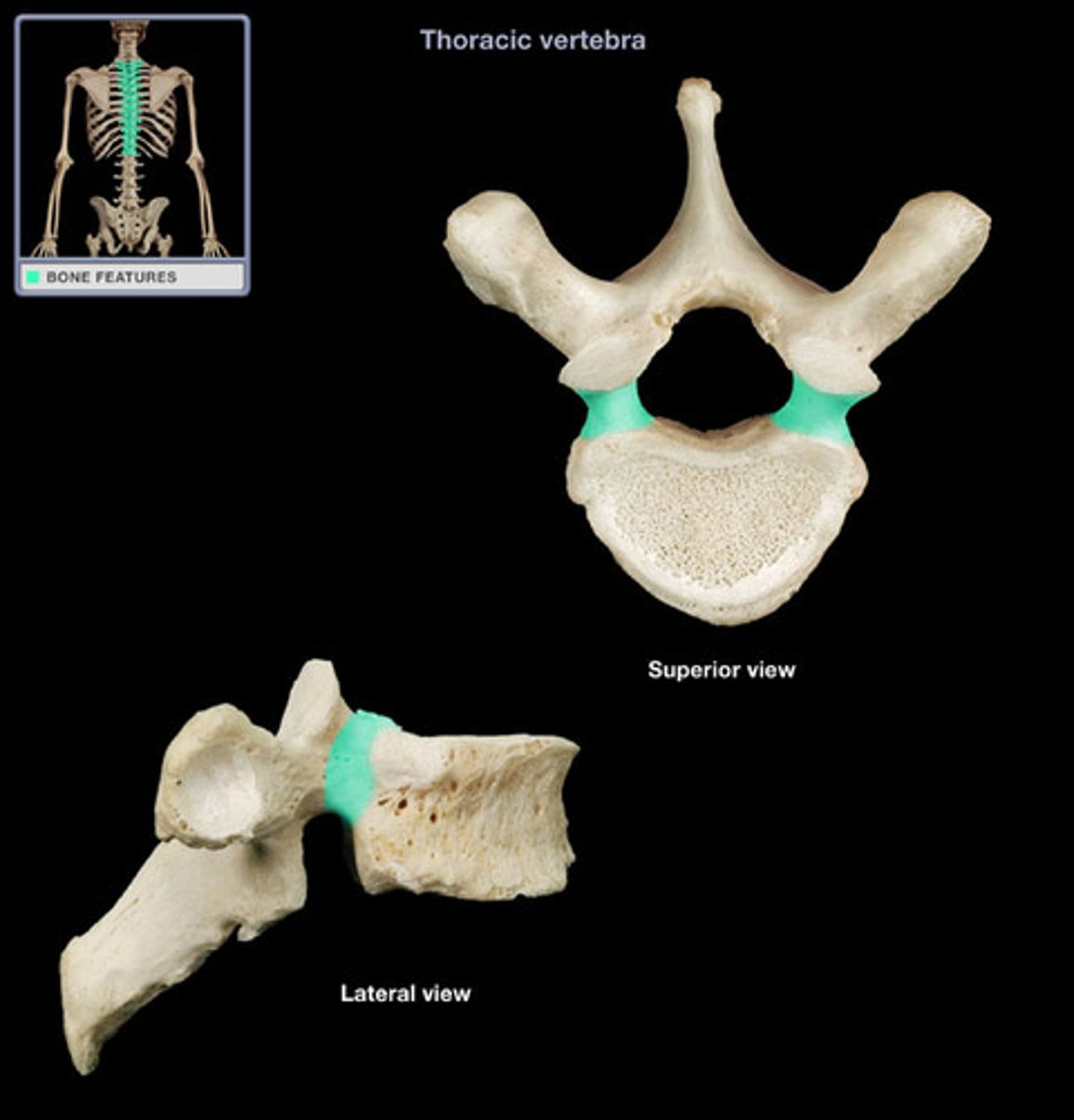
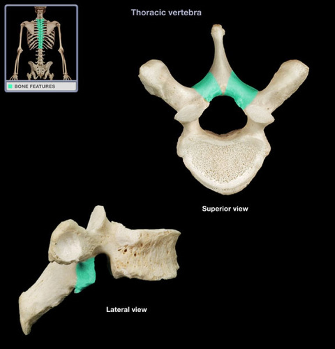
Pedicle of vertebra
Short, thick bony projection that connects the vertebral body to the vertebral arch.
Lamina of vertebra
Connects transverse process to spinous process.
Vertebral processes
A group of structures extending from the vertebral body.
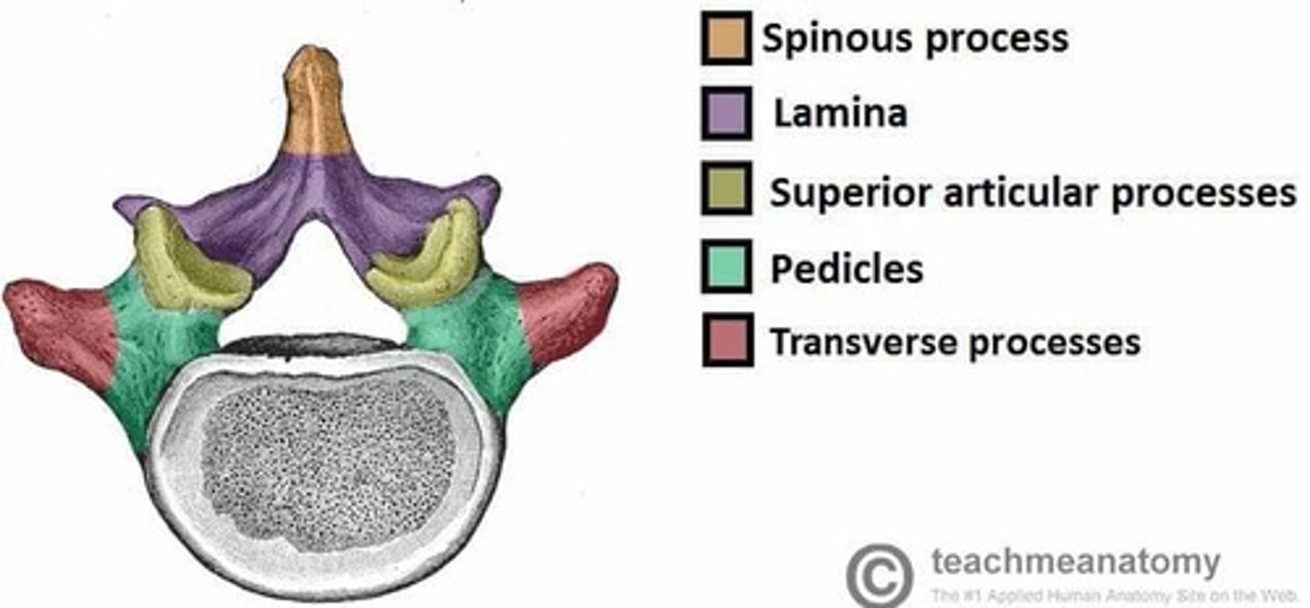
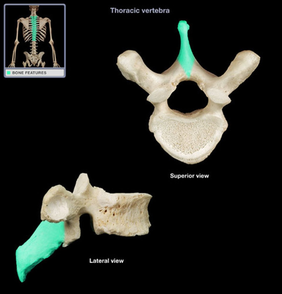
Spinous process
Sharp, slender projection.
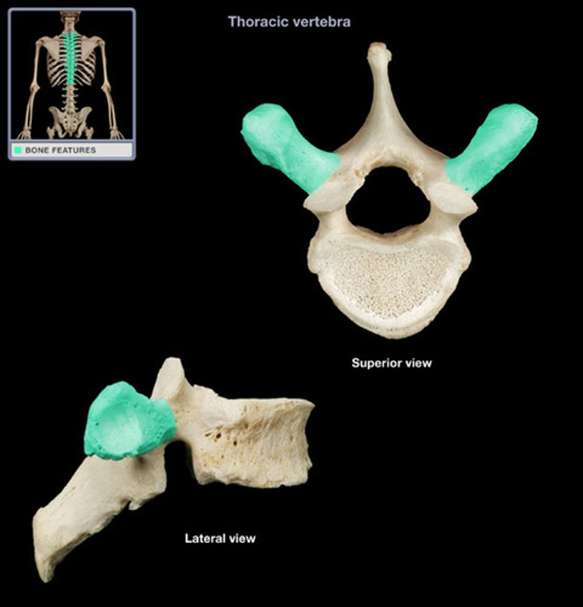
Transverse process
Two lateral projections from the vertebral arch.
Articular process
Bony projection from the vertebral arch that forms the facet joints with adjacent vertebrae, allowing for controlled movement of the spine. Has superior and inferior parts.
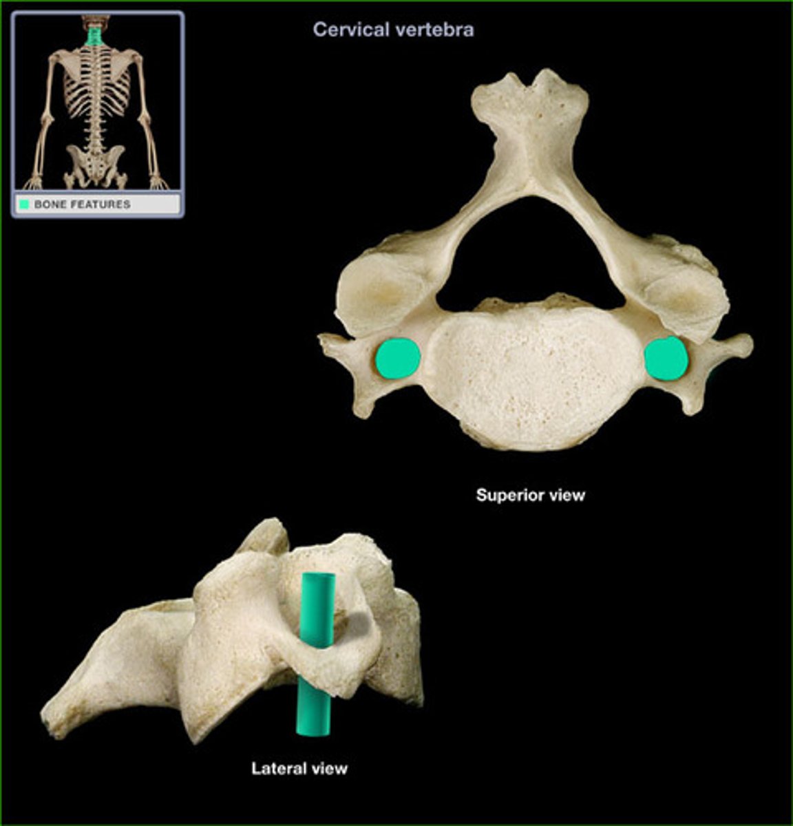
Foramen transversarium
Passageway for the vertebral artery through the cervical vertebrae.
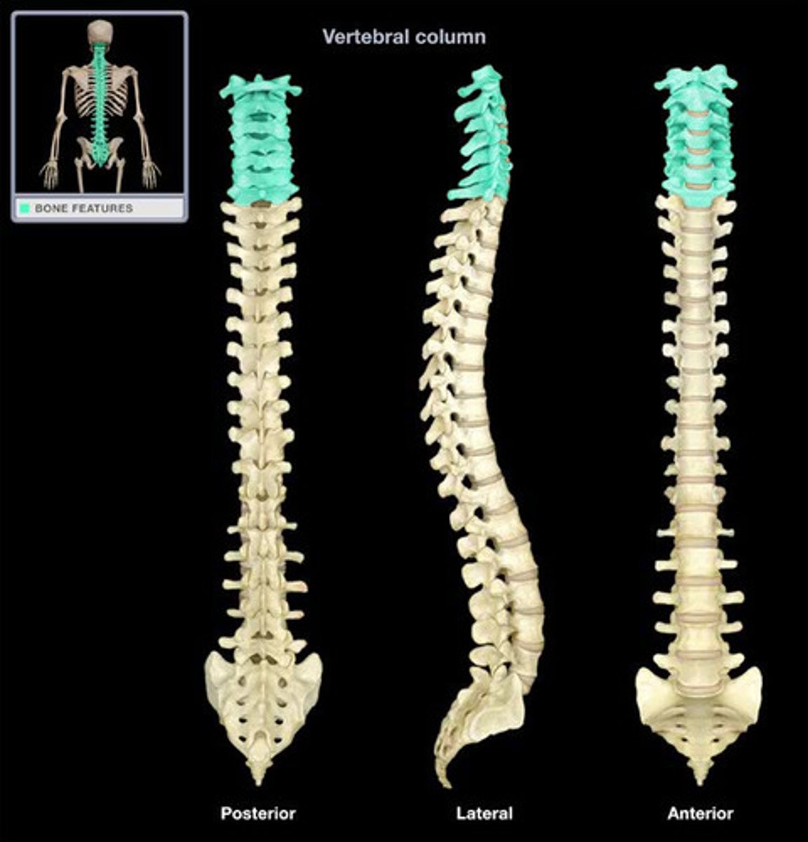
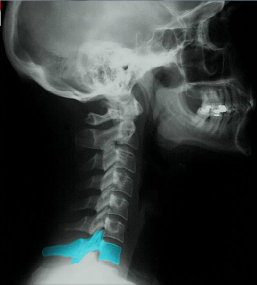
Cervical vertebrae
Vertebrae of the neck.
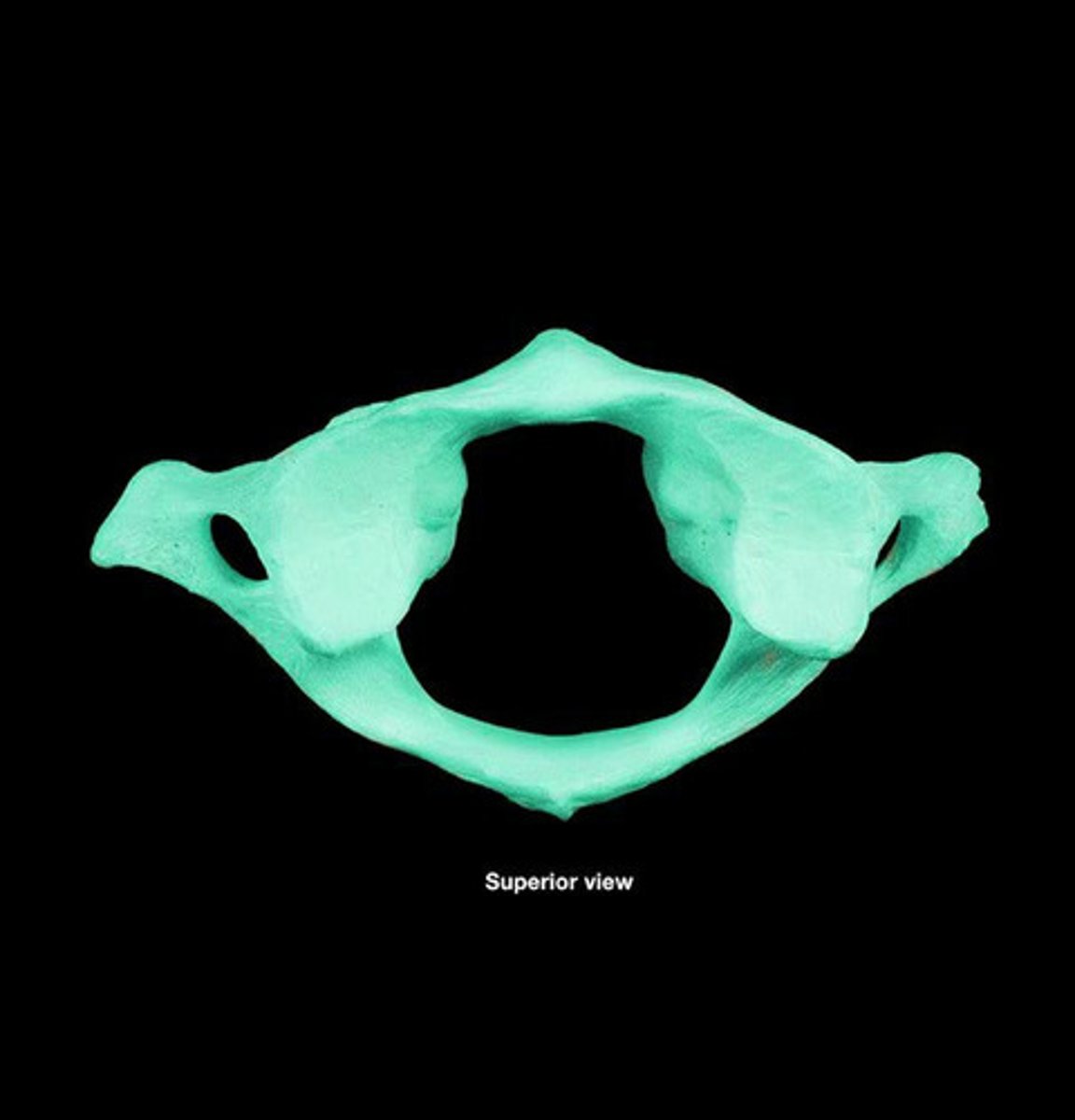
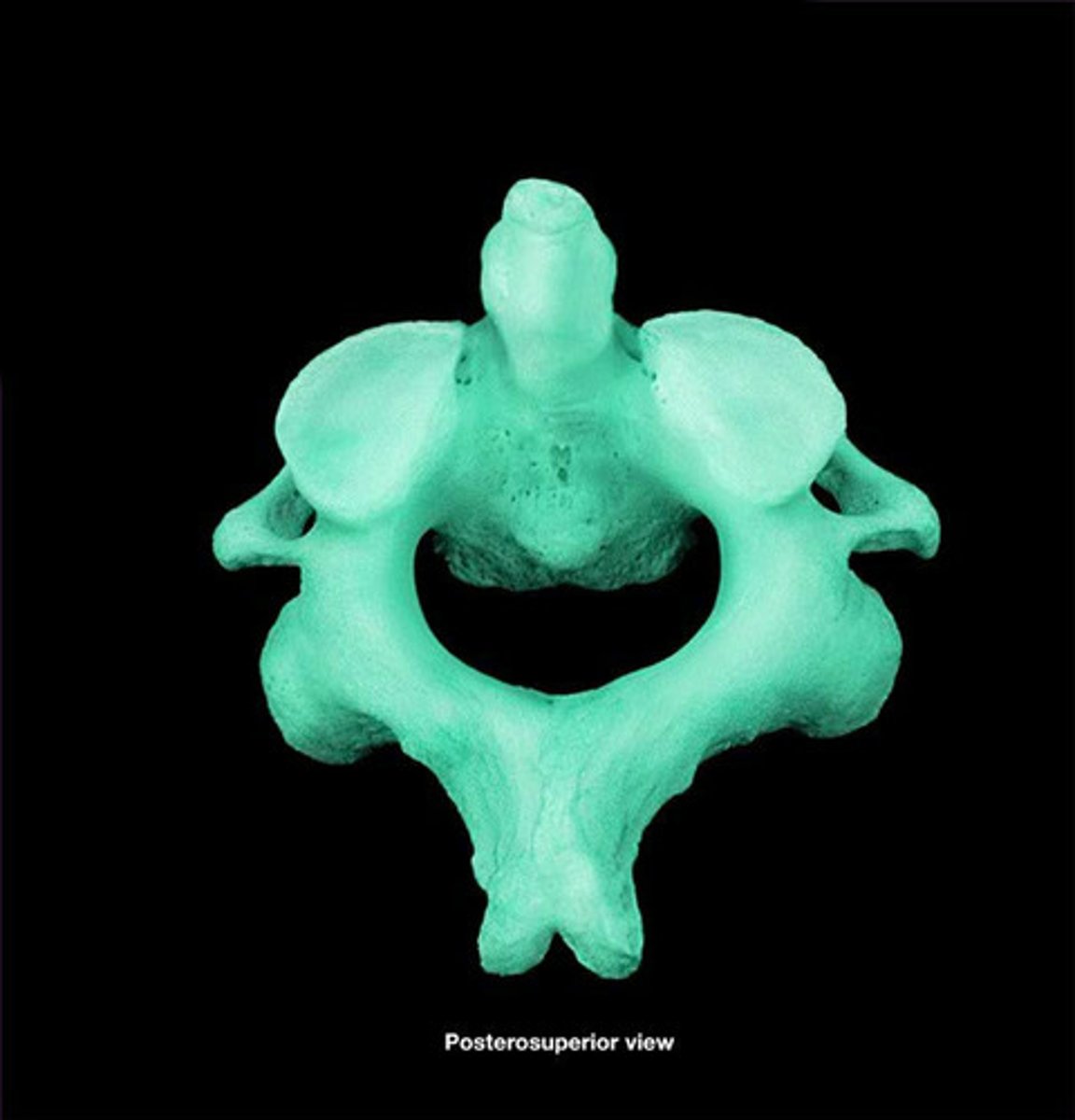
C1 (Atlas)
Lacks body and spinous process, allows head to nod.
C2 (Axis)
Has a dens/odontoid process projecting superiorly from the body, allows head to rotate.
C7 (vertebra prominens)
Prominent bump on back of neck, with a spinous process not bifid and especially long.
Thoracic vertebrae
Goes from T1-T12.
T1
Characterized by a heart-shaped body, a long spinous process that points downward, and a facet for the head of the first rib on its body.
Lumbar vertebrae
Goes from L1-L5.
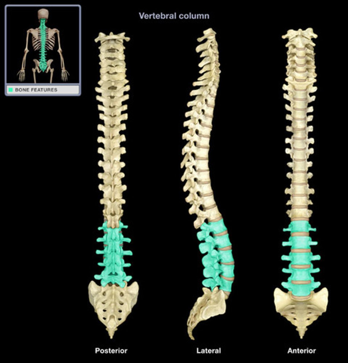
L5
Characterized by the largest body of all vertebrae, a thick spinous process, and large transverse processes to support the weight of the upper body.
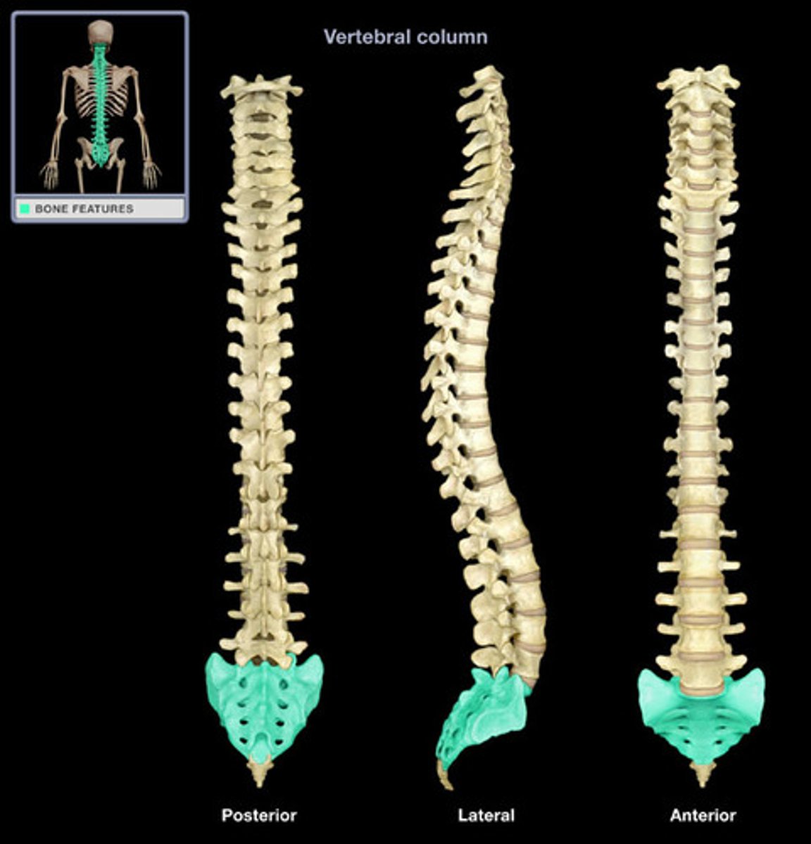
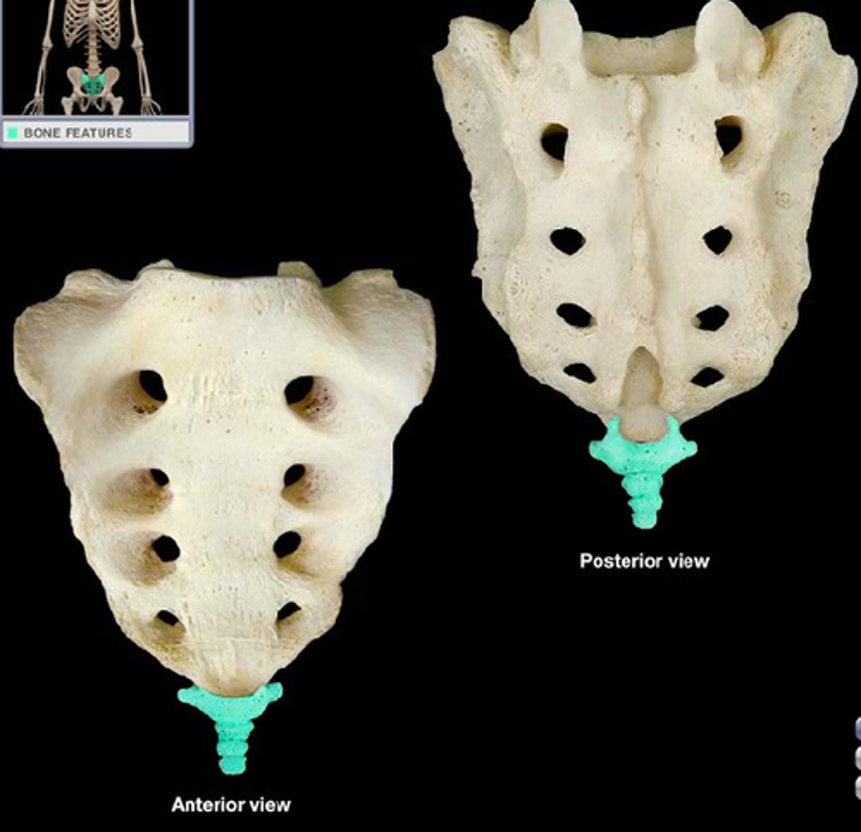
Sacral vertebrae
These are 5 vertebrae which are fused to form the sacrum in the pelvis.
Coccygeal vertebrae
They make up the tailbone.
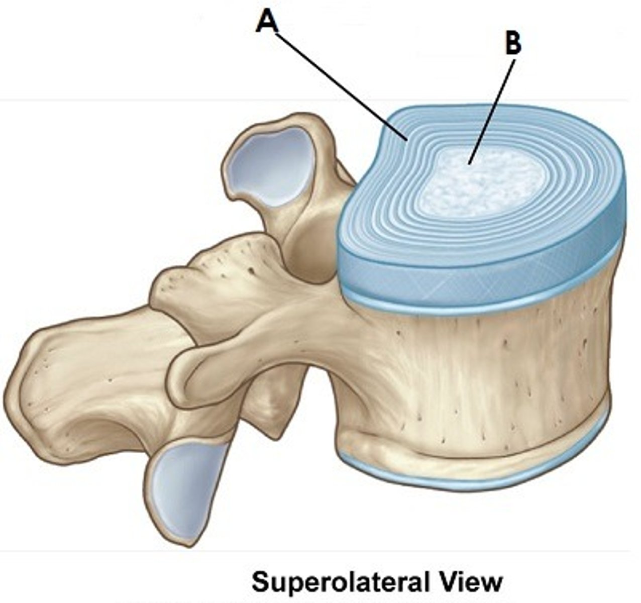
Intervertebral disc
Mass of fibrocartilage between adjacent vertebrae.
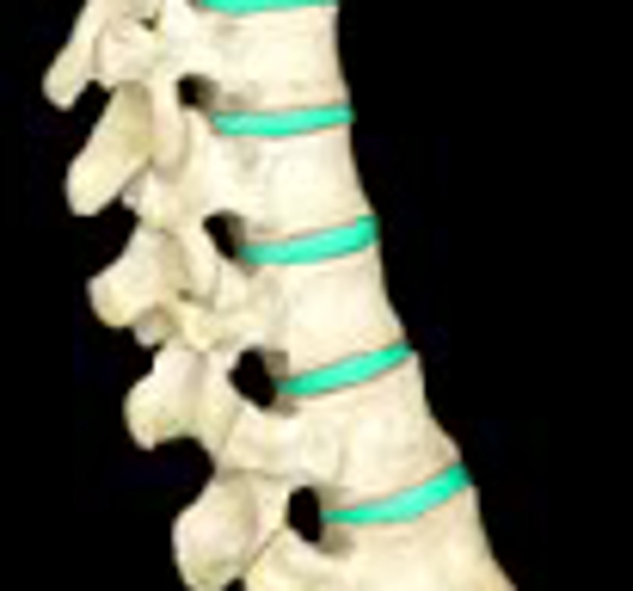
Anulus fibrosis
Outer collar composed of collagen and fibrocartilage. Letter A.

Nucleus pulposus
The soft, fibrocartilaginous, central portion of intervertebral disk. Letter B.