WK1: Cardiac Masses + Sources of Embolism & Systemic Diseases
1/69
There's no tags or description
Looks like no tags are added yet.
Name | Mastery | Learn | Test | Matching | Spaced |
|---|
No study sessions yet.
70 Terms
Define Cardiac Masses
Abnormal lesions/structure(s) w/in o around the heart.
Very rare.
Types of Cardiac Masses
tumors
Thrombus
Vegetations: endocarditis
Normal structures or artifacts mistaken for masses
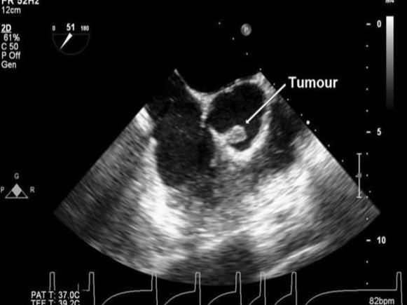
Which are more common, benign or malignant cardiac tumors?
Benign
Most common benign cardiac Tumor:
Myxoma
Most common malignant cardiac tumor:
Angiosarcoma

Traits of Papillary Fibroelastomas:
Benign cardiac tumor
common on aortic valve & papillary muscles
Usually on arterial side of AoV
Small, mobile, pedunculated
May appear like Lambl’s excrescences

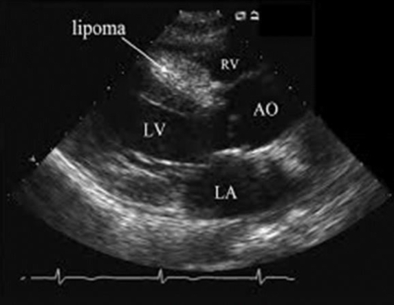
Traits of Lipomas
benign cardiac tumor
Well-encapsulated
Can occur in any heart layer
Mostly sessile (NOT pedunculated/attached by a stalk)

Traits of rhabdomyomas
benign cardiac tumor
Most common tumor in children
Yellowish-gray color
Invades ventricular myocardium
May cause arrhythmias
Associated w/ tuberous sclerosis
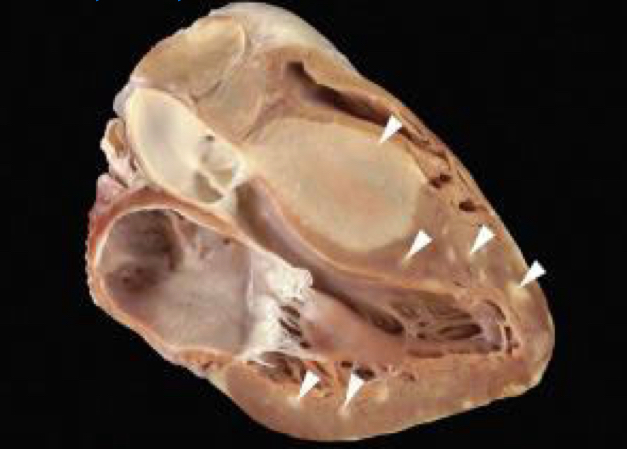

Traits of Fibromas
benign cardiac tumor
Bulky
Embedded in the myocardium of any chamber
Usually ventricular
Usually intramural
Age range: 2 - 57
Difficult to resect d/t size in symptomatic pts
Typically results in transplant
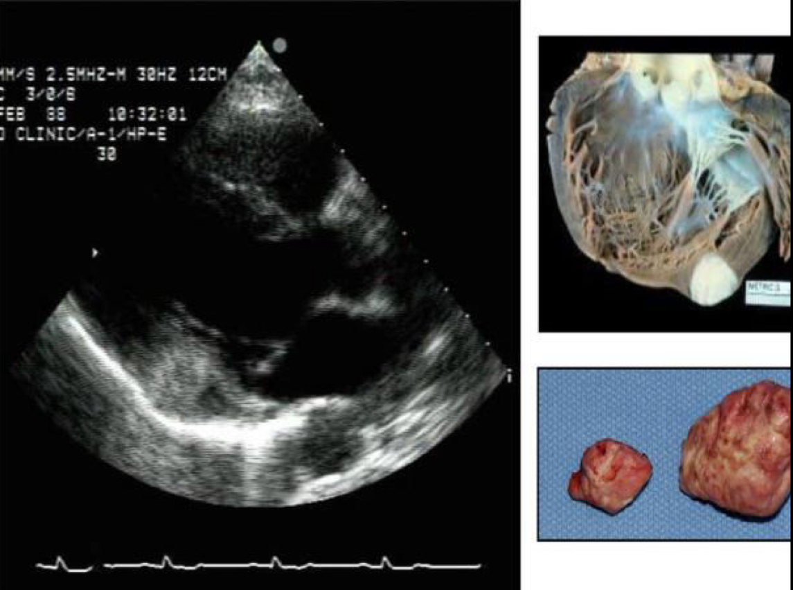
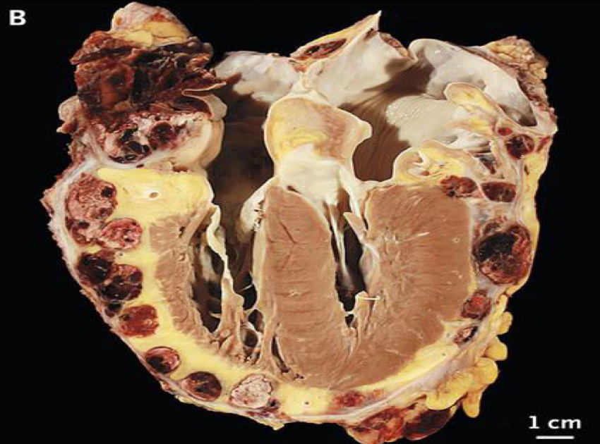
Traits of Angiosarcomas
malignant cardiac tumor
Most common primary malignant tumor
Mostly occur in the RA
Infiltrates into cardiac tissue
Large, mural mass
May extend into the pericardium

Traits of rhabdomyosarcomas
malignant cardiac tumor
2nd most common primary malignant tumor
Can occur in any layer of the heart
25% found in those <20 y/o
Primary benign cardiac tumors:
papillary fibroelastoma
Lipoma
Rhabdomyoma
Fibroma
Primary malignant cardiac tumors
angiosarcoma
Rhabdomyosarcoma
Other (rare) ones: fibrosarcoma, lymphoma, sarcoma, mesothelioma
Non-primary/secondary cardiac tumors are also called _________.
Metastatic
Which type of cardiac tumor is more common, primary or secondary?
Secondary are 20-40x more common than primary tumors
Where do secondary cardiac tumors most often metastasize to?
Which side of the heart do they occur most often (right or left)?
The pericardium (w/ associated effusion). May also have myocardial infusion & intracavitary extension
They occur most often in the Right heart.
Examples of secondary cardiac tumors:
renal cell carcinoma (enter the RA via the IVC)
Carcinoid: a deposition of serotonin on the endocardium, causing stiffness. Mostly on the right heart.
Ways that metastatic cancer can enter the heart:
lymphatic system
Pulmonary veins to the LA
IVC to the RA
Indications & Signs/Symptoms of cardiac tumors:
Family hx
Carney Syndrome (myxomas)
Dyspnea, syncope, palpitations, chest pain, fever, weight loss, TIA, dizziness
Malignant tumors:
Obstruction to blood flow
Interference of heart function
Local invasion: arrhythmias/effusions
Embolisms
Where is cardiac thrombus most often located?
LV
LAA
IVC/RA
What are some potential causes of cardiac thrombus?
reduced LV function
Afib
Hemodynamic stasis:
Dilated CMO
MI
Aneurysm
Foreign medical device
Pacer wires
PICC lines
Typical appearance of cardiac thrombus:
layered/laminar: distinct layers of different echogenicities
Single
Multilobulated
Pedunculated
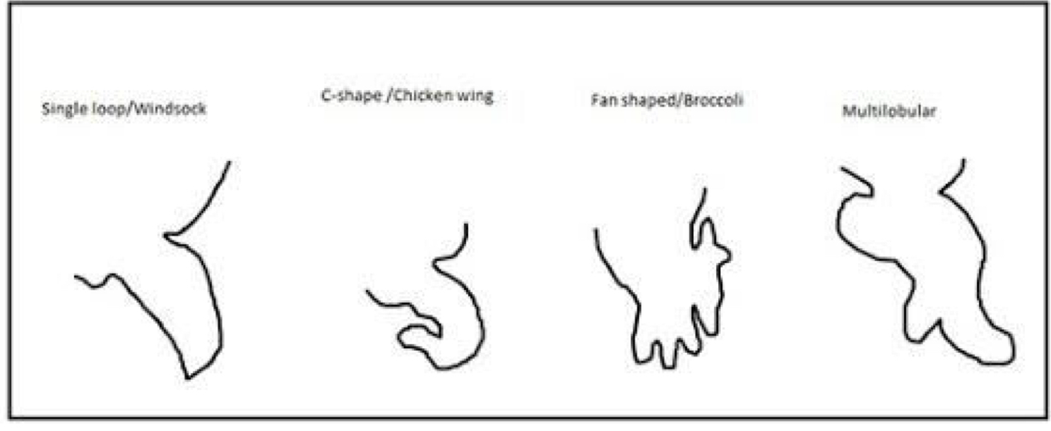
Different LAA shapes:
Treatments for cardiac thrombus:
anti-coagulation
NOAC/DOAC: apixiban
Heparin
Thrombolysis
EKOS: Ultrasound assisted catheter-delivered thrombolysis. Thrombus “opens up” d/t ultrasound and allows more medication into the thrombus
Oral
Thrombectomy
Angiovac
Surgical
What are some indications to use EKOS?
When should it be turned off/not used?
most commonly used for pulmonary embolisms
Can be used for critical DVTs
MUST be turned off during echocardiograms
Complications of cardiac masses:
risk stratification for surgeries/procedures
Embolism
Progression of malignant processes
Hemodynamic consequences
Predisposing conditions for a cardiac source of embolism:
PFO
IAS aneurysm
Apical aneurysm
Prosthetic valves
Aortic atheroma
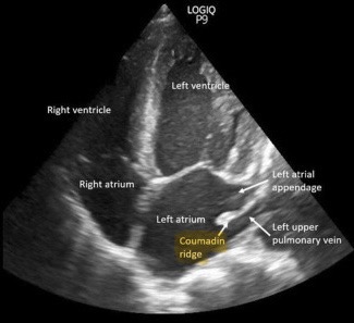
What is the Ridge of Coumadin?
a part of the left atrium that lies between the left atrial appendage
Amyloid is what type of particle?
Hormone
Protein
Amino Acid
Body Cell
Protein
What is amyloidosis?
A heterogenous disease (multiple etiologies)
The deposition of extracellular proteins in various organs, including the myocardium
Types of amyloidosis
AL (Primary): abnormal antibody production in bone marrow
AA 9secondary): triggered by an inflammatory response to disease
Hereditary (Familial): Inherited, most commonly from the liver making abnormal proteins
Senile: amyloid deposition d/t advanced age
Consequences of amyloidosis on the heart:
thickened myocardium
Diastolic dysfunction (early)
systolic dysfunction (late)
Conduction disorders
Embolic events
Signs & Symptoms of amyloidosis:
symptoms of congestive heart failure: SOB, edema
Peripheral neuropathy
Other cardiomyopathies
Arrhythmias
Hepatomegaly
Amyloidosis Treatments
Heart failure medications
Organ transplant
Chemotherapy
Anti-inflammatory meds
Stem cell transplant
Cardiac transplantation
What is the typical strain pattern for amyloidosis? Is it specific for this disease?
Apical Sparing: a preservation of contractility in the apical segments, with a decrease in the basal & mid segments
It is NOT specific for amyloidosis
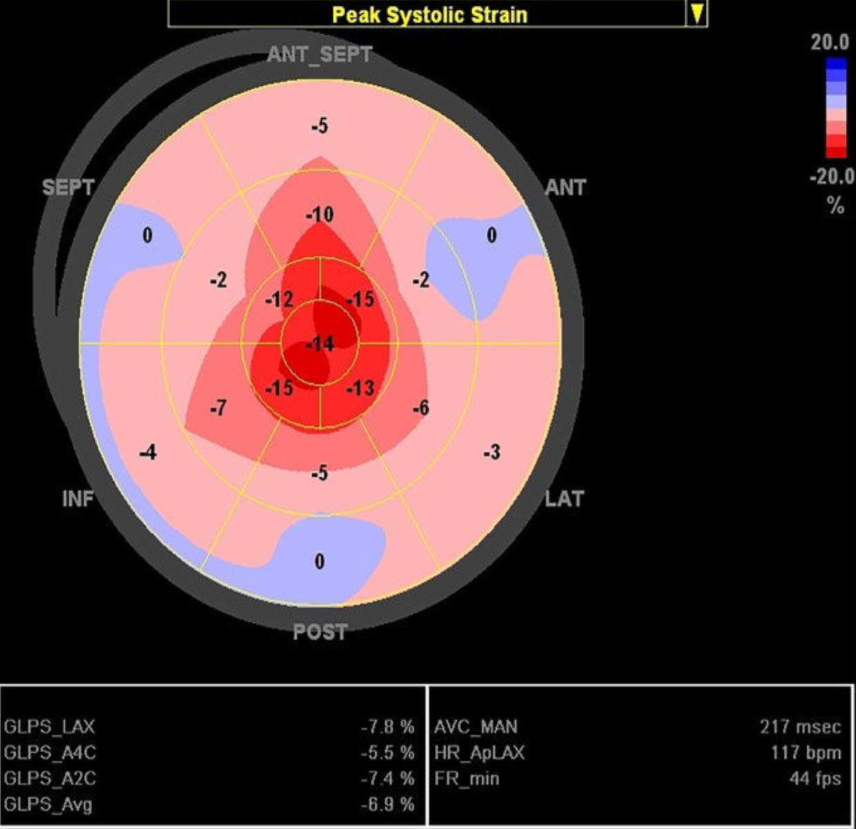
Echo features of amyloidosis
increased ventricle wall thickness
→ Ground glass or granular myocardium (sparkling appearance)
Atrial dilation (diastolic dysfunction)
Pericardial/pleural effusion
Significant diastolic dysfunction
→ Altered strain pattern (apical sparing)
Thickened valves w/ regurgitation (usually mild)
Thickened IAS
Thickened papillary muscles
What is carcinoid heart disease (CaHD)
generally malignant, metastatic endocrine tumor
Cardiac manifestations
Hepatic metastases release tumor substances to the right heart
Fibrous white place deposits lead to fibrosis & valve dysfunction
Predominance on the right heart (TV, PV)
Which valve is most commonly involved in CaHD?
the TV
Etiology of CaHD
when neuroendocrine tumors metastasize to the liver & begin secreting serotonin
Usually originate in the small intestine & appendix
Some originate in the lungs
50% of carcinoid tumors have cardiac involvement
Worsened prognosis
Echo features of CaHD
TV involvement: severe TR w/ minimal TS
Restricted TV movement
Varying PI & PS
Usually PS
R Heart Failure (volume overload)
Potential MV & AoV Involvement
Myocardial Metastases
Pericardial effusion
Clinical Presentation of CaHD:
Carcinoid Syndrome:
Face flushing
Hypotension
Diarrhea
Bronchospasm
Wheezing
Heart failure (late)
Treatments of CaHD
chemotherapy
Surgical removal of tumor
Valve replacement
Medications to decrease serotonin levels
Heart failure meds
Which organ system is primarily affected in sarcoidosis?
Lungs (pulmonary)
What is sarcoidosis?
Inflammatory disease; abnormal collections of inflammatory cells, congregate into masses (granulomas)
Invade organ tissue, can cause dysfunction/failure
Most commonly affects the lungs
May impact the heart & skin
Unknown etiology (idiopathic)
Clinical presentation of cardiac manifestations of sarcoidosis:
asymptomatic
Depends on size & quantity of granulomas
Sudden death
Arrhythmias & conduction abnormalities
LV dysfunction & CHF
Typically presents before age 40
Echo features of sarcoidosis:
enlarged LV
Decreased systolic function
RWMA
RV & RA enlargement/hypertrophy
PHTN from lung sarcoidosis
Septal wall thinning
W/ or w/out aneurysm
Diastolic dysfunction
Pericardial effusion
Treatments for sarcoidosis:
None, if asymptomatic
Anti-inflammatory meds (corticosteroids)
Immune suppression (methotrexate)
Pacemaker/ICD implantation for arrhythmias
Transplant
What is hyperesinophilic syndrome?
abnormal amount of eosinophils in the blood stream
Infiltrative disease, may impact multiple organs
Cardiac, skin, renal
Confirmed by blood testing (>1500 eosinophils/mm³ for >6mo)
What are the stages of hypereosinophilic syndrome?
3 stages
Necrotic Stage: eosinophils damage the endocardium
Thrombotic Stage: thrombus forms on the damaged endocardium
Concern w/systemic embolization.
Fibrotic Stage: the endocardium becomes fibrotic & scarred.
LV filling becomes restrictive
What is Loeffler’s Cardiomyopathy?
A form of restrictive CMO
Caused by significant endocardial damage from hypereosinophilic syndrome
Echo features of hypereosinophilic syndrome
normal LV/RV size & function
Atrial enlargement
Restrictive physiology (decreased LV compliance)
Treatment of hypereosinophilic syndrome
treat CHF
Anticoagulants
Treat the hypereosinophilic (steroids)
Endocardial stripping for fibrosed tissue
What is hemochromatosis?
Excess deposition of iron in the blood, affection organ/tissue function
Rare before age 20
Peaks in the 5th decade
Types of Hemochromatosis:
Primary (hereditary): excess absorption of dietary iron
Secondary (acquired): transfusion, iron therapy
Clinical Presentation of hemochromatosis
males >40 y/o
Liver & heart failure
Hyperpigmentation of the skin
Echo appearance of hemochromatosis
dilated cardiac chambers
Normal wall thickness
Global systolic dysfunction
Progressive diastolic dysfunction
What is HIV/AIDS? What is its involvement in the cardiovascular system?
A virus that attacks the immune system that reduces its effectiveness & leads to susceptibility to disease and infections
Develop intramuscular plaque at a greater rate
2x risk of cardiovascular events
Although rare, with the advent of antiretrovirals - this is a common finding with late stage AIDS:
Kaposi sarcoma
Skin rush
Systemic HTN
Thrush
Kaposi sarcoma: a type of cancer that forms in the lining of blood vessels and lymph vessels
Cardiac concerns in patients w/ HIV/AIDS:
DCM
Pericarditis
RV dysfunction
Endocarditis
CAD
Cardiac tumors (Kaposi’s sarcoma)
Echo features in pts w/ HIV/AIDS:
dilated LV and/or RV
Systolic dysfunction
Varying diastolic dysfunction
MR
Endocarditis (bacterial & NBTE)
Pericardial effusion
Pleural effusion
Constrictive pericarditis
Cardiac neoplasm
PHTN
Treatments for HIV/AIDS
Antiretroviral therapy
Cause lower risks of complications
CHF medications
Antibiotics (endocarditis)
What is Rheumatoid Arthritis (RA)?
What is the clinical presentation?
What is its cardiac involvement?
Chronic inflammatory disease primary impacting the joints
Clinical Presentation:
Common in women
Fatigue, myalgias, fever, limited joint motion
Effusion, MI, valvular disease
What is antiphospholipid syndrome?
What is its clinical presentation?
What is its cardiac involvement?
A hypercoagulability syndrome.
Can cause PHTN from PE
Causes cardiac dysfunction w/ potential thrombosis of coronary arteries
What is scleroderma?
What is its clinical presentation?
What is its cardiac involvement?
Excessive connective tissue that accumulates in various body tissues.
Can cause PHTN, systemic HTN
Presents w/ conduction abnormalities
What is systemic lupus erythematosus (SLR)?
What is its clinical presentation?
What is its cardiac involvement?
an autoimmune disease
Prevalent in women
Cardiac manifestations:
Valvular: Libyan-sacks endocarditis (w/ regurgitation)
Pericardial effusion
Global LV dysfunction
PHTN w/ pulmonary involvement
Diffuse leaflet thickening
What is Marfan’s Syndrome?
What are common cardiac manifestations?
What are common echo features?
Hereditary (autosomal dominant) connective tissue disorder
Most common cause of death is Ao dissection
MV disease
Dilated Asc Ao
AI
Ao Dissection
Echo features:
Ao root dilatation
Dilated asc Ao
Annuloaortic ectasia
Ao dissection
Myxomatous MV w/ prolapse
Types of vasculitis that may have cardiac involvement:
Giant cell Arteritis: Ao aneurysms & dissections
Takayasu Arteritis: granulamatous pan arteritis of large vessels (AR, Ao Dilatation)
Kawasaki Disease: acute systemic vasculitis of unknown origin
Churg-Strauss Syndrome: Pericardial effusion, valve regurgitation
Wegner’s Granulomatosis: pericarditis, mass lesions
Kawasaki Disease is also called….
Mucocutaneous Lymph Node Syndrome
Causes swelling of mucous membranes & lymph node glands
What is Kawasaki’s Disease? What are common cardiac findings?
acute systemic vasculitis of unknown origin
Seen <5 y/o
Cardiac involvement:
Coronary artery dilation/aneurysm
LV dysfunction
MR
Pericardial effusion
Endocrine diseases with potential cardiac involvement:
hyperthyroidism
Hypothyroidism
Pheochromocytoma: excessive catecholamines
Acromegaly: secretion of excessive growth hormone (usually pituitary adenoma)