L10 Action potentials
1/18
There's no tags or description
Looks like no tags are added yet.
Name | Mastery | Learn | Test | Matching | Spaced | Call with Kai |
|---|
No analytics yet
Send a link to your students to track their progress
19 Terms
What generates the resting membrane potential?
What causes the inside of the cell to be negative?
Efflux of K⁺ down its concentration gradient through open K⁺ channels.
K+ out now so inside negative
What role does permeability play in resting potential?
What happens if the membrane becomes more permeable to Na⁺?
Resting potential mainly depends on K⁺ permeability, as PK⁺ ≈ 20× PNa⁺.
The membrane potential becomes less negative (depolarizes).
What is an action potential?
What is the time course of an AP?
A rapid, transient reversal of the membrane potential once a critical depolarization (threshold) is reached.
<5 ms in neurons, ~10 ms in skeletal muscle, ~250 ms in cardiac muscle.
What are the 7 steps of the AP (7) RDRPRHR
1. Resting: Leak potassium channels open, voltage gated sodium and potassium channels closed
2. Depolarization to threshold: initial depol open some sodium channels -> sodium influx -> positive feedback -> all sodium channels open
3. Rising phase: rapid sodium influx drives Vm towards sodium equilibrium around +40mV
4. Peak: sodium channels inactive via inactivation gate
5. Falling phase (repolarization): Deplayed opening of voltage gated potassium channels -> potassium efflux
6. After hyperpolarization: Both potassium leak and voltage gated sodium channels active resulting in Vm dipping below rest toward -90mV (potassium equilibriu,)
7. Return to rest: Potassium channels close and sodium channels back to rest
What are the two states of voltage-gated K⁺ channels?
Resting and activated.
What determines the shape of the AP?
Changes in Na⁺ and K⁺ conductance (gNa and gK).
Why is there an after-hyperpolarization?
K⁺ channels open more slowly and stay open longer than Na⁺ channels.
How does stimulus intensity affect APs?
Stronger stimuli increase firing frequency, not AP size. More Aps means more neurotransmitter release and higher concentration of neurtransmitters
What is the "all-or-none" principle?
Once threshold is reached, an AP always occurs with the same amplitude.
Explain propagation of APs step by step
1. AP generated at a spike generating zone (axon hillock), sodium enter cell through voltage gated sodium channels making inside positive
2. Adjacent region to the AP is still at resting potential. Voltage difference cause current to flow: potassium inside the axon moves toward the depolarized region, sodium outside the axon moves toward the depolarized region, chloride/another anions may move in diff directions producing equivalent positive current flow
3. Local depol spreads passively and quickly along the axon and if membrane potential in the next segment reaches threshold VGSC open
4. VGSC opening move sodium in creating new local AP, inside positive which continues the process along the axon
What prevents backward propagation of the AP?
The refractory period caused by Na⁺ channel inactivation. The RF period is a time during which another AP cannot be initiated
How does myelination improve AP conduction?
- Prevents potassium and sodium flowing out and increases conduction speed by insulating axons, reducing current loss and allowing APs to jump between nodes of Ranvier which has a high density of sodium channels
What is saltatory conduction?
The "jumping" of APs from node to node along a myelinated axon.
How does axon diameter affect conduction speed?
Larger diameter → lower internal resistance → faster conduction.
What are the 4 nerve fibre types, size, myelinated or unmyelinated?, speed and what do they detect (add diagram)
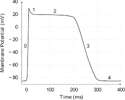
What are the steps of ventricular cardiac myocyte AP (6)
1. Resting potential: -90mV close to potassium equilibrium
2. Rapid depol: VGSC open, sodium in, inside positive
3. Initial repol: Sodium channels inactive, transient potassium channels open resulting in brief potassium efflux
4. Plateau: L Type calcium channels open-> sodium in balanced potassium out-> prolonged depol maintaining contraction
5. Repolarization: Calcium channels close, delayed rectifier potassium channels open, membrane back to -90mV
6. Back to resting
How do cardiac APs differ from neuronal APs?
They last longer and involve Ca²⁺ entry that sustains depolarization.
What are autorhythmic cells?
Cells that spontaneously generate APs (e.g. SA node).
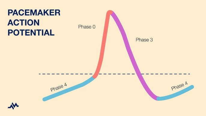
Step by step of pacemaker (SA) node AP (3)
Phase 4: I_f (HCN) Na⁺ in, + I_Ca,T T type calcium channels→ to threshold.
Phase 0: I_Ca,L Ca²⁺ in → upstroke.
Phase 3: I_K delayed rectified moves K⁺ out → repolarize → I_f reactivates.