immunologic laboratory tests
1/8
Earn XP
Description and Tags
Name | Mastery | Learn | Test | Matching | Spaced |
|---|
No study sessions yet.
9 Terms
SAMPLE COLLECTION AND HANDLING
Nearly all serologic tests require serum or plasma as the sample. Whole blood should not be sent to the diagnostic laboratory when serum or plasma is specified. The most practical method of collection is the Vacutainer System (Becton Dickinson, Franklin Lakes, NJ), which is commonly available from many veterinary and medical supply companies. A red-topped vacuum tube is used when serum is required, and a lavender-topped tube is used to collect plasma; if heparinized plasma is specifically requested, a green-topped tube is used. If a blood sample is to be collected in a syringe, then a 5 ml syringe and 20 gauge needle combination should be used, because it causes the least hemolysis
HANDLING SEROLOGIC SAMPLES
When serum is to be submitted, the blood sample is allowed to dot for 20 to 30 minutes at room temperature, and it is then centrifuged for 10 minutes at a speed of no faster than 1500 rpm. If little serum has separated after centrifuging, "rimming" the tube with a wooden applicator stick to loosen the clot may help; however, this also may cause hemolysis. If plasma is desired, the sample may be centrifuged immediately After centrifugation, a small pipette is used to aspirate the serum or plasma (i.e., the upper layer) off of the packed erythrocytes. The aspirate is placed into a transfer tube or another sealable test tube and clearly labeled. The serum or plasma may be tested immediately or frozen or refrigerated for later use. Once thawed, samples cannot be re-frozen for later testing without compromising the sample. Samples for most serologic tests do not need to be frozen, but they should be shipped cold, especially during hot weather. The major problem with shipping tubes is breakage. The tubes must be packed firmly in place with packing material so that they do not move around when the package is jarred. Each sample must be clearly and correctly labeled and the pertinent paperwork enclosed to facilitate proper reporting of the results from the laboratory.
TESTS OF HUMORAL IMMUNITY (antibodies)
Enzyme immunoassay, latex agglutination, immunodiffusion, and rapid immunomigration are methods available for use in veterinary practice laboratories. These methods have been validated for testing to identify a large number of specific antigens. Additional immunoassays that incorporate these methods can detect certain blood components, such as coagulation factors,
Enzyme-Linked Immunosorbent Assay (ELISA)
has been adapted to many agents commonly tested for in the veterinary laboratory. With monoclonal antibodies, the specificity of ELISA is high, meaning little cross-reactivity occurs with other agents. This phenomenon makes ELISA an accurate way to detect specific antigens (e.g., viruses, bacteria, parasites, hormones) in serum. ELISA may also be used to test for an antibody in the serum, in which case the test kit contains the specific antigen. Some of the available ELISA kits detect heartworms, feline leukemia virus, feline immunodeficiency virus, canine parvovirus, and progesterone. For the ELISA antigen- detection system, monoclonal antibody is bound to the walls of the wells in a test tray, to a membrane, or to a plastic wand. Antigen, if present in the sample, binds to this antibody and to a second enzyme-labeled antibody that is added to help with the detection of the antigen. This is followed by rinsing. This is a critical step in the process. If the wells are not thoroughly washed, false-positive results can occur. When a chromogenic (color-producing) substrate is added to the mixture, it reacts with the enzyme to develop a specific color, thereby indicating the presence of antigen in the sample. If the sample contained no antigen, the entire enzyme-labeled antibody would be washed away during the rinsing process, and no color reaction would develop.
Competitive Enzyme-Linked Immunosorbent Assay (CELISA)
when used to test for patient antigen, involves the use of an enzyme-labeled antigen as well as monoclonal antibodies. Patient antigen, if present, competes with enzyme-labeled antigens for the antibodies that coat the test wells. Color developer reacts with the enzyme to produce a color. The intensity of the color produced varies with the concentration of the patient antigen. Equine infectious anemia antibodies may be detected in horse serum with a CELISA test.
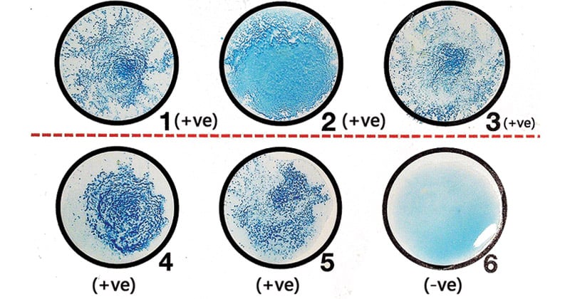
Latex Agglutination
makes use of small, spherical, latex particles that are coated with antigen and suspended in water. If serum that contains the corresponding antibody is added to the mixture, the formation of antibody-antigen complexes causes agglutination (clumping). Agglutination changes the appearance of the latex suspension from smooth and milky to clumpy because the latex particles have clustered and cross-linked together. If no antibody is present in the sample, the mixture of latex and serum remains evenly dispersed. The strength of reaction can be graded (i.e., +1, +2, +3, or +4) to provide an indication of the amount of antigen present. One common test that uses this method is the test for bovine brucellosis antibodies. It is important to note that false negatives can occur when excessive amounts of antigen or antibody are present in the sample. In the presence of excess antibody, it is possible that each antibody molecule binds with only one or two antibodies and does not cross-link so that agglutination does not occur. This is known as the prozone phenomenon. Excess antigen can result in lack of cross-linking when the excess antigen surrounds any small clumps that may form. This is known as the postzone phenomenon. It is important to note that prozone and postzone phenomena can occur due to the characteristics of the patient sample or can occur due to errors in test performance such as improper pipetting techniques that result in inappropriate quantities of reagent added to the test system.
Lateral Flow Immunoassay Rapid Immunomigration and Immunochromatography
has also been described as rapid immunomigration (RIM) or immunochromatography. The signal-generating components of these tests are colloidal gold, enzymes, and color reagent or agglutinated latex particles. All three types of components create a positive result. In this type of procedure, the signal-generating componenti5 conjugated to antibodies specific for the antigen being tested. These conjugated antibodies are present in the membrane of the test cassette where the patient sample is applied. If antigens are present in the patient sample, they bind to the conjugated antibodies, and the antibody-antigen complexes migrate along the membrane to another area of the cassette, where the results are read. Buffer may be added to help with the migrational flow of the antibody-antigen complexes. In the reading area, a second antibody is present in the membrane. If antigen is in the sample, it is captured-along with the first antibody and the conjugate-by the second antibody. The accumulation of conjugate in that area causes a color change. To ensure quality results, control antigen is present in another area of the membrane strip. The conjugated first antibody binds to the antigen in the control area. Its accumulation also causes a color change and occurs whether or not antigen is present in the patient sample. A positive patient result shows two areas of color change: one for the patient and one for the quality control antigen. If the control antigen area does not change color, then the test is considered invalid, regardless of any color change in the patient area.
IMMUNOLOGY ANALYZERS
Automated analyzers are available for use in the veterinary practice laboratory. In some cases, the analyzer is required to run certain tests. Large reference laboratories often have automated analyzers for the performance of immunoassays. These analyzers are capable of performing tests on numerous patients simultaneously. Most are completely automated and require only the patient information entered in the analyzer's computer and the patient sample loaded. Small in-house immunology analyzers are generally of the type that only read test results. The technician prepares the sample and the test device and places the device on the analyzer. The results are read by the analyzer at the appropriate time.
Chemiluminescence
Many automated analyzers utilize the principles of chemiluminescence to detect and quantify specific antigens. The principle is similar to the ELISA method except that the test uses a substrate that reacts to produce light rather than an enzyme that reacts to produce color. The light produced can be detected by a photomultiplier, and the amount of light produced can be quantified. The chemiluminescent immunoassay (ChLIA) has many applications both in medicine and in other industries. In addition to immunology testing, ChLIA has been used for detection and quantification of other substances, including thyroid hormones, cortisol, pancreatic lipase, progesterone, and testosterone.