1/66
Looks like no tags are added yet.
Name | Mastery | Learn | Test | Matching | Spaced |
|---|
No study sessions yet.
Learning Objectives
1.3 Body Fluid Compartments
-discuss the distribution of total body water into intracellular fluid (ICF) and extracellular fluid (ECF), 2 components of the ECF, and barriers for fluid movement
14.1 Renal Functions
-state the 6 general functions of the kidneys
14.2 Structure of the Kidneys and Urinary System
-describe the tubular and vascular anatomy of the kidneys and the urinary system
-list the 3 layers that make up the glomerular filtration barrier
-identify the structures that form the juxtaglomerular apparatus
14.3 Basic Renal Processes
-understand that filtration, reabsorption, and secretion are the 3 basic processes that lead to the formation of urine
-describe the 4 forces that determine net glomerular filtration pressure and state if they favor or oppose filtration
-describe how the regulation of the number of membrane channels and transporters can alter the ability to reabsorb and secrete substances
What is the most abundant substance in the body?
water
What function does water perform in the body?
solvent for all dissolved constituents in the body
What is total body water (TBW) comprised of?
-1/3 Extracellular fluid (ECF)
-2/3 Intracellular fluid (ICF)
What is ECF made up of?
-3/4 interstitial fluid
-1/4 plasma
What is the vital role of kidneys?
maintain the volume and composition of the body fluids constant despite wide variations in daily intake of water and solute
-plasma purification (ensure ECF is pure)
What are the functions of the kidney?
-regulate water and inorganic-ion balance = osmolarity;
ECFV = BP
-acid-base balance
-eliminate metabolic waste products (urea, uric acid, creatine)
-eliminate foreign compounds (drugs, toxins, pesticides)
-gluconeogensis
-secrete hormones
1.Erythropoietin = Hematocrit
2.Renin = RAAS
3.1-a hydroxylase = 1, 25-dihydroxy Vitamin D3
Can normal renal function be maintained when 1 kidney is removed?
Yes, because of …
-enhanced growth factor
-hypertrophy
-hyperplasia
-vasodilation
-hyperfiltration
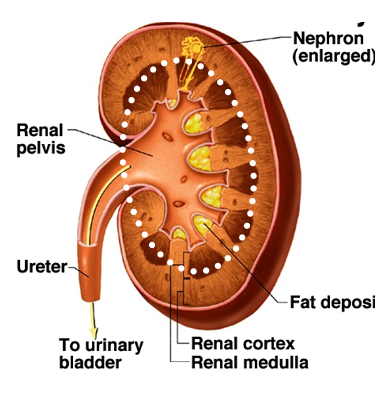
What is the outer layer (cortex) of the kidney responsible for?
-site of glomerular filtration and convoluted tubules

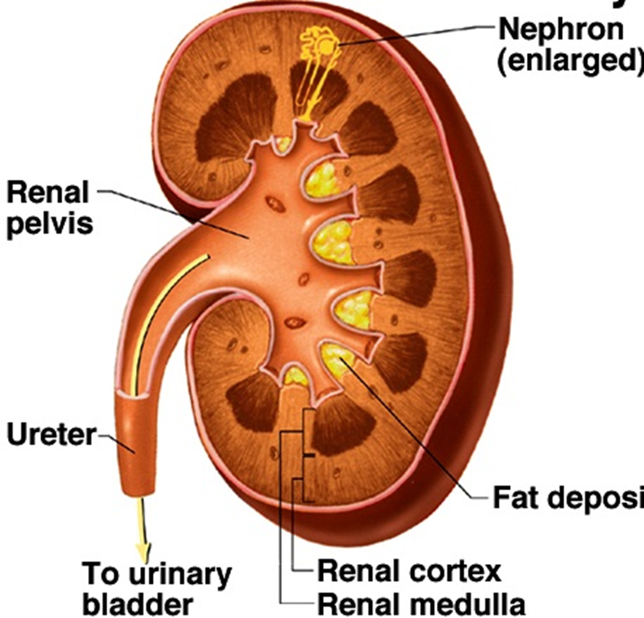
What is the inner part (medulla) of the kidney responsible for?
-location of longer loops of Henle
-drainage of collecting ducts into renal pelvis and ureter

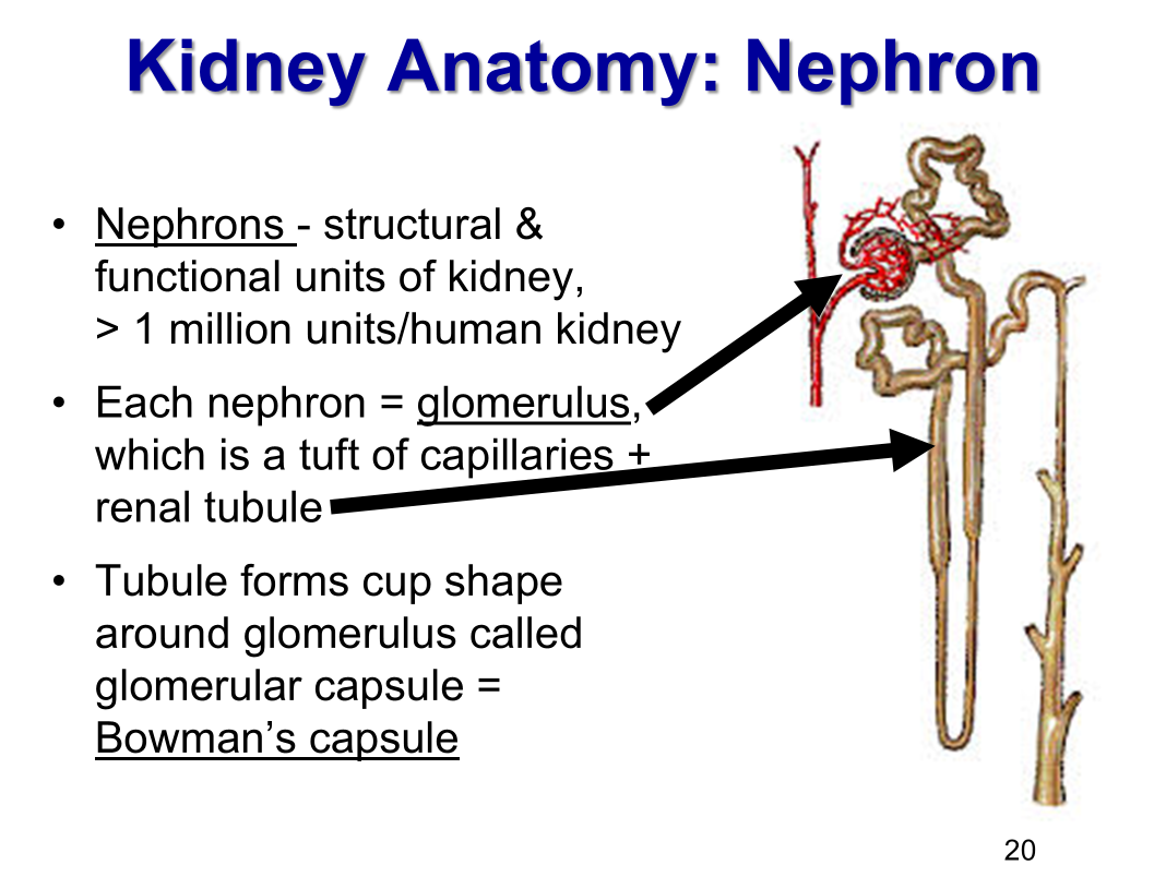
How many nephrons in a kidney?
1 million (number assigned at birth)

Basic structure of a nephron
1.Renal corpuscle (reference point of all tubules)
2.Proximal (near) tubule (hollow tube)
3.Loop of Henle: ascending limb, descending limb
4.Distal (distant) tubule
5.Collecting duct
AA-Carries blood into the glomerulus
EA-carries blood away from the glomerulus
What is a nephron?
the structural and function unit of a kidney
What is each nephron made of?
-glomerulus (a tuft of capillaries)
-renal tubule

What is the cup shape around the glomerulus formed by the tubule called?
Bowman’s capsule
What are the two type of nephrons?
-Cortical nephrons
most nephrons are in this category
in short loops of Henle
-Juxtamedullary
in long loops of
involves in concentration of urine

What 2 fluid filled spaces make up the renal corpuscle?
-vascular space
glomerular capillaries
contains plasma, RBCs, WBCs, proteins, electrolytes
-urinary space
Bowman’s space
contain ultrafiltrate of plasma
filtrate passes from vascular into tubule system
1st step in urine formation
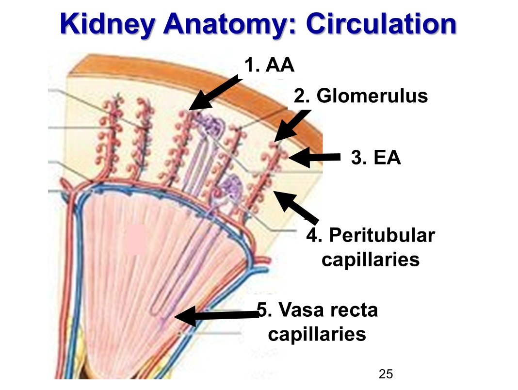
What is the order of circulation for the kidney?
Afferent arteriole (smooth muscle; can contract and relax)
Glomerulus → capillaries
Efferent arteriole (smooth muscle; can contract and relax)
Peritubular capillaries
Vasa recta capillaries

What are the functions of each capillary bed in renal circulation?
-glomerular capillaries → filtration
-peritubular capillaries → reabsorption, secretion
-vasa recta capillaries → concentrating
What is the result of the glomerular capillary being fed and drained by arterioles?
Allows glomerular capillary hydrostatic pressure to be veyr high, which forces fluid and solute out of blood into Bowman’s capsule by bulk flow
What is the significance of the peritubular and vasa recta capillaries?
most of filtrate is reabsirbed by renale tuble cells and returned to blood through peritubular and vasa recta capillaries
What is the order of renal plasma flow?
100% of plasma volume enters via renal artery
20% of plasma flitered from glomerular capillaries into Bowman’s space
80% of pslama travels in efferent arterioles
solute and water reabsorbed in peritubular and vasa recta capillaries
99.9% of plasma volume exits via renal vein

What are the basic renal processes?
-filtration (CV)
Where: Occurs in the renal corpuscle (glomerulus + Bowman’s capsule)
What: Blood flows into the glomerulus; Filtration occurs when water and small solutes (like ions, glucose, amino acids, urea) pass through the filtration membrane into Bowman’s capsule, forming filtrate.
Purpose: Start urine formation by filtering plasma.
-reabsorption (GI)
Where: Along the renal tubule ( mainly proximal convoluted tubule)
What: The body reclaims useful substances (water, glucose, amino acids, Na+, Cl-, HCO₃⁻) from the filtrate and returns them to the blood in the peritubular capillaries.
Purpose: Prevent loss of essential substances; maintain fluid and electrolyte balance.
-secretion (GI)
Where: Mainly in the distal convoluted tubule and collecting duct.
What: The nephron actively adds substances (H+, K+, NH₄+, creatinine, drugs) from the blood into the tubular fluid.
Purpose: Eliminate waste not filtered at the glomerulus and regulate acid-base and electrolyte balance.

What is the flow of the basic renal processes?
-Filtration
Glomerular capillary lumen → Bowman’s spcae (bulk flow)
-Tubular reabsorption
tubular lumen → peritubular capillary plasma
-Tubular secretion
peritubular plasma (capillary lumen) → interstitial space → tubular cell → tubular lumen
How do you calculate amount excreted in urine?
Amount filtered + Amount Secreted - Amount Reabsorbed
(F+S-R = E)
What are the layers of the glomerular filtration barrier?
glomerular capilalry endothelium (inner layer) (allows plasma through, but blocks blood cells) - vascular side
basement membrane (middle layer) (charge and size barrier) - shared
podocytes of Bowman’s capsule (outer layer) (filtration slits) - tubular side

What does the Glomerular filtration barrier exclude?
-proteins
-blood cells
-large molecules
Which structure in the glomerular filtration barrier is primarily responsible for allowing large volumes of solute-rich fluid to pass while preventing cells from leaving the bloodstream?
Fenestrated capillaries in the glomerular capillary endothelium (allowing plasma to pass but blocking blood cells)
What is the composition and function of the basement membrane in the glomerular filtration barrier?
-Produced by endothelial and podocyte cells (shared)
-repels large, negatively charged proteins
Blood enters the glomerulus through the ______ and exits through the ______.
Afferent arteriole; efferent arteriole
The filtrate formed after passing through the glomerular capillary wall is typically: ____________
Solute-rich but protein-poor
-b/c Filtrate is solute-rich (contains ions, glucose, etc.) but low in protein since large molecules cannot cross the capillary wall.
Damage to the basement membrane that removes its negative charge would most likely result in:
Proteinuria (protein in urine)
-b/c the loss of charge selectivity allows negatively charged plasma proteins (like albumin) to pass into the filtrate.
The foot processes of podocytes primarily function to:
Surround the basement membrane and form filtration slits
What are filtration slits?
Gaps (clefts) between podocyte foot processes where filtrate enters Bowman’s capsule.
What would most likely occur if podocyte foot processes were damaged or fused together?
Protein leakage into the urine (proteinuria)
-b/c damage to the podocytes disrupts the slit diaphragms, allowing large proteins to pass into the filtrate.
Which substances can pass through the filtration slits into Bowman’s space?
Water, glucose, amino acids, urea
-b/c the filtration barrier allows only small molecules
How would you describe podocytes in the kidney?
They are specialized epithelial cells of Bowman’s capsule that wrap around glomerular capillaries.
The foot processes (podocyte termination [ends]) of podocytes primarily function to:
Surround the basement membrane and form filtration slits.
Descrive filtration slits
Gaps between podocyte foot processes where filtrate enters Bowman’s capsule
What would most likely occur if podocyte foot processes were damaged or fused together?
Protein leakage into the urine (proteinuria)
-b/c damage to the podocytes disrupts the slit diaphragms, allowing large proteins to pass into the filtrate.
The capsular space of Bowman’s capsule is:
The region where filtrate collects after passing through podocyte filtration slits
-b/c filtrate moves through the three filtration layers and enters the capsular space, where it is collected before passing into the renal tubule.
What is the role of podocytes?
They form the final barrier that selectively allows passage of small solutes into the filtrate.
What determines the net filtration pressure (NFP) across the glomerular capillary wall?
Hydrostatic pressure (P) and Oncotic pressure (π) opposing one another
What is oncotic pressure?
-(π; mmHg)
-pressure generated by large molecules trapped (especially proteins) in solutions → “pulling pressure”
-Osmotic force caused by proteins in a fluid (mainly plasma proteins like albumin).
-In the glomerulus, this pulls fluid back into the capillaries, opposing filtration
What is hydrostatic pressure?
-(P; mmHg)
-pressure exerted by liquids → “pushing pressure”
-The physical pressure of fluid against a wall.
-In the glomerulus, this comes from blood pressure pushing fluid out of the capillaries.
Why does filtration at the glomerus occur?
because of the hydrostatic pressure generated by the heart
The glomerular capillaries are fenestrated (full of pores) and have slit pores between podocyte foot processes.
The blood pressure (hydrostatic pressure) inside these capillaries pushes fluid out of the blood and into Bowman’s space.
This movement is one-way — no absorption occurs back into the glomerular capillaries here (unlike in systemic capillaries).
Locations of pressures in glomerular capillary wall
PGC → glomerular capillary blood pressure→ favors filtration
PBS → fluid pressure in Bowman’s space → opposes filtration
πGC → glomerular capillary oncotic force → opposes filtration
πBS → nonexistent (zero) → favors filtration, but minimal

What happens to the glomerular filtration rate (GFR) when PGC is decreased?
GFR decreases
-b/c PGC is the main force driving filtration, so a decrease in it would reduce filtration rate
What is the significance of the Net Filtration Pressure (NFP =PGC−PBS−πGC) being positive?
positive pressure means filtration occurs continuously across the glomerulus
What is the main driving force for filtration?
Glomerular capillary pressure (PGC)
Describe the role of PBS
It resists filtration by pushing against incoming filtrate.
-The fluid already present in Bowman’s capsule creates a back-pressure (PBS) that opposes filtration
Which force is normally negligible in the glomerulus under healthy conditions?
πBS (Bowman’s space oncotic pressure)
-Under normal conditions, there are virtually no proteins in Bowman’s space, so πBS equals zero and does not significantly affect filtration
If the efferent arteriole constricts, what happens to PGC and the filtration rate?
PGC increases; filtration rate increases
-b/c constricting the efferent arteriole causes blood to “back up” into the glomerulus, raising PGC and enhancing filtration pressure
Which combination favors filtration?
↑ PGC and ↓ πGC
What change would increase the glomerular filtration rate (GFR)?
Dilation of the afferent arteriole
-b/c afferent dilation is an increase of blood flow into glomerulus, which is an increase in PGC , which increases filtration
Which condition would decrease filtration at the glomerulus?
increased plasma protein concentration
-b/c ↑ plasma proteins → ↑ oncotic pressure (πGC) → more force pulling fluid back into capillaries → ↓ filtration.
An obstruction in the renal tubule would most directly lead to:
increased PBS and decreased filtraton
-b/c Obstruction causes fluid backup into Bowman’s space, raising PBS which opposes filtration
Which combination favors fluid movement into Bowman’s space (filtration)?
↑ PGC, ↓ πGC, ↓ PBS
Which situation would most likely oppose filtration at the glomerulus?
Increase in glomerular capillary oncotic pressure
-b/c ↑ πGC pulls fluid back into the capillary → opposes filtration
If the efferent arteriole dilates, what happens to filtration pressure and GFR?
PGC decreases → GFR decreases
-b/c Dilation allows blood to exit easily → ↓ PGC → ↓ filtration rate
What effect does a decrease in plasma protein concentration have on filtration?
Decreases πGC; favoring filtration
-b/c ↓ plasma proteins → ↓ oncotic pressure (πGC)→ less force pulling water back into capillaries → more filtration.
If hydrostatic pressure in Bowman’s capsule (PBS) increases significantly, which of the following occurs?
Filtration rate decreases
-b/c ↑ PBS from blockage or backup) directly opposes filtration → GFR decreases.
Afferent arteriole constriction (less blood entering glomerulus) results in:
↓ PGC → ↓ GFR
Severe blood loss or low blood pressure results in:
↓ renal perfusion → ↓ PGC → ↓ GFR
Why does no reabsorption occur at the glomerulus?
Glomerular capillary pressure remains high along the entire capillary length
-b/c PGC remains greater than the opposing pressures throughout the glomerular capillaries, so filtration dominates with no reabsorption
What are nephrons?
hollow tubes with filtered plasma in the lumen and capillary at the epithelial cell base
