Muscle Anatomy and Function
1/122
Earn XP
Description and Tags
These flashcards cover the key muscles of the head, neck, back, thorax, and abdomen, focusing on their identification, functions, and actions.
Name | Mastery | Learn | Test | Matching | Spaced | Call with Kai |
|---|
No analytics yet
Send a link to your students to track their progress
123 Terms
Orbicularis muscles
Circular muscles around openings.
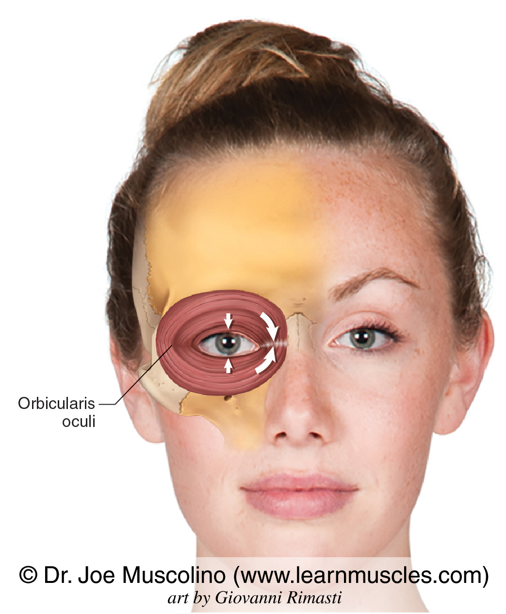
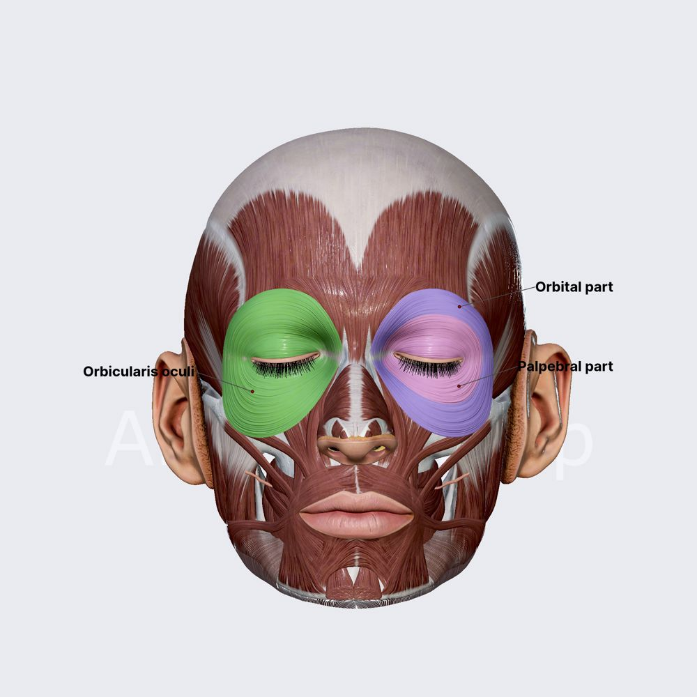
Orbicularis oculi
Muscle surrounding the eye socket that closes eyelids.
Why does contracting the right external oblique cause a rotation in the left side?
Origin: ribs (top)
Insertion: pelvis (bottom)
👉 It pulls:
ribs down + toward left pelvis
So your upper body:
➡ twists LEFT✔ = contralateral rotation
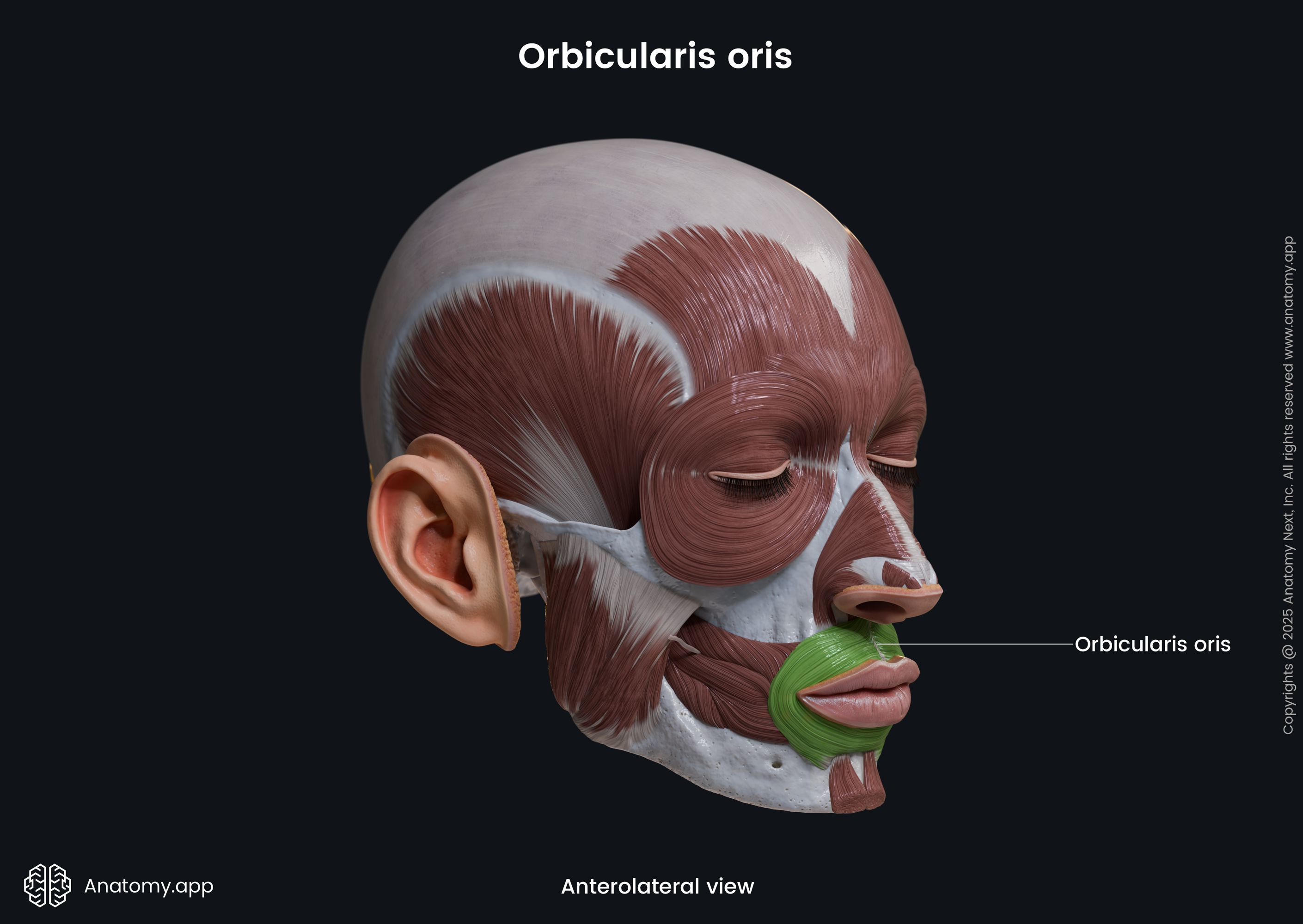
Orbicularis oris
Muscle around the lips that closes lips.
Levator costae
Muscle that elevates the ribs, aiding in inhalation.
Frontalis
Flat muscle covering the forehead that raises eyebrows.
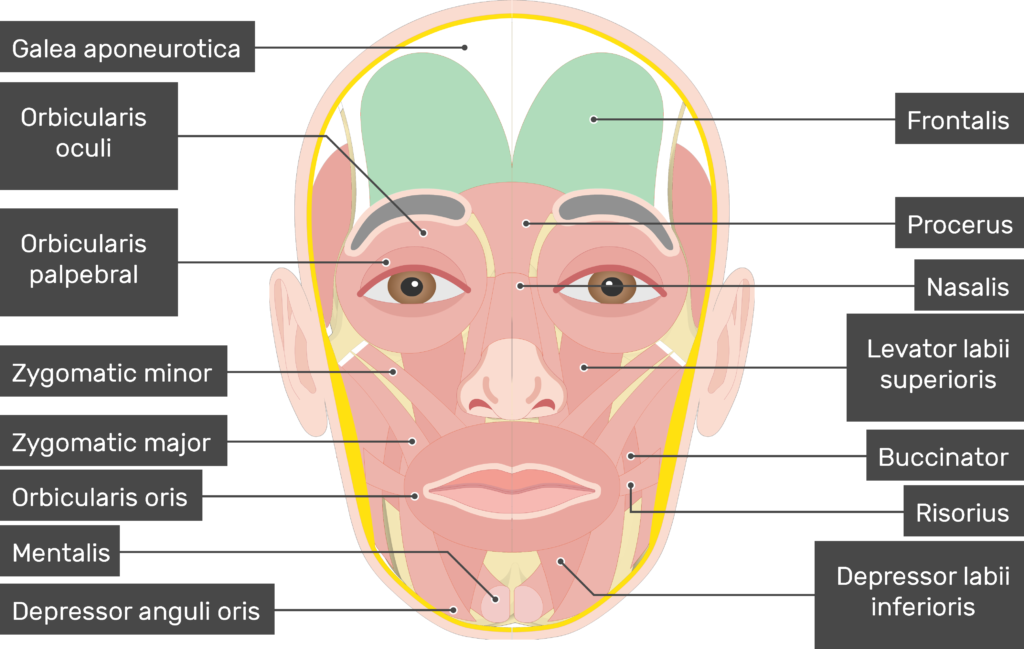
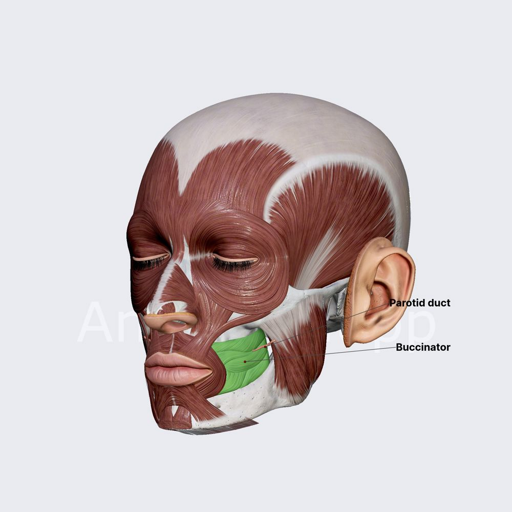
Buccinator
Flat muscle in the cheek wall that keeps food between teeth during chewing.
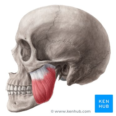
Masseter
Large rectangular muscle on side of jaw that elevates mandible. Origin: zygomatic arch
Insertion: lateral surface of the mandibular ramus
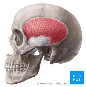
Temporalis
A large fan-shaped muscle on the side of the skull that elevates and retracts the mandible.
origin: floor of the temporal fossa
Insertion: coronoid process of the mandible
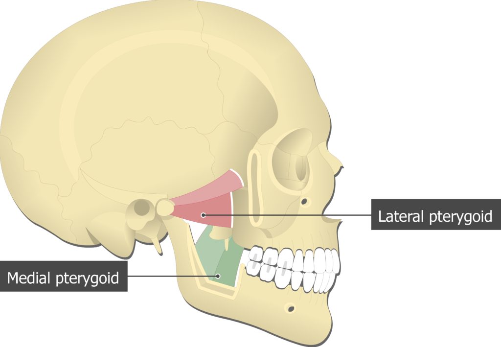
Pterygoid muscles
Deep muscles near the TMJ responsible for jaw protrusion and side-to-side movement.
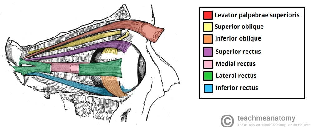
Extrinsic eye muscles
Muscles controlling eye movement; there are 6, including 4 rectus and 2 oblique.
Rectus muscles
Muscles that run straight from the back of the orbit to the eye.
Oblique muscles
Muscles that run diagonally across the eyeball.
Superior rectus
Muscle that elevates the eye.
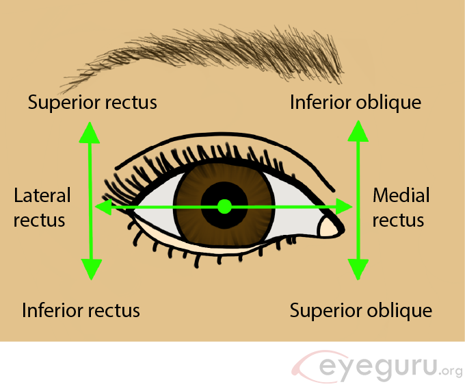
Inferior oblique
extorsion (outward rotation), elevation and abduction.
Inferior rectus
Muscle that depresses the eye.
Superior oblique
Primary action: intorsion
Secondary: depression
Tertiary: abduction (helping with lateral movement)
Lateral rectus
Muscle that abducts the eye.
Medial rectus
Muscle that adducts the eye.
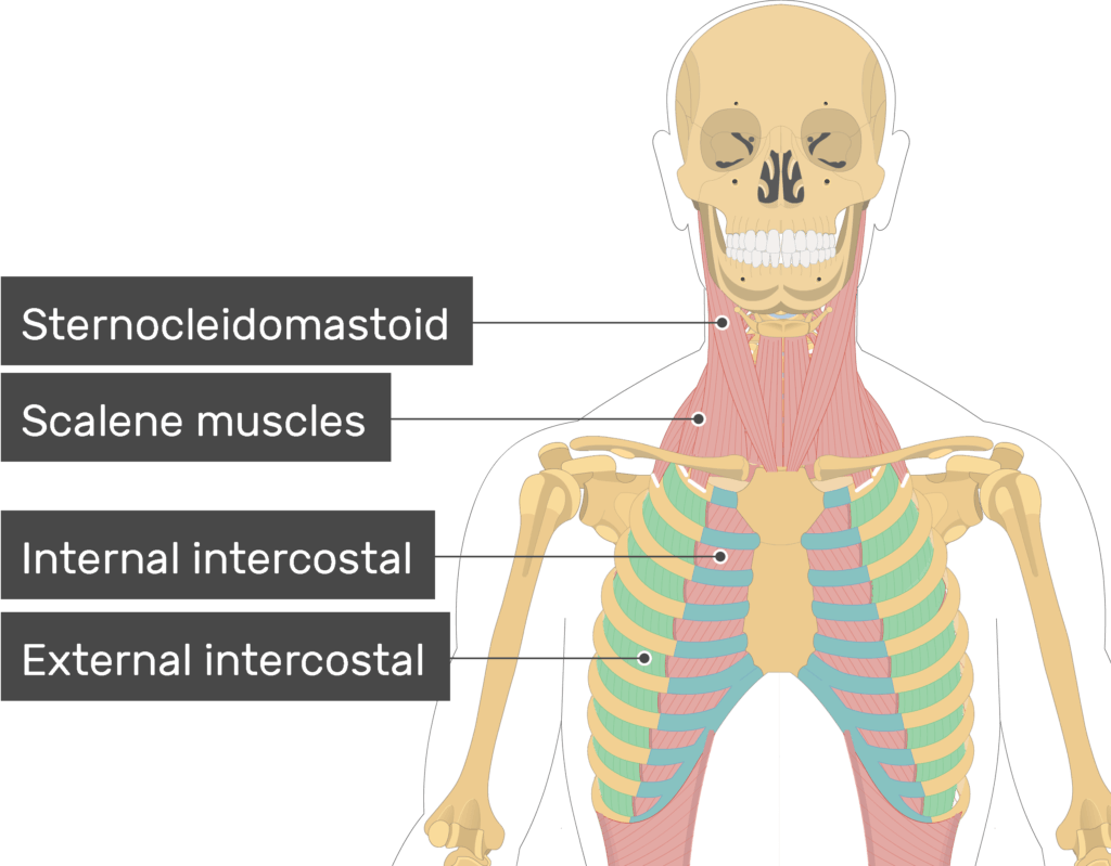
Sternocleidomastoid (SCM)
Large diagonal muscle running across the side of the neck.
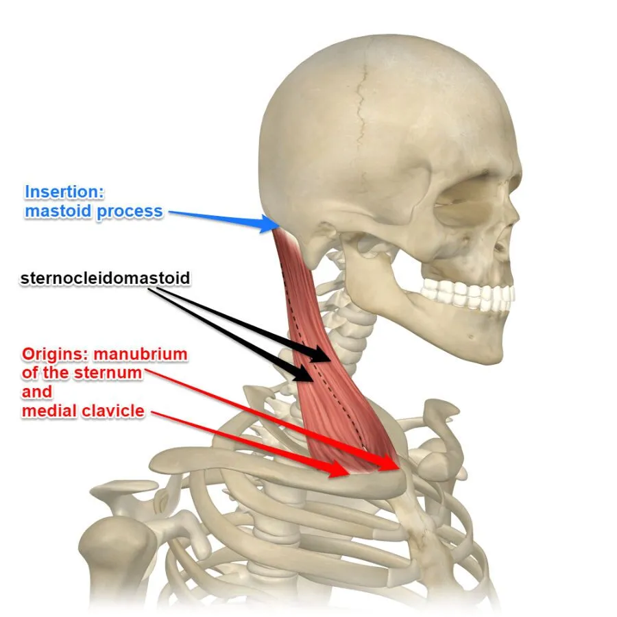
SCM inerstion and origin
Attaches to sternum, clavicle, and mastoid process.
Bilateral SCM contraction
Flexes the neck (the head is pulled forward)
Unilateral SCM contraction
Two options: contralteral rotation of the head (makes the face move in the opposite direction) or ipsilateral lateral rotation
Contraction of the msucles makes the origin and insertion…
move more closely together
Suprahyoid muscles
Muscles that elevate the hyoid bone.
Stylohyoid
Muscle that runs from the styloid process of the temporal bone to the hyoid bone, assisting in elevating the hyoid during swallowing.
Digrastic
muscle with two muscle bellies that helps to depress the mandible and elevate the hyoid bone.
Infrahyoid muscles
Muscles that depress the hyoid bone.
Scalene muscles
Muscles located on the sides of cervical vertebrae.
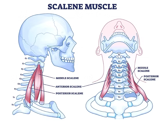
Lateral flexion of the scalene muscles
refers to the bending of the neck to the side, facilitating movements such as turning the head.
Bilateral flexion of the scalene muscle
elevates the first and second ribs
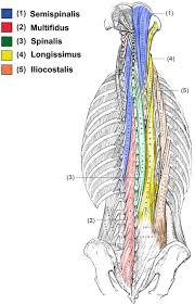
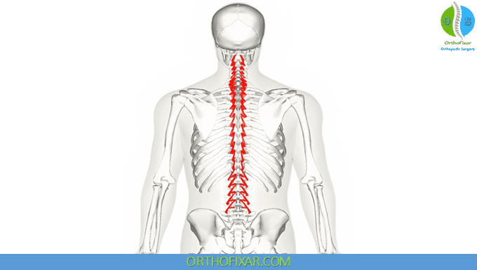
Intrinsic back muscles
Deep muscles that stabilize and move the vertebral column.
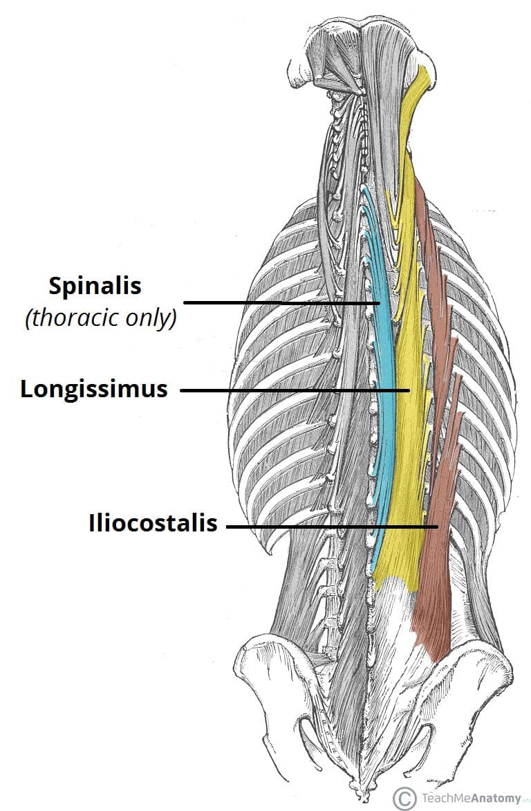
Erector spinae
The largest intrinsic back muscle group responsible for extending the spine.
Unilateral contraction of the erector spinae
involves lateral flexion and rotation of the vertebral column toward the same side.
Transversospinales
Muscle group stabilizing and rotating vertebrae. Semispinalis, multifidus, rotatores.
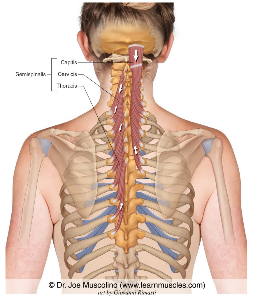
Semispinalis
The semispinalis is a muscle of the transversospinales group, primarily responsible for extending and rotating the vertebral column. Capitis, Cervicis, Thoracis
Splenius capitus
is a muscle in the splenius group that extends and rotates the head and neck.
Spinotransversales group
A group of muscles including the splenius capitis and splenius cervicis, responsible for extending and rotating the head and neck.
Splenius cervicus
is a muscle in the splenius group that extends and rotates the head and neck, similar to the splenius capitis.
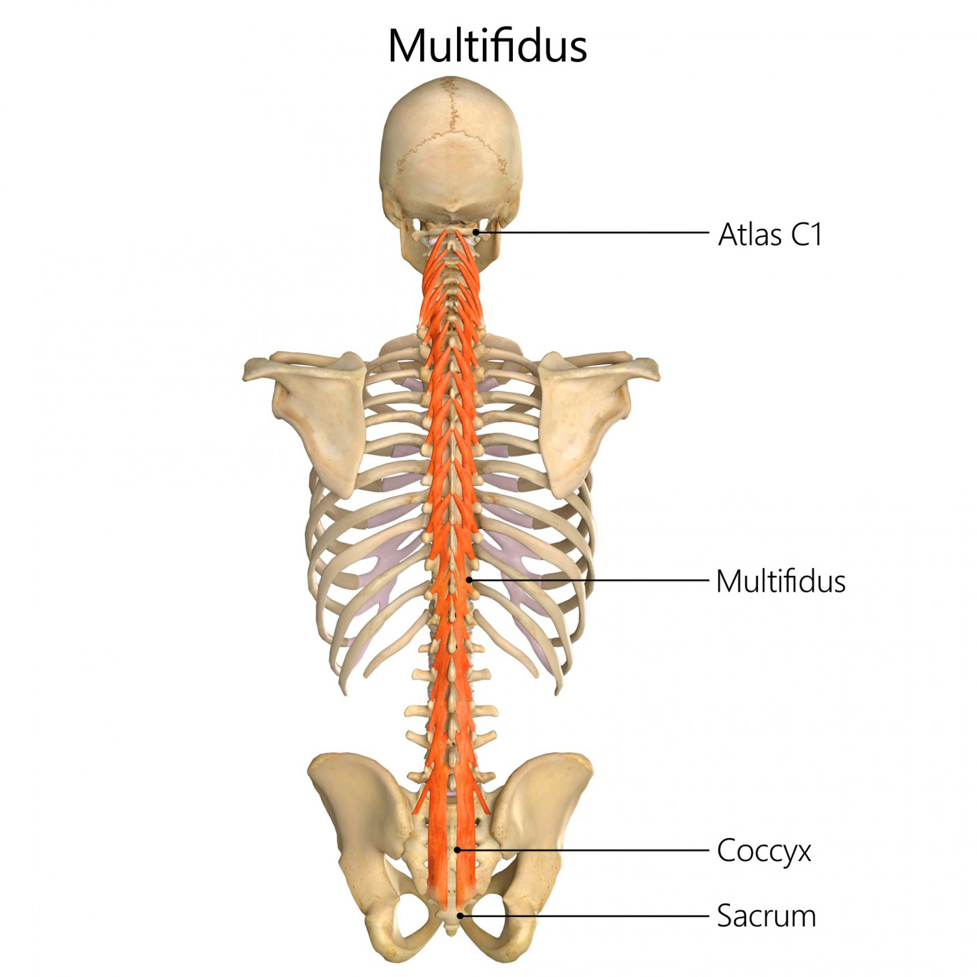
Multifidus
A muscle of the transversospinales group, the multifidus stabilizes the vertebral column and assists in the rotation and extension of the spine.
Rotatores
The rotatores are a group of muscles within the transversospinales, located between the vertebrae and primarily involved in stabilizing and rotating the spine.
Intercostal muscles
Muscles located between ribs that aid in breathing.
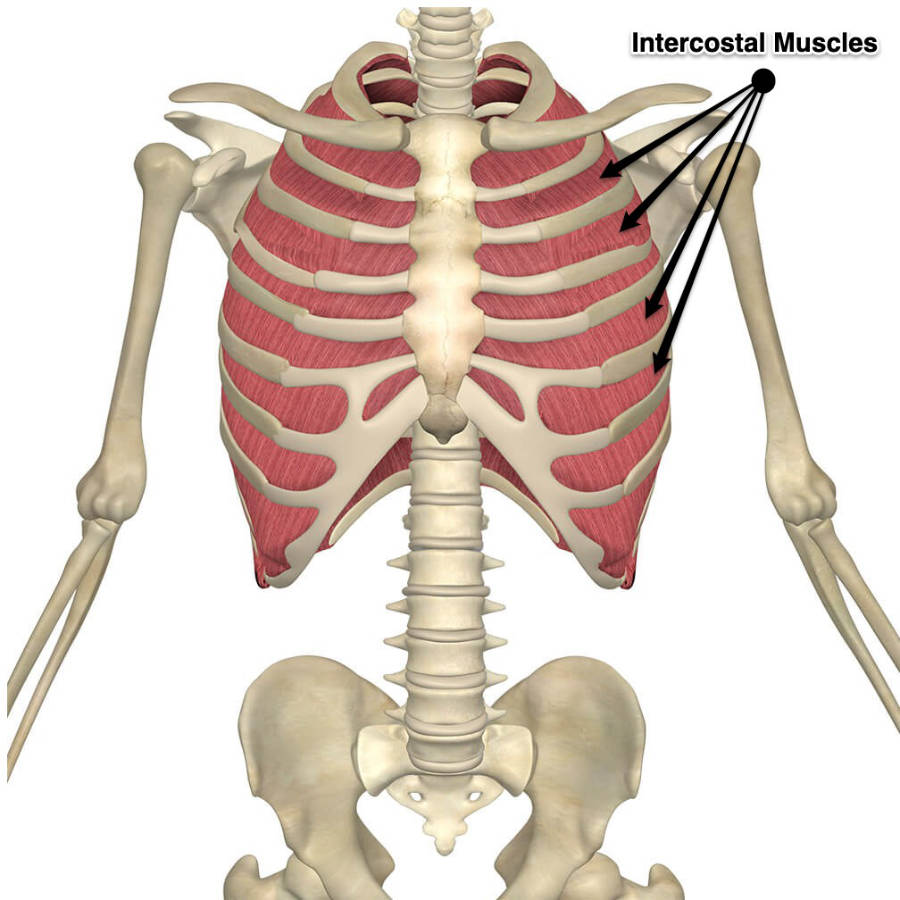
External intercostals
Muscles that elevate ribs during inhalation.

Internal intercostals
Muscles that depress ribs during exhalation.
Innermost intercostals
Muscles located beneath the internal intercostals that assist with forced exhalation.
Serratus posterior superior
Muscle that elevates ribs.
Serratus posterior inferior
Muscle that depresses ribs.
Abdominal muscles
Muscles responsible for flexing the trunk, compressing the abdomen, and assisting in breathing.
Thoracic diaphragm
The main muscle of breathing that separates the thoracic and abdominal cavities.
Pelvic diaphragm
Muscles that support pelvic organs and form the pelvic floor.
Modiolus
A fibromuscular node at the corner of the mouth where many facial muscles attach.
Facial nerve (CN VII)
Nerve that innervates most facial expression muscles.
Aponeurosis
A flat sheet-like tendon connecting muscles.
Chewing
Another term for mastication.
Temporomandibular joint (TMJ)
Joint that mastication muscles move.
Trigeminal nerve (CN V)
Nerve that innervates mastication muscles.
Iliocostalis
Lateral column of the erector spinae.
Longissimus
Middle column of the erector spinae.
Spinalis
Medial column of the erector spinae.
Splenius
Intrinsic back muscle found in the posterior neck responsible for extending and rotating the neck.
Levator ani
Muscle that along with coccygeus forms the pelvic diaphragm.
Coccygeus
Muscle that supports pelvic organs and completes the pelvic diaphragm.
Food between the teeth
Function of the buccinator during chewing.
Zygomaticus major
Muscle that raises the corners of the mouth (smiling).
Epicranius
Muscle with two bellies connected by an aponeurosis.
Levator palpebrae superioris
Muscle that elevates the upper eyelid.
Occipitalis
Muscle that forms the back of the epicranius.
Lateral flexion
Movement achieved by unilateral contraction of the SCM or intrinsic back muscles.
Bilateral contraction
Result in flexion or extension of the neck or spine.
Muscle architecture
Determines the movement and force a muscle can produce.
Line of action
Direction a muscle pulls between its attachments.
Facial expression muscles
Muscles that move the skin of the face to produce facial expressions.
Hyoid bone
Bone separating suprahyoid and infrahyoid muscles.
Shoulder girdle
Not discussed explicitly, but connects to neck and upper limb muscles.
Fibre direction of external oblique
Fibres run downwards like putting hands in pockets.
Fibre direction of internal oblique
Fibres run upwards and medially.
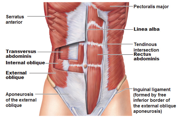
Fibre direction of transversus abdominis
Fibres run horizontally.

Inspiration
Breathing in, involving several muscles including scalene.
Expiration
Breathing out, primarily involving internal intercostals.
Absence of muscle contraction
Results in relaxation of thoracic cavity during exhalation.
Muscle contraction
Increases pressure in the thoracic cavity.
Facial movement coordination
Function of the modiolus.
Common exam focus
Patterns of shape, position, and fibre direction rather than minute details.
Pelvic support
Function of the pelvic diaphragm muscles.
Transversospinales group
Includes semispinalis, multifidus, and rotatores.
Flex trunk
Main function of abdominal muscles.
Ribs attached by scalenes
Ribs 1 and 2.
Muscles attached to hyoid
Suprahyoid elevate, infrahyoid depress.
Bilateral contraction of the erector spinae
is a mechanism that extends the vertebral column, allowing for upright posture and stability.
Recti vs. Obliques
Difference in muscle alignment affecting eye movement.
Rectus definition
Means straight.
Oblique definition
Means diagonal or angled.
Attachment angles
Influence the movement of oblique muscles.
Muscle innervation
Determines function and control of muscles.
Facial expression coordination
Affected by the modiolus and facial nerve.
Prevention of cheek collapse during chewing
Role of the buccinator.
Facial movements role
Controlled primarily by facial expression muscles.
Muscle group function
Muscles categorized by location and action.
Hyoid elevation
Function of suprahyoid muscles.
Hyoid depression
Function of infrahyoid muscles.