Cerebral Cortex
1/41
There's no tags or description
Looks like no tags are added yet.
Name | Mastery | Learn | Test | Matching | Spaced |
|---|
No study sessions yet.
42 Terms
What are the 4 domains of cognition
Executive function
attention
selective → choosing what to focus on
sustained → sustain that focus
divided → can I put my focus on multiple things
memory
working memory
declarative memory
episodic
semantic
orientation
Cognitive Domain – Executive Function
• Definition: Higher order cognitive process that enables a person to engage in independent,
purposeful and goal-directed behaviour
• Includes volition, planning, evaluating consequences, purposeful action, shifting between
information sets, multi-tasking, monitoring and updating working memory and inhibition
• Involved in the control of attention and working memory
• Neurology:
• Dorso-lateral prefrontal cortex and anterior cingulate cortex
• Role in both cognitive and motor networks in the brain
• Problems in executive function:
• Ineffective self-monitoring and difficulty with self-correction
• Difficulty applying information to new or unfamiliar situations
Cognitive Domain - Attention
Definition: Number of different processes that are related to how the brain becomes
receptive to stimuli and how it begins processing these stimuli
• Ability to select and attend to a specific stimulus while simultaneously suppressing
extraneous stimuli
• 3 processes: selective, sustained, divided
• Prefrontal cortex
• Problem in attention:
• Decreased ability to process and assimilate new information
1. Selective attention •
Ability to select relevant stimuli and the concurrent suppression of irrelevant stimuli and suppression of distractors
2. Sustained attention
Ability to maintain focus on a task over a period of time
3. Divided attention
Ability to perform more than one task at once, shifting attention from one task to the other
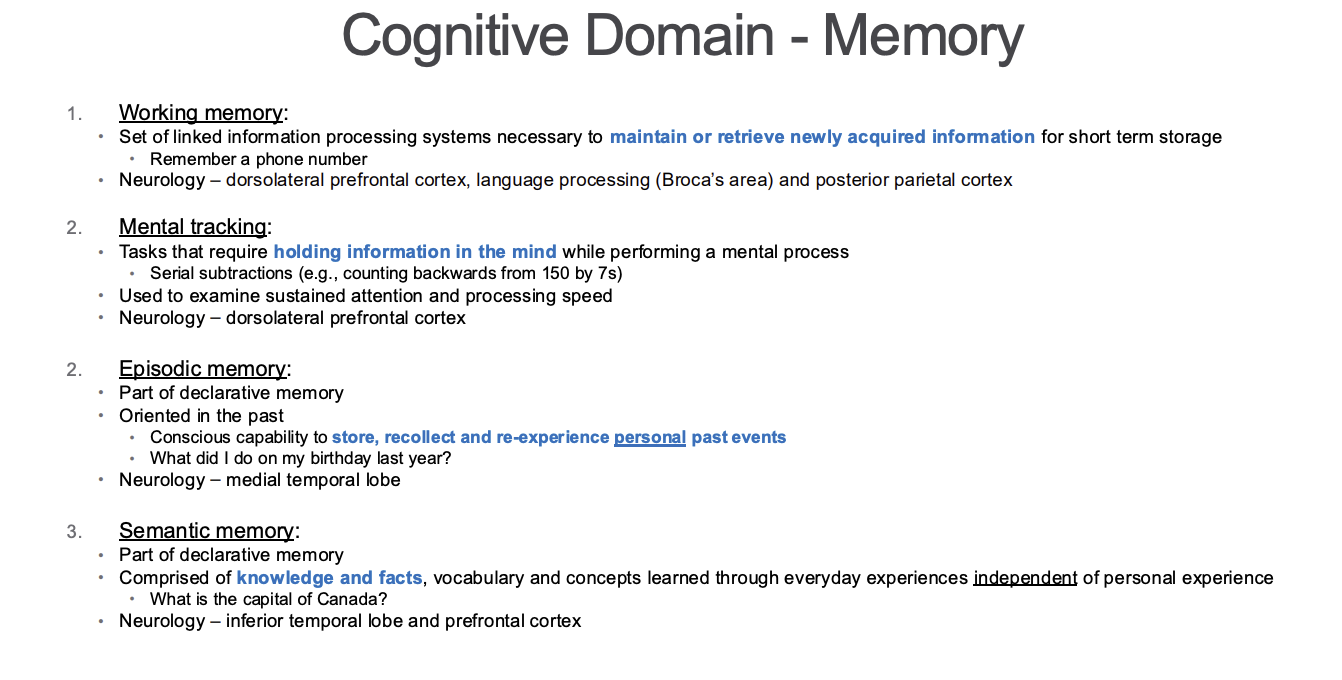
Cognitive Domain - Memory
• Definition: Ability to store experiences and perceptions for later recall
• Registration, encoding, storage, recall and retrieval of information
• Need memory for motor learning!
• Neurology – temporal lobes, especially hippocampus
The 3 R’s of memory
Registration – Must focus attention and ignore distractions for information to get into memory
Also dependent on motivation and ability to associate it meaningfully with information already in memory
Retention – Making information stick in your memory once it is registered
Consolidation of information happens in the hippocampus
Makes information stable for long term memory
Long term retention of memories involves structural changes in neurons (neuroplasticity)
Retrieval – Pulling information out of your mental library (long term memory)
Most accurate when retrieved in the same context it was created • Can be distorted as memories are stored in multiple sites
Cognitive Domain – Orientation
• Definition: Knowledge related to person, place & time
• Neurology – frontal parietal association areas
• Problem in orientation:
• Disoriented
• Person, place or time
Memory Flow Chart
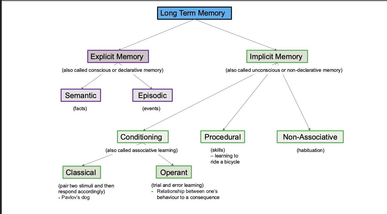
Explicit Learning
Conscious – requires attention and awareness
Learning using explicit memory can be expressed in declarative sentences
“First I button the top button, then the next one.”
Need to have the ability to verbally express the process that is being performed
Cannot be used with patients with cognitive or language deficits that impair their ability to recall and express knowledge
Example – Sit to stand
Teach a specific sequence
First move to the edge of the chair
Lean forward
Then stand up
Advantage of declarative learning is that it can by practiced in many ways
Most importantly – mental rehearsal
Mental rehearsal activates the same regions of the brain as active movement
Declarative learning requires intact sensory association areas (somatosensory, visual and auditory), medial temporal lobe, hippocampus
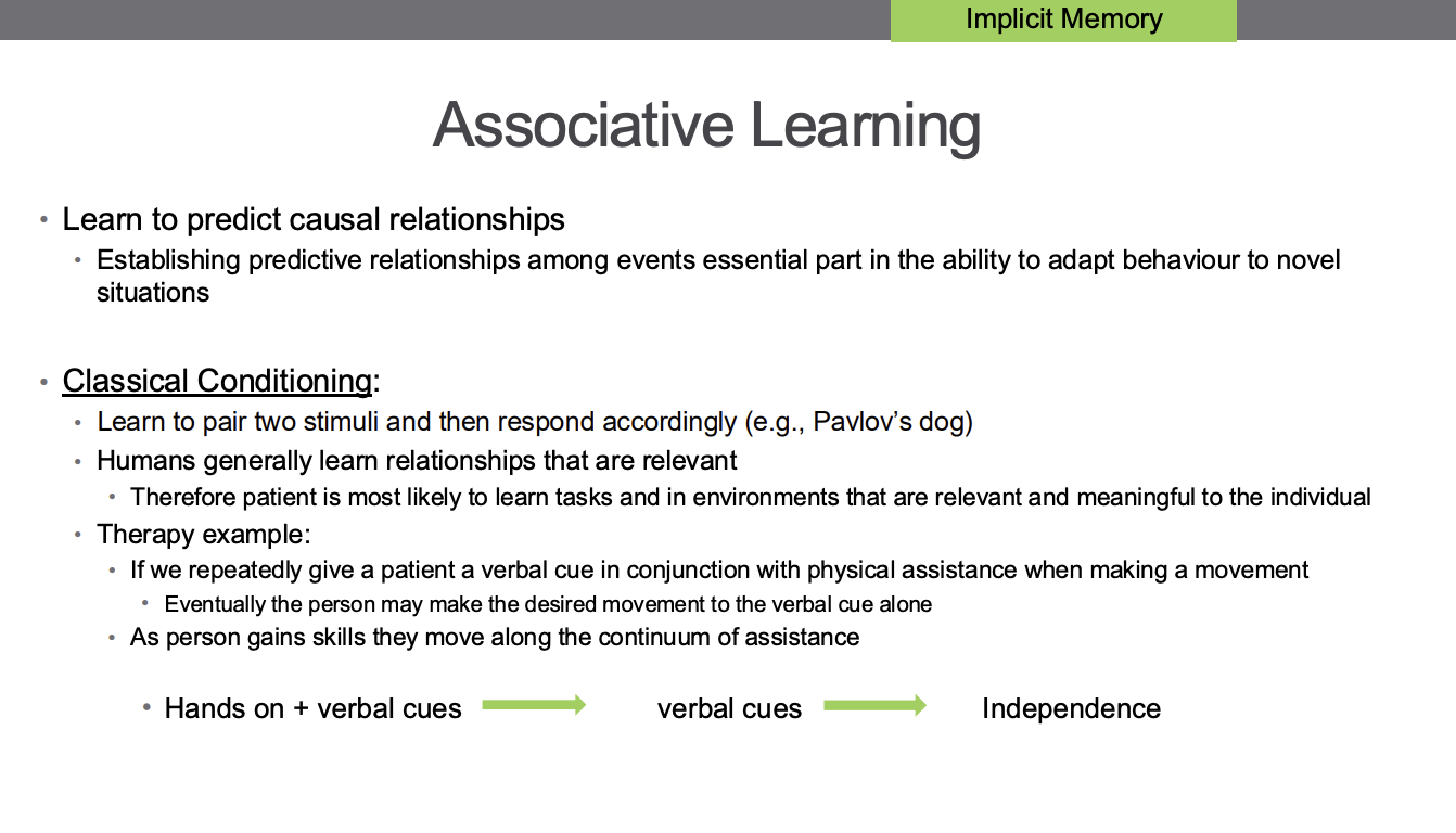
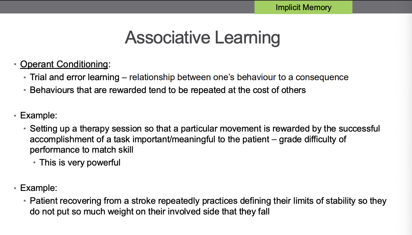
Associative Learning
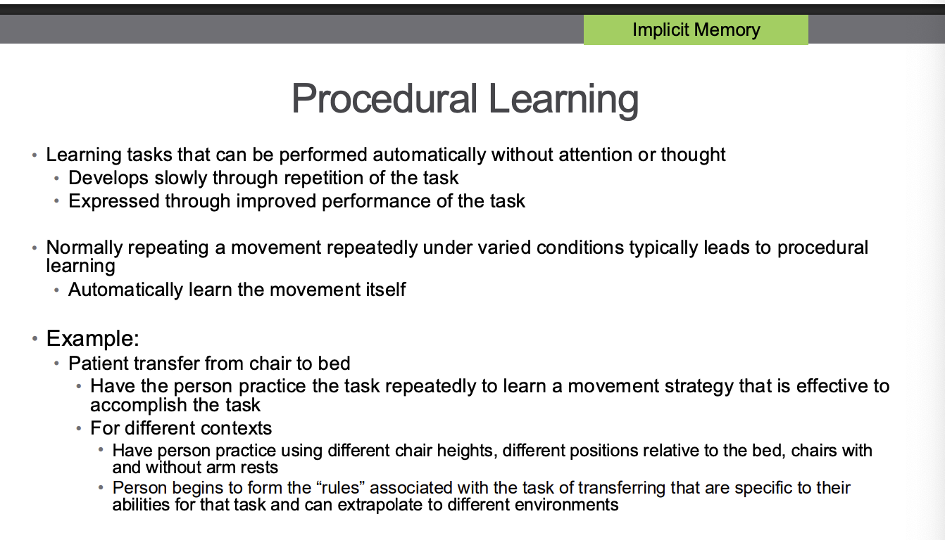
Procedural Learning
Motor Learning (what is it, and what are the 4 concepts)
• Motor learning focuses on the acquisition and/or modification of movement
• Occurs through practice or experience leading to relatively permanent changes in the
capability for producing skilled movement.
• Normal development seen in infants and children
• Reacquisition of skills lost through injury
• 4 concepts:
1. Process of acquiring the capability for skilled movement
2. Learning results from experience or practice
3. Learning cannot be measured directly • It is inferred through behaviour 4. Learning produces relatively permanent changes in behaviour • So short term alterations are not considered learning
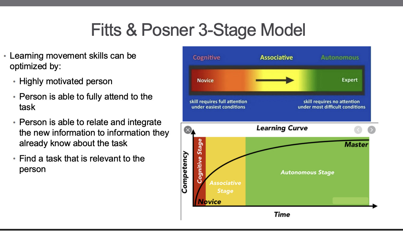
Fitts and Posner 3-stage model
1st Stage – “Cognitive Stage of Learning”
• Learner is concerned with understanding the nature of the task, developing strategies
that could be used to carry out the task
• These require a high degree of cognitive resources
• Especially attention
• Brain intensive activities – can be fatiguing because of this
• Performance tends to be variable
• Many strategies for performing the task are being applied
• Improvements can be quite large in this stage
2nd Stage – “Associative Stage”
• Person has selected the best strategy for the task and now begins to refine the skill
• Less variability in performance
• Improvement is slower than the first stage
• Length of time in this stage depends on
• The performer and intensity of practice
3 rd Stage – “Autonomous Stage”
• Automaticity of the skill and low degree of attention required for its performance • Paying too much attention to elements of the task in this phase results in a decrease in performance • Person can begin to devote attention to other aspects of the skill • Walking as the skill • Scanning environment for obstacles • Performing a secondary task
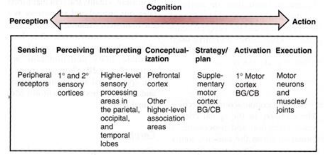
Cognition perception → action continuum
Sensory Areas of the Cerebral Cortex
Primary Somatosensory Cortex:
Located within the central sulcus and the postcentral gyrus (parietal lobe)
Receives information from tactile and proprioceptive receptors via DCML
Receive pain and temperature information as well, though that is widely distributed beyond just the primary somatosensory cortex
• Crude awareness of somatosensation occurs in the thalamus
Neurons in primary somatosensory cortex identify the location of stimuli and discriminate among various shapes, sizes and textures of objects
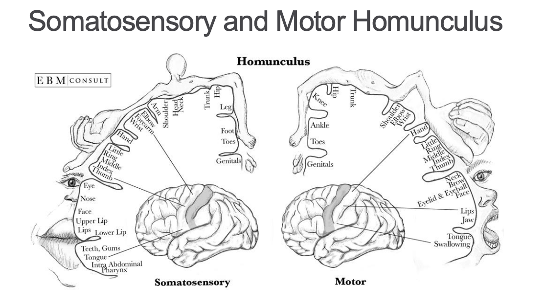
Primary Somatosensory Cortex
• Gyrus (postcentral) posterior to central sulcus in parietal lobe
• Somatotopic representation – homunculus
• The amount of primary somatosensory cortex devoted to a body part is not directly proportional
to the absolute size of that body surface
• Proportional to the density of cutaneous sensory receptors on that body part.
• Density of cutaneous sensory receptors on a body part generally indicates degree of sensitivity of tactile stimulation
experienced at that body part
• Contralateral representation
Sensory Areas of the Cerebral Cortex
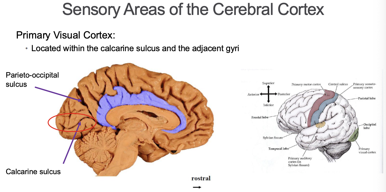
Primary Auditory Cortex:
Located within the lateral fissure and the adjacent superior temporal gyrus • Receives information from the cochlea of both ears via a pathway that synapses through the thalamus before reaching the cortex • Provides conscious awareness of the intensity of sounds
Primary Vestibular Cortex:
• Located posterior to the primary somatosensory cortex • Receives information about head movements and head position relative to gravity • Vestibular nuclei in brainstem send information via thalamus • From thalamus to primary vestibular cortex
Secondary Sensory Areas
Sensory Loss
Complete loss of sensation = anaesthesia
Abnormal sensory phenomena = paraesthesia
Radicular pain
Dysesthesia = unpleasant, abnormal sensation
Allodynia = pain produced by normally non-painful stimuli
Hyperalgesia = enhanced pain to normally painful stimuli
Caused by lesions anywhere in somatosensory pathways
Peripheral nerves, nerve roots, DCML, spinothalamic pathway, thalamus, thalamocortical white matter, primary somatosensory cortex
Ascending Long Tracts – Conscious
DCML: proprioception, vibration, fine, discriminative touch
Spinothalamic tract: pain, temp, crude touch
crosses immediately
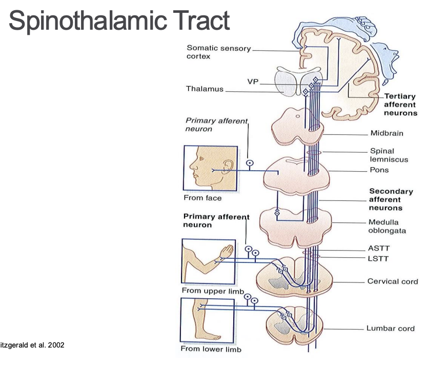
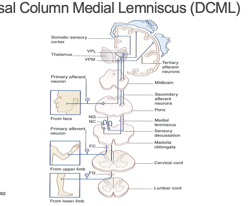
crosses at medial lemniscus
Lesions in DCML
Test integrity of DCML
two point discrimination
romberg sign: positive = severe swaying with feet together or in tandem stance with eyes closed
sensation loss: ipsilateral below level of sensory decussation
contralateral above level of sensory decussation
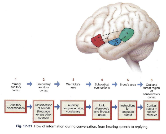
Function of communication in the cerebral cortex
in 95% of people the cortical areas responsible for understanding language and producing speech
language - a communication system based on symbols
speech - verbal output
different regions of the brain are responsible for each
comprehension of spoken language - wenickes area
instructions for language output - brocas area
planning the movements of speech
reading required intact vision, secondary visual areas for visual recognition of written symbols and connections with an intact wernicke are for interpreting the symbols
What do the contralateral areas corresponding to Wernicke’s and Broca’s areas in right hemisphere do?
In most people
areas are associated with noverbal communication
gestures, facial expressions, ton of voice and posture convey meanings in addition to the verbal message
R hemisphere area corresponding to wernickes area
vital for interpreting non verbal signals from other people
R hemisphere area corresponding to brocas area
provides instructions for producing nonverbal communication, including emotional gestures and intonation of speech
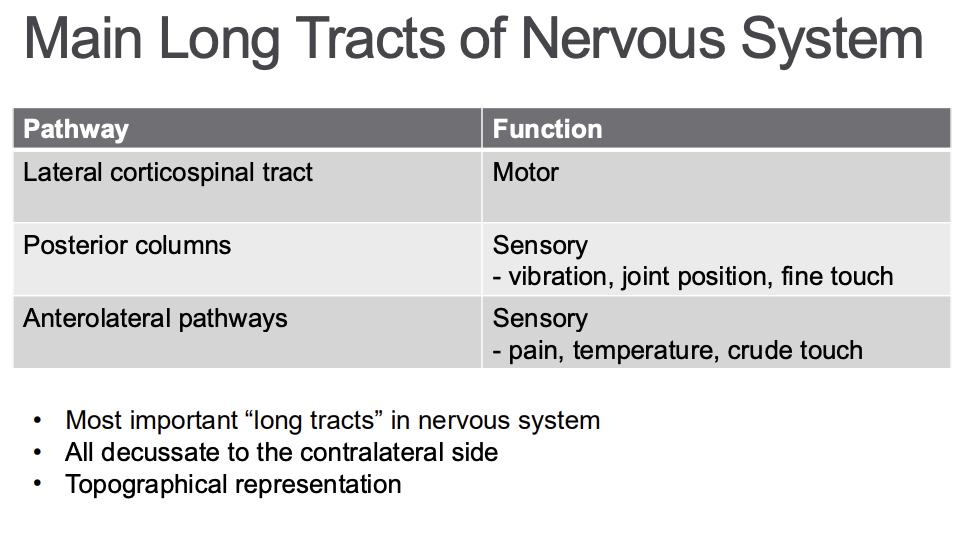
Main long tracts in the nervous system
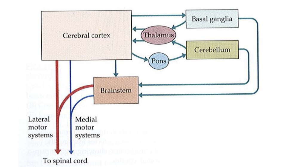
General organization of motor systems
Primary Motor Cortex
• Gyrus anterior to the central sulcus • Lateral and medial surface of frontal lobe • Contains distinctive Betz cells (pyramidal neurons) • Do not compose entire motor output from primary motor cortex (3%) • Marker for the primary motor cortex • Descending via the corticospinal tract • Connections with supplementary motor cortex, premotor cortex • Sensory inputs play an essential role in motor control • Participate in motor circuits and feedback loops
Motor function in the cortex (location and name)
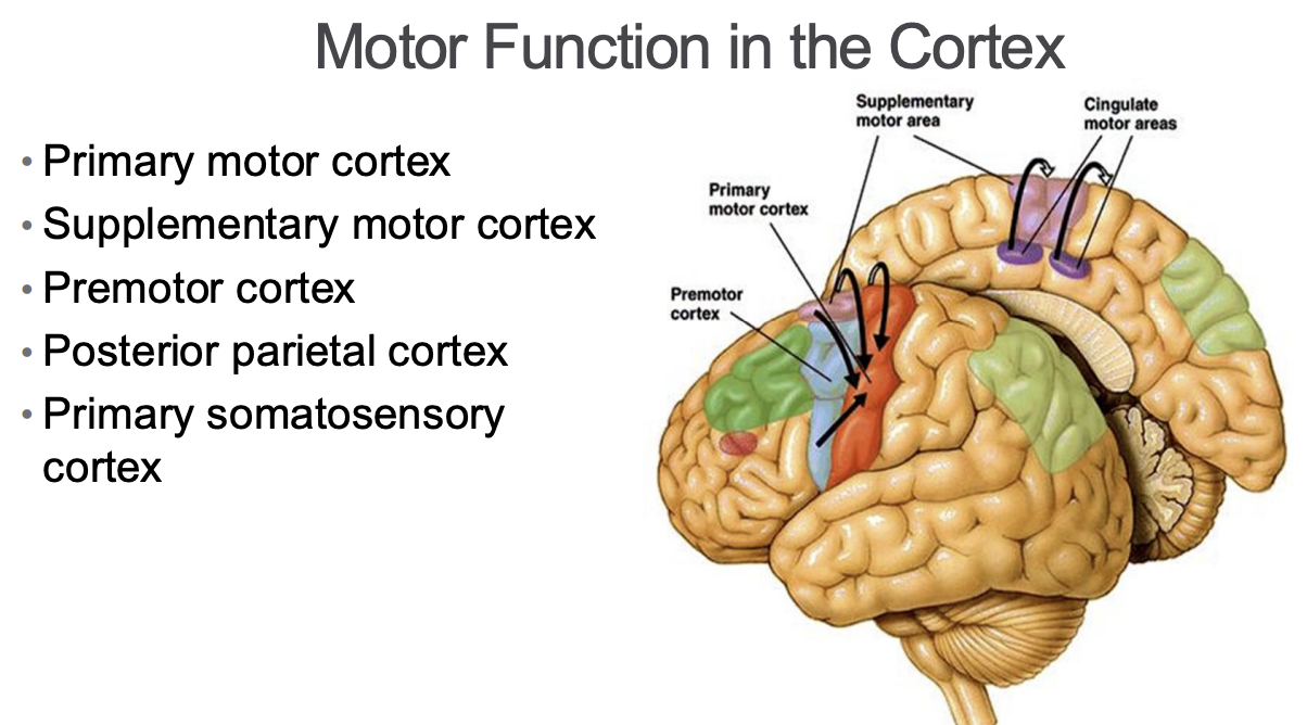
Supplementary Motor Area
• Located on superomedial surface of frontal lobe anterior to the primary motor cortex • Connections to primary motor cortex • Direct projections to spinal cord • Functions: • Preparation of internally initiated movements • Bilateral coordination • Postural stabilization (stance & gait)
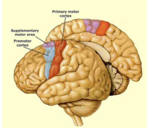
Premotor area
• Located on lateral surface of frontal lobe anterior to the primary motor cortex • Connections: • primary motor cortex, supplementary motor cortex, prefrontal cortex, parietal cortex • Direct projections to spinal cord – corticospinal tract • Functions: • Preparation of spatial & sensory guided movements • Rules to perform specific tasks
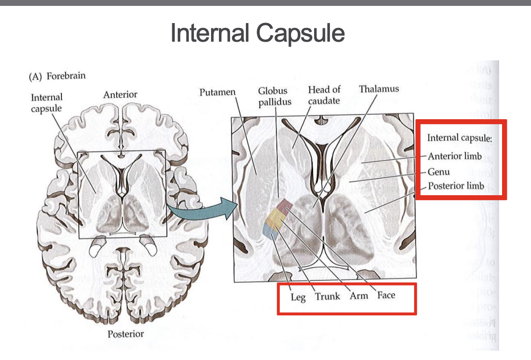
Cingulate Motor Area
Medial surface of brain, inferior to supplementary motor area
part of cignulate gyrus
Function;
motor selection based on reward evaluation
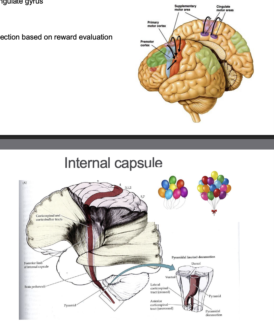
Upper Motor Neurons
cell bodies in cerebral cortex
carry motor systems information to lower motor neurons in spinal cord and brainstem
project to muscles in the periphery
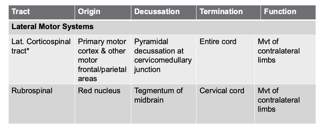
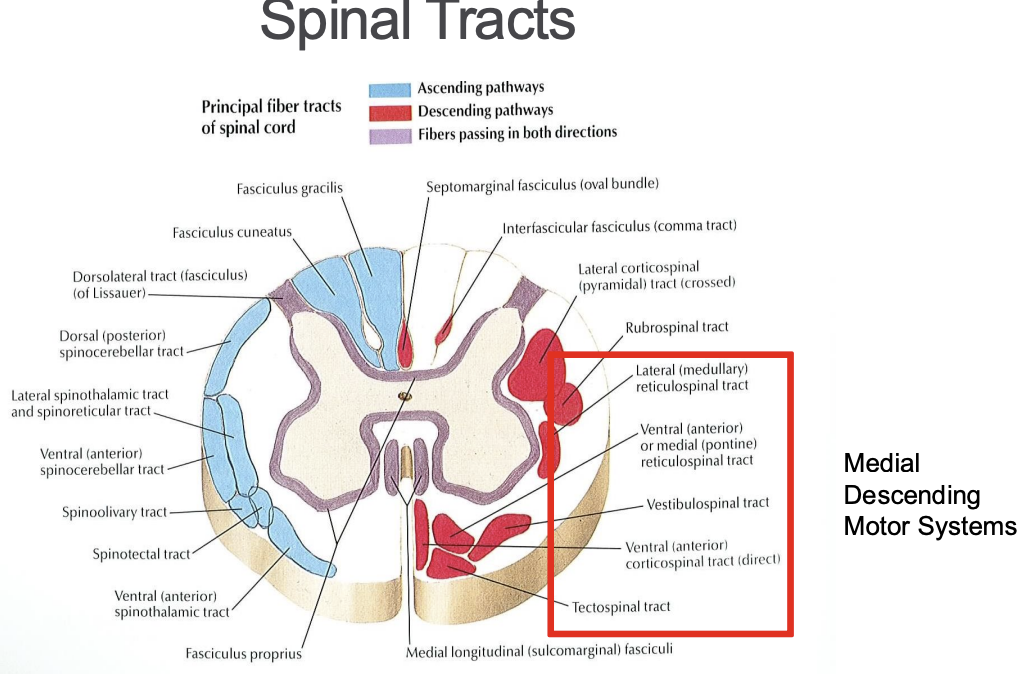
based on location of traits in spinal cord can be divided into:
lateral motor system
medial motor system
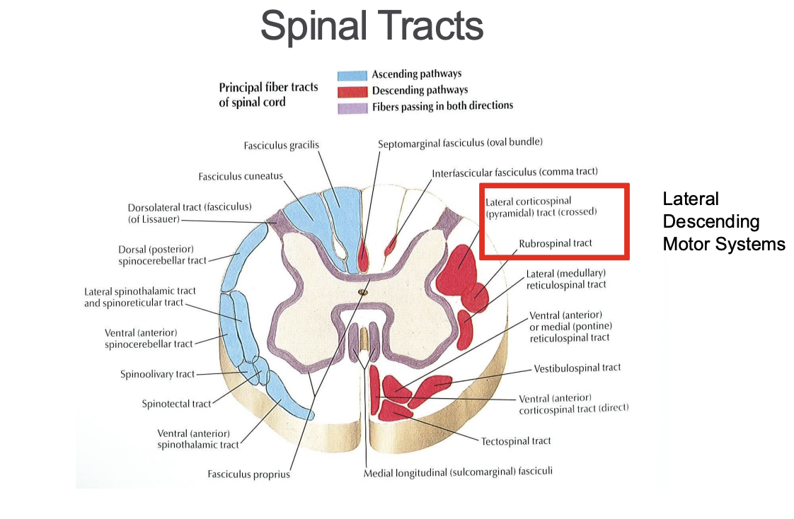
Lateral Descending motor system
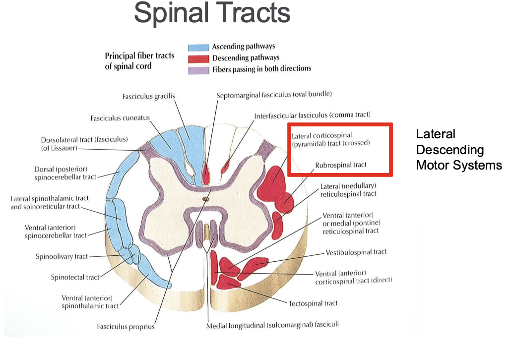
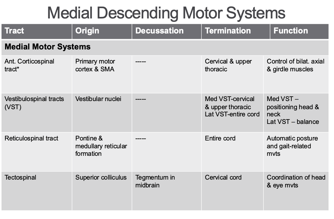
Medial descending motor systems
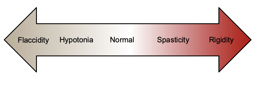
Muscle tone - definition
The sensation of resistance felt as one manipulates a joint through a range of motion, with the patient attempting to relax (passive movement)
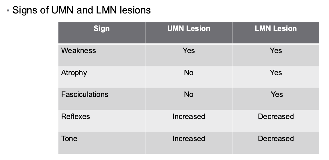
Upper motor neuron vs. lower motor neuron lesions
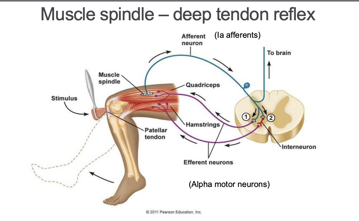
Defining Spasticity
motor disorder characterized by a velocity-dependent increase in tonic stretch reflexes (muscle tone) with exaggerated tendon jerks (DTR), resulting from hyper-excitability of the stretch reflex, and is one component of the upper motor neuron syndrome
Terms used in loss of muscle function
Denoting severity: • -paresis = weakness • -plegia = no movement • Paralysis = no movement • Palsy = imprecise term for weakness or no movement
Denoting location: • Hemi = one side of body • Para = Both legs • Mono = one limb • Di- = Both sides of body equally affected • Quadri- or tetra- = all four limbs
Apraxia
Disorder of motor planning
Inability to execute learned purposeful movements
Not limited by lack of the desire or the physical capacity to perform the movements
Not caused by incoordination, sensory loss, or failure to comprehend simple commands •
Common types: 1. Ideomotor apraxia • Inability to plan or execute actions based on semantic memory • i.e. pretend to brush your hair • Able to do automatically
2. Ideational apraxia • Inability to select and carry out appropriate motor command
Consciousness
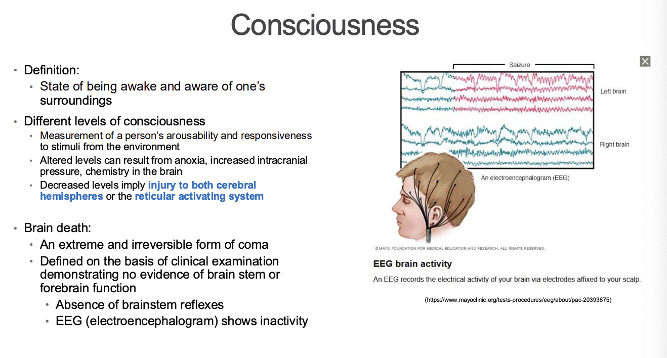
Coma and brainstem reflexes (6 reflexes)
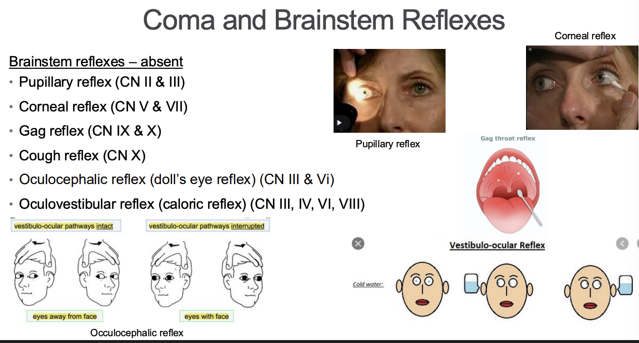
Coma and the other states an individual can be in
Coma – unarousable unresponsiveness in which the patient lies with eyes closed • Result of catastrophic brain injury – common trauma or anoxia • Result of dysfunction in two possible locations • Bilateral widespread regions of the cerebral hemispheres • Upper brainstem activating systems • Unlike brain death, brainstem reflexes occur in coma • Absence of meaningful or purposeful responses mediated by the cerebral cortex are absent • Cerebral metabolism is reduced by at least 50% • EEG shows activity, but abnormal • Does not show variation in activity as would be seen in sleep • Generally not a permanent state – within 2-4 weeks people will deteriorate or progress to other states of less profoundly impaired arousa
• Less impaired states than coma include: 1. Vegetative state • Regain sleep-wake cycle and reflexes mediated through the brainstem but remain unconscious • Poor prognosis for recovery after 3-12 months 2. Minimally conscious state • Variable degree of responsiveness including ability to follow simple commands, say single words 3. Stupor • Deep sleep, person can be aroused only with vigorous and repetitive stimulation • Returns to deep sleep when not continually stimulated 4. Obtunded • Moderate reduction in alertness 5. Lethargy • Mild reduction in alertness 6. Alert • Appearance of wakefulness , awareness of self and environment
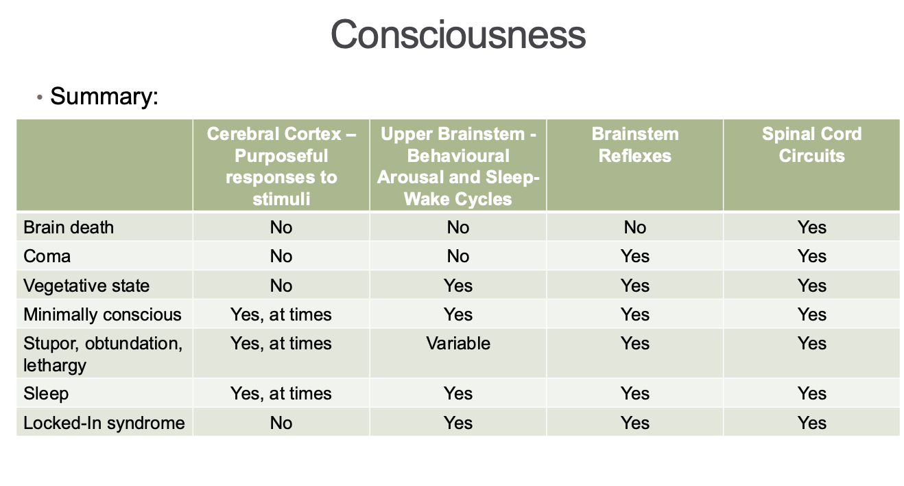
Glasgow Coma Scale
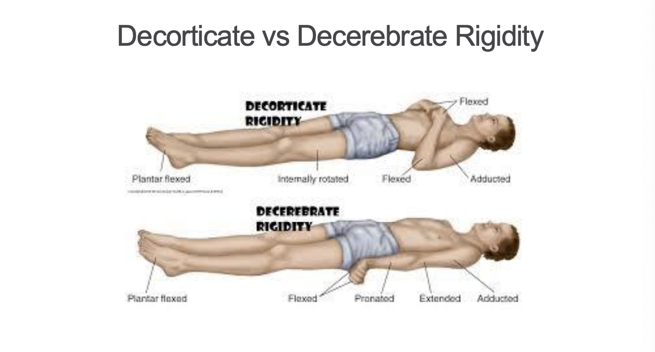
Decorticate vs decerebrate rigidity
