Motor System II EXAM 2
1/193
There's no tags or description
Looks like no tags are added yet.
Name | Mastery | Learn | Test | Matching | Spaced | Call with Kai |
|---|
No analytics yet
Send a link to your students to track their progress
194 Terms
medial and inferior vestibular nuclei
where does the medial VST originate
the medial VST
what part of the VST Originates in the medial and inferior vestibular nuclei
descends bilaterally
How does the medial VST descend?
the medial VST
which descending pathway of the vestibulospinal tract descends bilaterally
lateral vestibular nuclei
where does the lateral VST originate
descends ipsilaterally
how does the lateral VST descend
the lateral VST
which descending pathway of the vestibulospinal tract descends ipsilaterally
upper cord as part of medial longitudinal fasciculus (MLF)
where does the medial VST descend bilaterally to
medial laminae VII/VIII alpha motoneurons & interneurons
what is the target or termination site of the medial VST
the medial VST
what pathway of the VST descends bilaterally and terminates at the medial laminae VII/VIII alpha motoneurons & interneurons
- control of neck musculature
- head-on-trunk stabilization
what is the function of the medial VST
the medial VST
which pathway of the VST is responsible for the control of neck musculature and head-on-trunk stabilization
head-on-trunk stabilization reflex
what reflex is the medial VST responsible for
Descend ipsilateral to all cord in anterior funiculus
where does the lateral VST descend ipsilaterally to
medial laminae VII/VIII alpha neuron & interneurons
what is the target or termination site of the lateral VST
the lateral VST
what pathway of the VST descends ipsilaterally and terminates at the medial laminae VII/VIII alpha neuron & interneurons
paravertebral and proximal limb extensor
posture and balance
flexor
Lateral VST
Mainly excites motoneurons to _____ and _____ muscles = antigravity mm = control of _____ and _____
some inhibits _____ muscles
the lateral VST
what pathway of the VST is responsible for the excitation of paravertebral and proximal limb extensors for control of posture and balance
the lateral VST
which pathway of the VST can be found throughout the entire spinal cord
the medial VST
which pathway of the VST can be found throughout the upper spinal cord
1. Vestibular apparatus - excitatory
2. Fastigial nu - excitatory
3. Purkinje cells - inhibitory
what are the 3 inputs to the vestibulospinal tract (name if they’re excitatory or inhibitory)
the vestibular apparatus and fastigial nucleus
what inputs to the vestibulospinal tract are excitatory
Purkinje cells
what inputs to the vestibulospinal tract are inhibitory
the cortex
what part of the CNS has no direct input to the vestibular nucleus
any signal from the cortex has to pass through the thalamus first
why does the cortex have no direct input to the vestibular nucleus
- Coordination of head and body in space (vestibular input)
- Maintenance of body and limb posture (cerebellar/vestibular input)
what are the 2 main functions of the Vestibulospinal Tracts
pontine (oral and caudal) reticular nuclei
what is the origin of the medial reticulospinal tract
medial reticulospinal tract
what tract of the reticulospinal tract originates at the pontine (oral and caudal) reticular nuclei
ipsilateral
anterior funiculus
(a few may cross)
The descending pathway of the medial reticulospinal tract tends to be ________ in the _______
medial reticulospinal tract
what descending reticular pathway originates at the pontine (oral and caudal) reticular nuclei and descends ipsilaterally in the anterior funiculus
medullary (gigantocellular) reticular nuclei
what is the origin of the lateral reticulospinal tract
lateral reticulospinal tract
what pathway of the reticulospinal tract originates at the medullary (gigantocellular) reticular nuclei
bilateral with ipsilateral predominance in anterior funiculus (but a few decussate)
what is the descending pathway of the lateral reticulospinal tract
lateral reticulospinal tract
what descending reticular pathway originates in the medullary (gigantocellular) reticular nuclei and descends bilateral with ipsilateral predominance in anterior funiculus (but a few decussate)
anteromedial laminae VII/VIII interneurons
what is the target site for the descending reticulospinal tract
to regulate motor activity by controlling posture, balance, and locomotion in both axial and appendicular skeletal muscle
what is the function of the descending reticulospinal tract that terminates at the anteromedial laminae VII/VIII interneurons
bilateral
somatosensory
reticulospinal tracts activated by ______ descending cortical projections (corticoreticular) and ascending ______ (spinoreticular/pain fibers of ALS)
alpha motoneurons
what is influenced heavily by the reticulospinal tract to maintain posture and modulate muscle tone
Medial (pontine) RST
which reticulospinal pathway has excitatory effects to extensor and axial muscles
excitatory effects to extensors and axial muscles
what function is the Medial (pontine) RST responsible for
Lateral (medullary) RST
which reticulospinal pathway has inhibitory effects to extensor muscles of the neck and back
inhibitory effects to extensors, neck and back mm
what is the function of the Lateral (medullary) RST
when you want more posture and tone, cortex fires on reticular nuclei
when might the reticulospinal tract activate
diffuse activation of multiple inhibitory and excitatory neurons to muscles
What does a single pulse of stimulation to the reticulospinal tract result in and how does muscle respond
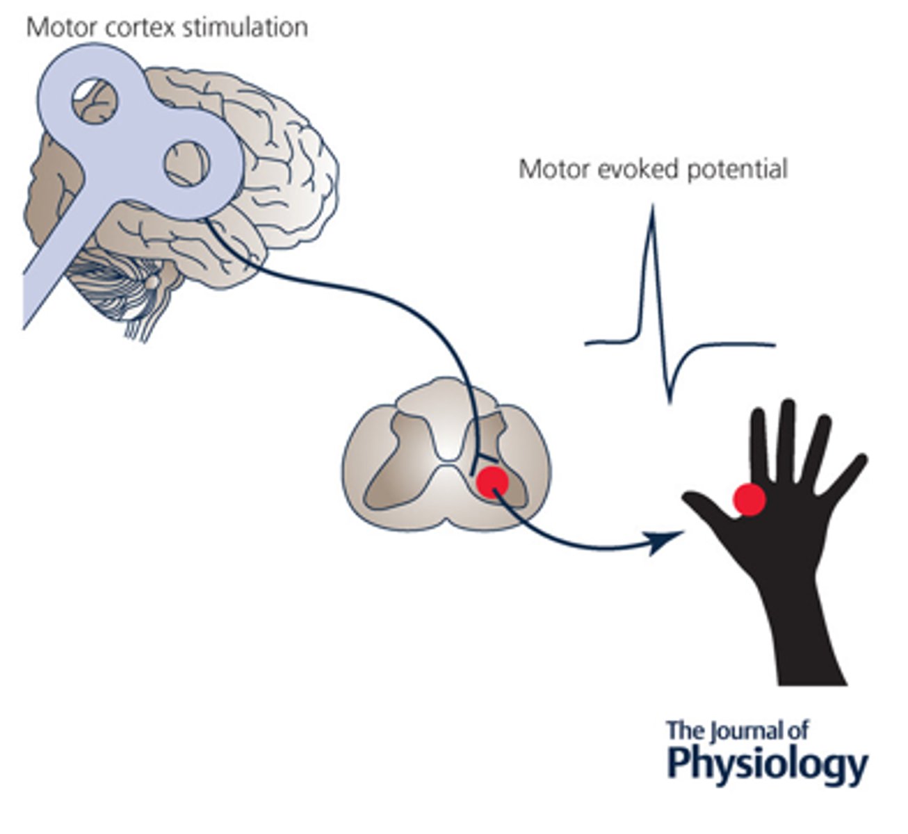
Motor evoked potential for normal motor cortex stimulation of the corticospinal
what is the outcome of a single pulse of stimulation to the corticospinal tract
Corticospinal stimulation
what type of stimulation
Reticulospinal stimulation
what type of stimulation
NE and serotonin
what 2 chemical messengers/neurotransmitters can increase the sensitivity of the reticulospinal tract
NE and serotonin increase the sensitivity of motor neurons in the reticulospinal tract
NE and serotonin have what type of effect on the reticulospinal tract
Activated by ipsilateral cortex (corticorubral) and contralateral cerebellum (cerebellorubral)
what activates the rubrospinal tract
the rubrospinal tract
what descending motor pathway is activated by ipsilateral cortex (corticorubral) and contralateral cerebellum (cerebellorubral)
Flexor motoneurons; supplement to corticospinal system
what are the cortical inputs to the rubrospinal tract
rubrospinal tract
what descending motor pathway has inputs from cortical influence of flexor motoneurons, and has supplemental input to corticospinal system
red nucleus
_________ receives many inputs from cerebellum, a circuitry for modifying motor performance or acquiring a new movement pattern
rubrospinal tract
what descending motor pathways receives inputs from the red nucleus and has a circuitry for modifying motor performance or acquiring new movement patterns stemming from the cerebellum
Excitation of UE flexors
what muscle action might the excitation of the rubrospinal tract result in
they’re "released" from their descending cortical control
what happens to spinal motor neurons during Decerebrate and Decorticate Lesions
RST and VST activity contribute to the spasticity or tonic extension of arms and legs seen in patients with damage to corticospinal tracts (CST) and RuST
If spinal motor neurons are "released" from their descending cortical control from Decerebrate and Decorticate Lesions, how does the reticulospinal and vestibulospinal tracts contribute to UMN signs
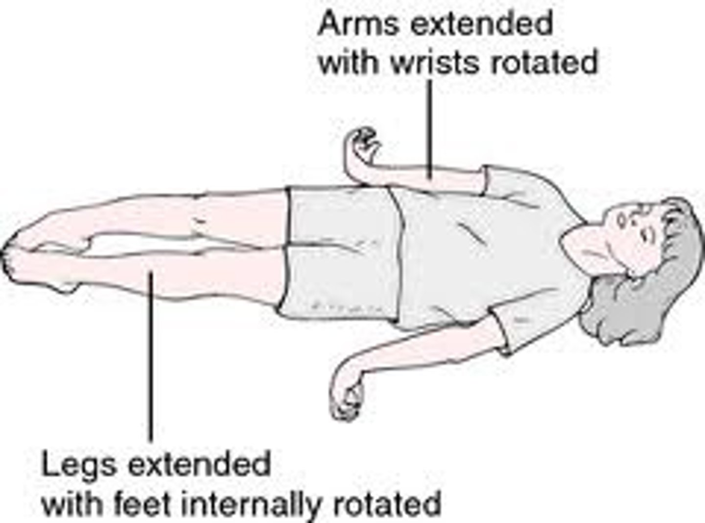
decerebrate rigidity
a severe neurological posture caused by damage to the brain stem (usually the midbrain) resulting in no input from the CST and RuST thus presenting with tonic extension of arms and legs
descending influences on cord motoneurons (CST,RuST) removed
what descending motor influences are lost during a decerebrate lesion
reticulospinal and vestibulospinal tracts
what 2 descending motor tracts are still intact with a decerebrate lesion
without cortical modulation from the CST and RuST, the reticulospinal and vestibulospinal tracts are constantly active having excitatory effects on extensor muscles for posture and balance
why do patients with a decerebrate lesion present with tonic extension of arms and legs
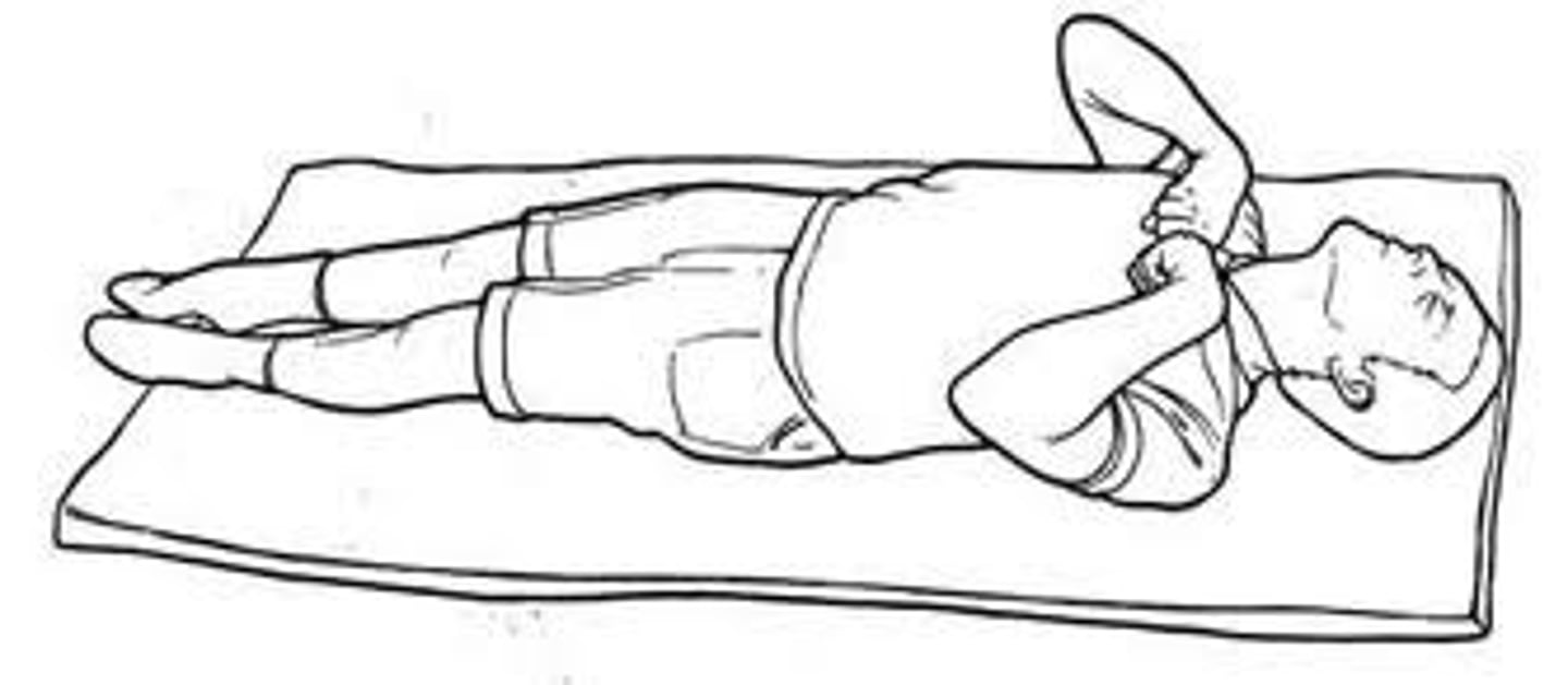
decorticate rigidity
an involuntary, abnormal reflex pose indicating severe damage to the brain's cerebral hemispheres, inner capsule, or thalamus. It is characterized by rigid, bent arms drawn toward the chest ("core"), clenched fists, and straight, internally rotated
removed cortical input to the Red nucleus (transaction of rostral to superior colliculus, brain injury or tumor)
decorticate rigidity results in:
the corticospinal tract
what descending motor pathway is cut off due to decorticate rigidity
- rubrospinal tract
- vestibulospinal tract
- reticulospinal tract
what 3 descending motor pathways are still intact with decorticate rigidity
red nucleus
what major brainstem structure of the rubrospinal tract is responsible for excitation of proximal UE flexors and is overactive in a decorticate lesion
increased flexor tone due to intact rubrospinal system that still activates proximal flexors of UE.
what happens to motor outputs as a result of loss of the upper corticospinal tract due to decorticate rigidity
Transtentorial herniation
what can cause decorticate posturing to progress to decerebrate posturing
Decerebrate posturing
what is this called?
decorticate posturing
what is this called?
layer V of cerebral cortex subserving motor function (topographically organized with large pyramidal neurons)
what is the origins of the corticospinal system
corona radiata and internal capsule down through the brainstem
through what pathways does cortical communication occur between cortical areas for motor activity
- 80-90% decussate in caudal medulla; descend in lateral funiculus
- 10-20% uncrossed, ipsilateral descent in anterior funiculus
once the descending motor cortical pathways of the corticospinal tract pass through the corona radiata and internal capsule down through the brainstem… what happens to the descending motor pathways
descend in lateral funiculus
through which funiculus do the corticospinal fibers that decussate in the caudal medulla descend
ipsilateral descent in anterior funiculus
through which funiculus do the corticospinal fibers that do not decussate descend
- interneurons in laminae IV-VII
- alpha motoneuron in lamina IX to distal flexor muscles
what are the 2 termination sites for the corticospinal tract
the cervical enlargement
in what part of the cervical spine does the corticospinal tract terminate
the lumbar enlargement
in what part of the lumbosacral spine does the corticospinal tract terminate
feet → legs → trunk → arms → hands → face
(medial to lateral) what is the topographical organization of the primary motor cortex (M1)
true
true or false: Deficits post-stroke match arterial & topographical arrangements
base of brainstem in medulla
where does decussation of the upper corticospinal tract occur
1. Anterior - anterior funiculus - ipsilateral descent
2. Lateral - lateral funiculus - contralateral descent
what are the 2 parts of the corticospinal tract on the spinal cord
anterior funiculus
within what funiculus does the anterior corticospinal tract descend
lateral funiculus
within what funiculus does the lateral corticospinal tract descend
ipsilateral
is the anterior corticospinal tract an ipsilateral or contralateral pathway
contralateral
is the lateral corticospinal tract an ipsilateral or contralateral pathway
anterior corticospinal tract
which pathway of the corticospinal tract is an ipsilateral pathway
lateral corticospinal tract
which pathway of the corticospinal tract is a contralateral pathway
Corticonuclear (corticobulbar) system
a two-neuron white matter pathway that carries motor signals from the cerebral cortex to cranial nerve nuclei in the brainstem.
voluntary control of muscles of the face, head, and neck, including chewing, facial expression, swallowing, and speech.
what is the function of the Corticonuclear (corticobulbar) system
layer V of cerebral cortex subserving motor function for the face (face motor cortex)
what is the origin of the Corticonuclear (corticobulbar) system
Corticonuclear (corticobulbar) system
what corticospinal pathway originates in layer V of cerebral cortex subserving motor function for the face (face motor cortex)
The genu of internal capsule and descend into the pons and medulla (located medial to the corticospinal tract)
what pathway does the Corticonuclear (corticobulbar) system take from layer V of cerebral cortex subserving motor function for the face to the motor nuclei or through nearby reticular formation interneurons to nuclei of V, VII, IX and X (both via nu ambiguus), XI and XII (i.e. motor nuclei)
Corticonuclear (corticobulbar) system
what corticospinal pathway passes Through the genu of internal capsule and descend into the pons and medulla (located medial to the corticospinal tract) to the motor nuclei responsible for facial movement
motor nuclei responsible for facial movement
what is the target for the Corticonuclear (corticobulbar) system
nuclei of V, VII, IX and X (both via nu ambiguus), XI and XII (i.e. motor nuclei)
what motor nuclei are responsible for facial and head movements and are the target nuclei of the Corticonuclear (corticobulbar) system
reticular formation interneurons
if the Corticonuclear (corticobulbar) system takes the indirect rout through the genu of internal capsule and descend into the pons and medulla (located medial to the corticospinal tract)…..what structure does it pass through before reaching the nuclei of V, VII, IX and X (both via nu ambiguus), XI and XII (i.e. motor nuclei)
- trigeminal nucleus
- facial nucleus
- nucleus ambiguous
- hypoglossal nucleus
- spinal accessory nucleus
what motor nuclei are part of the corticobulbar tract