a level biology, histology
1/29
There's no tags or description
Looks like no tags are added yet.
Name | Mastery | Learn | Test | Matching | Spaced |
|---|
No study sessions yet.
30 Terms
turn

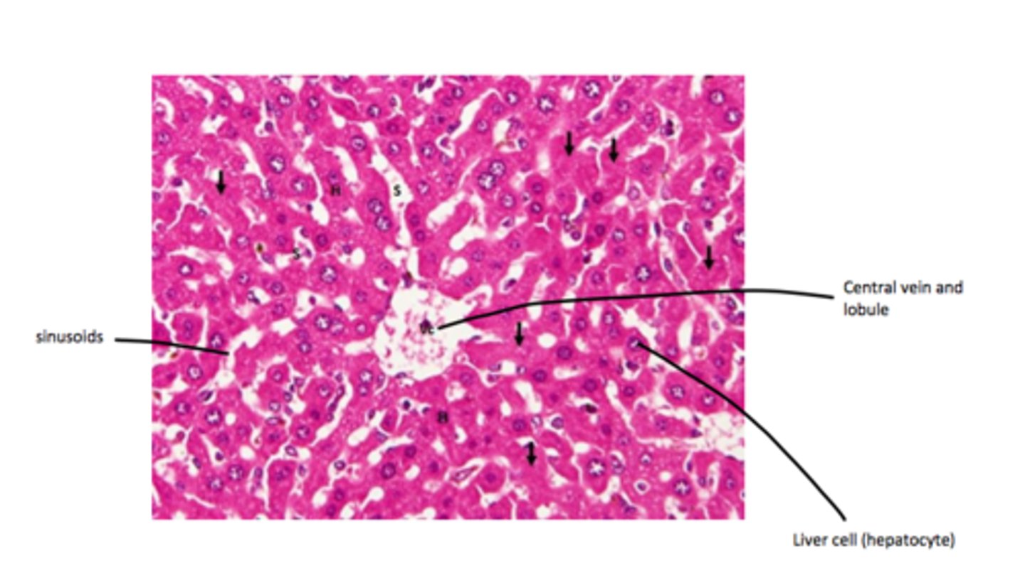
liver tissue
label each part
turn
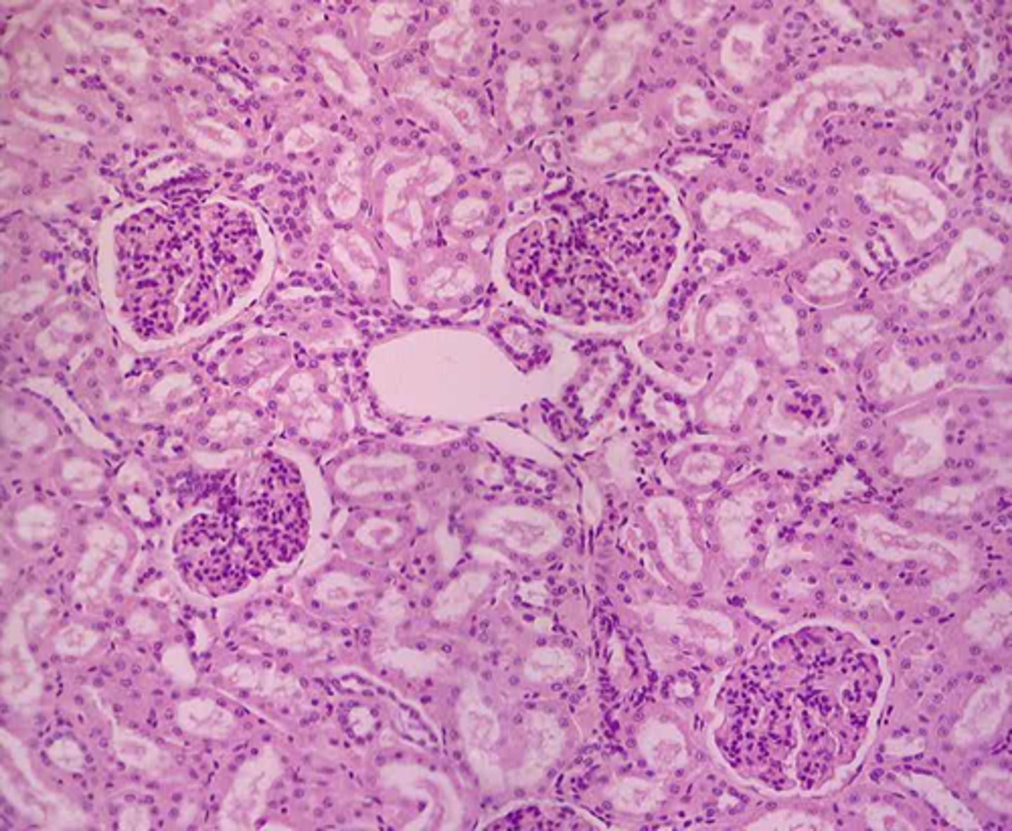
nephron
label each part
turn
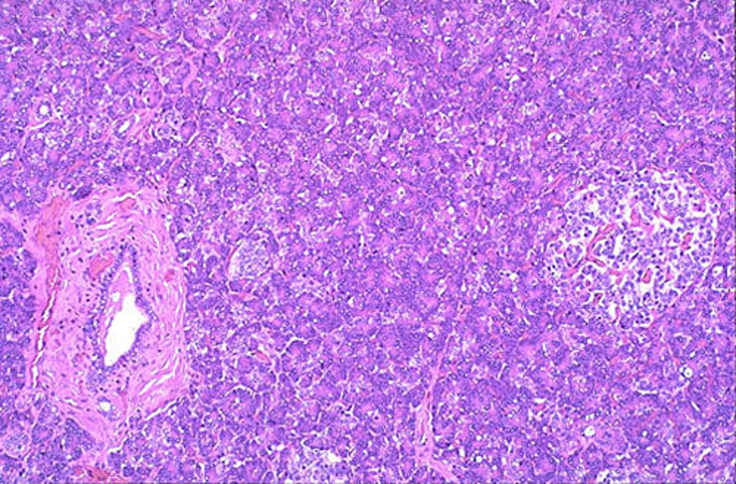
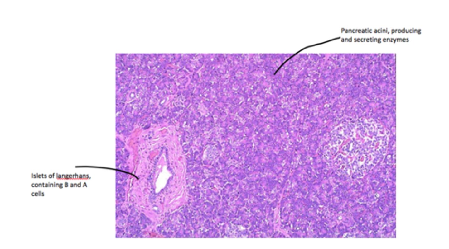
pancreas
label each part
turn

tracheae
what can you see in this micrograph?
walls of chitin,spiraclesandtrachae

turn
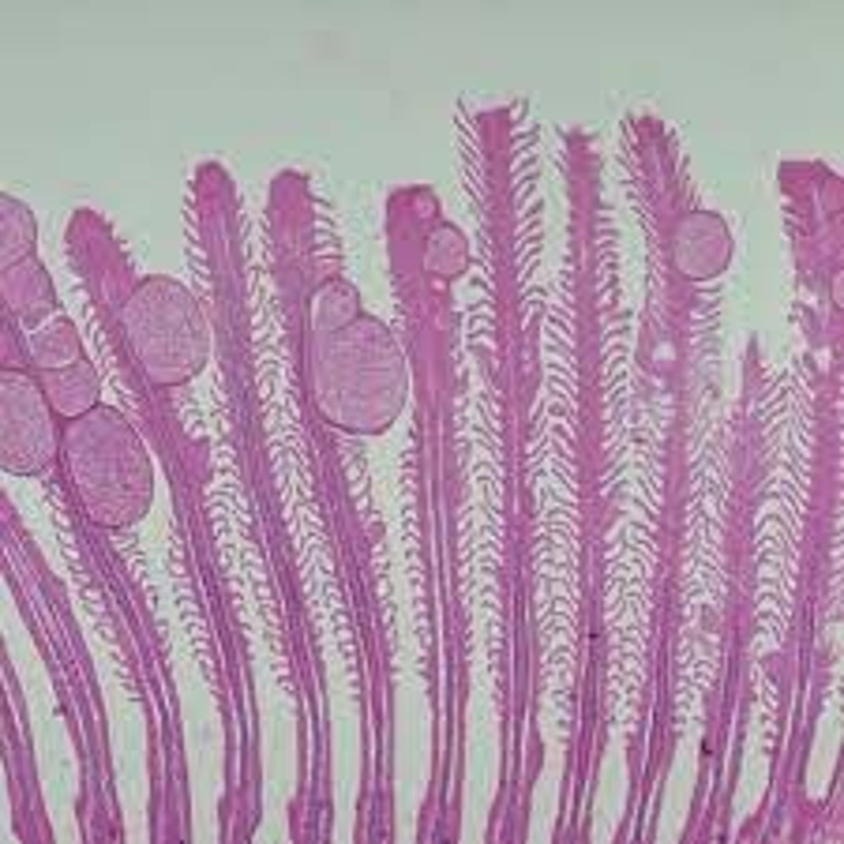
gills
turn
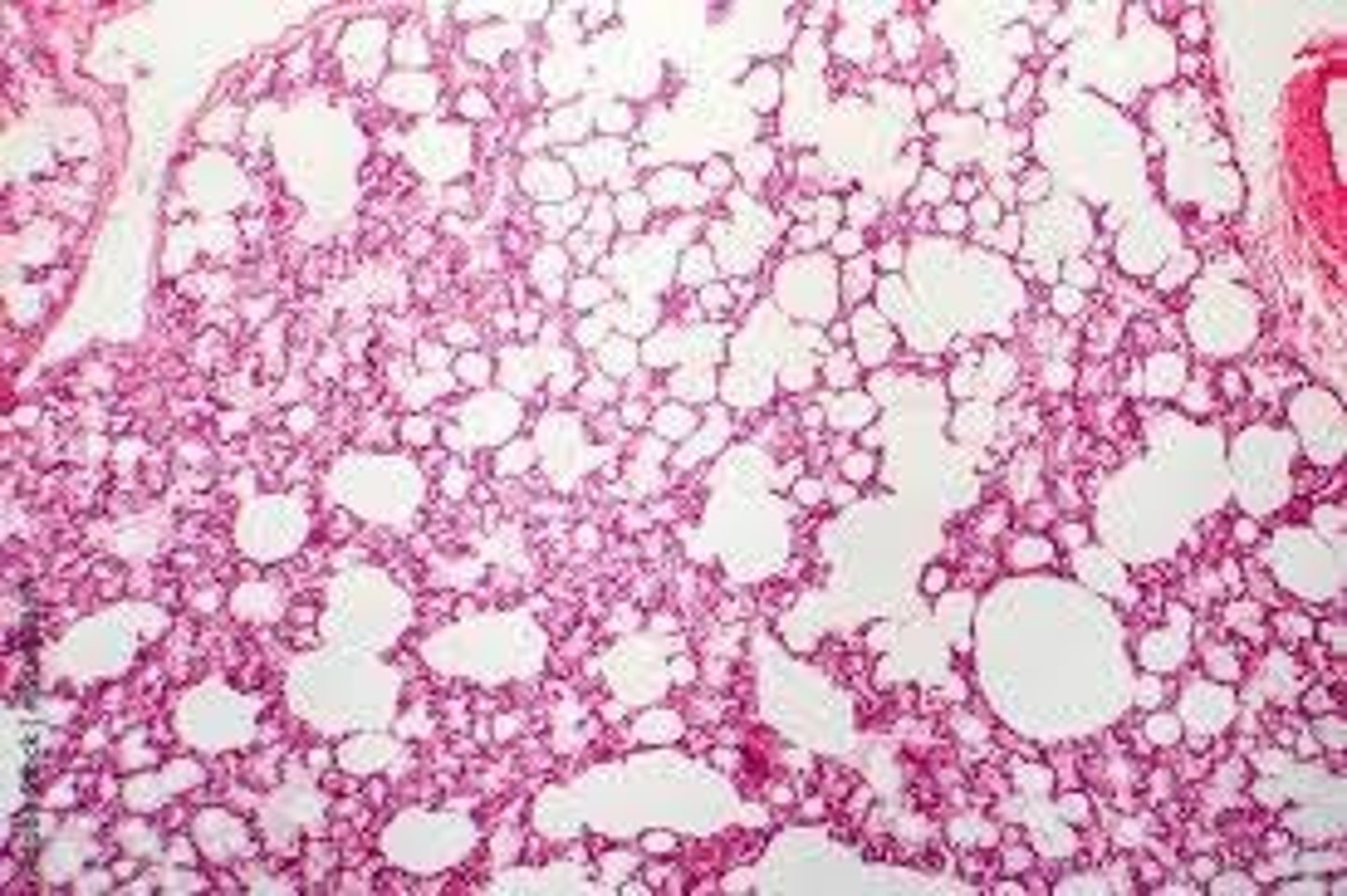
alveoli - the small air sacs
turn
cardiac muscle
turn
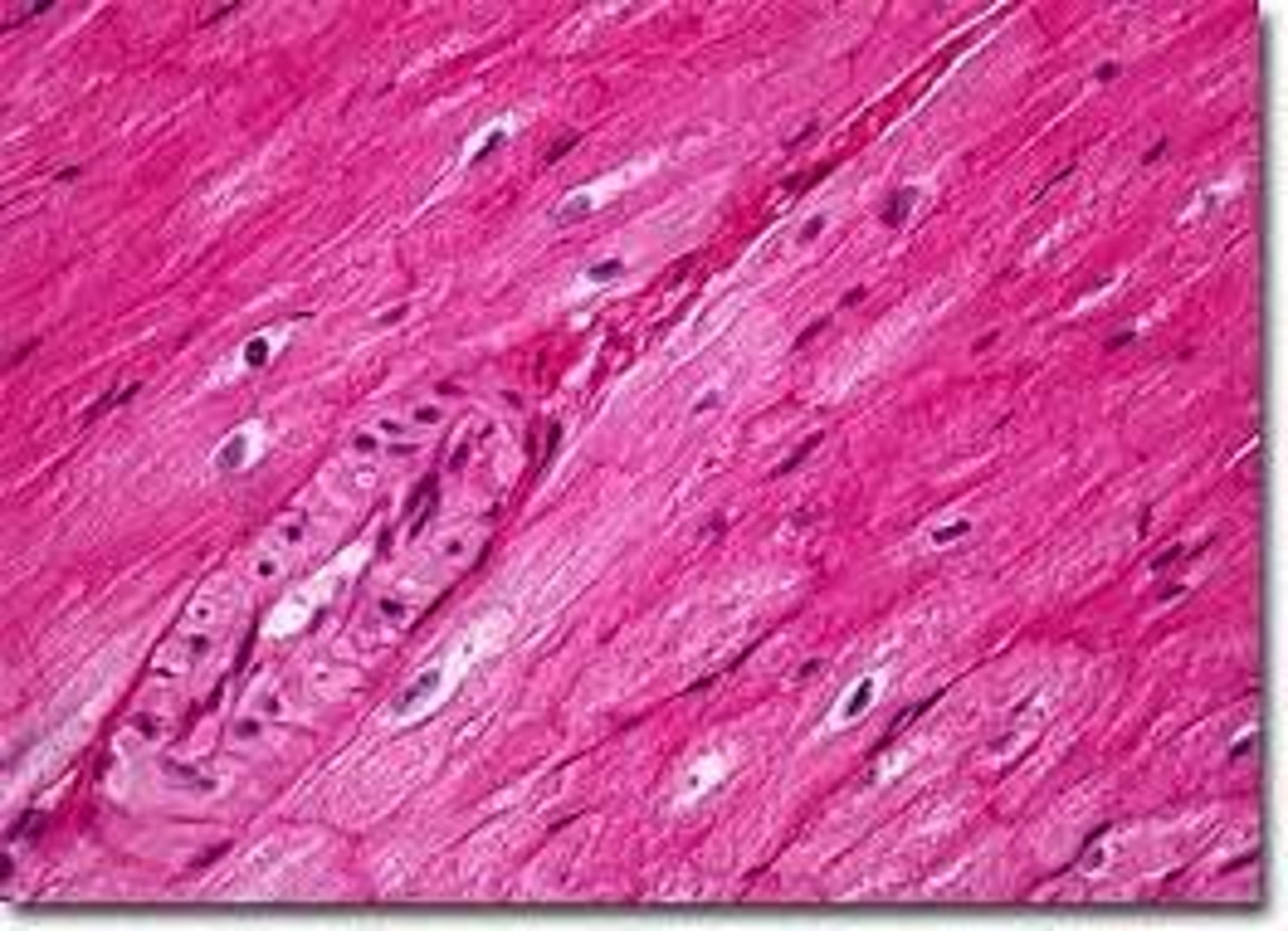
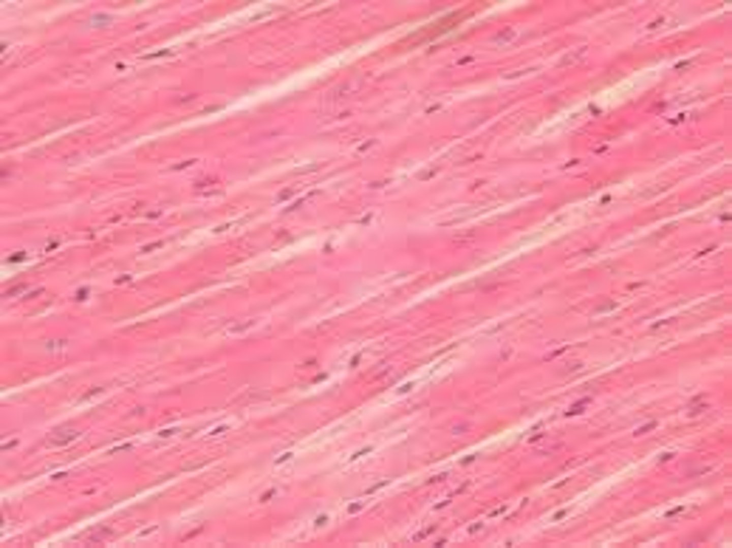
involuntary muscle
turn
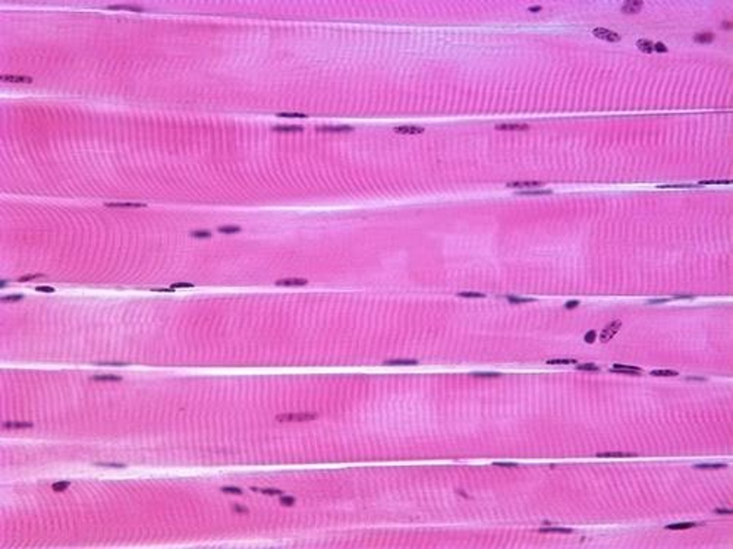
skeletal muscle
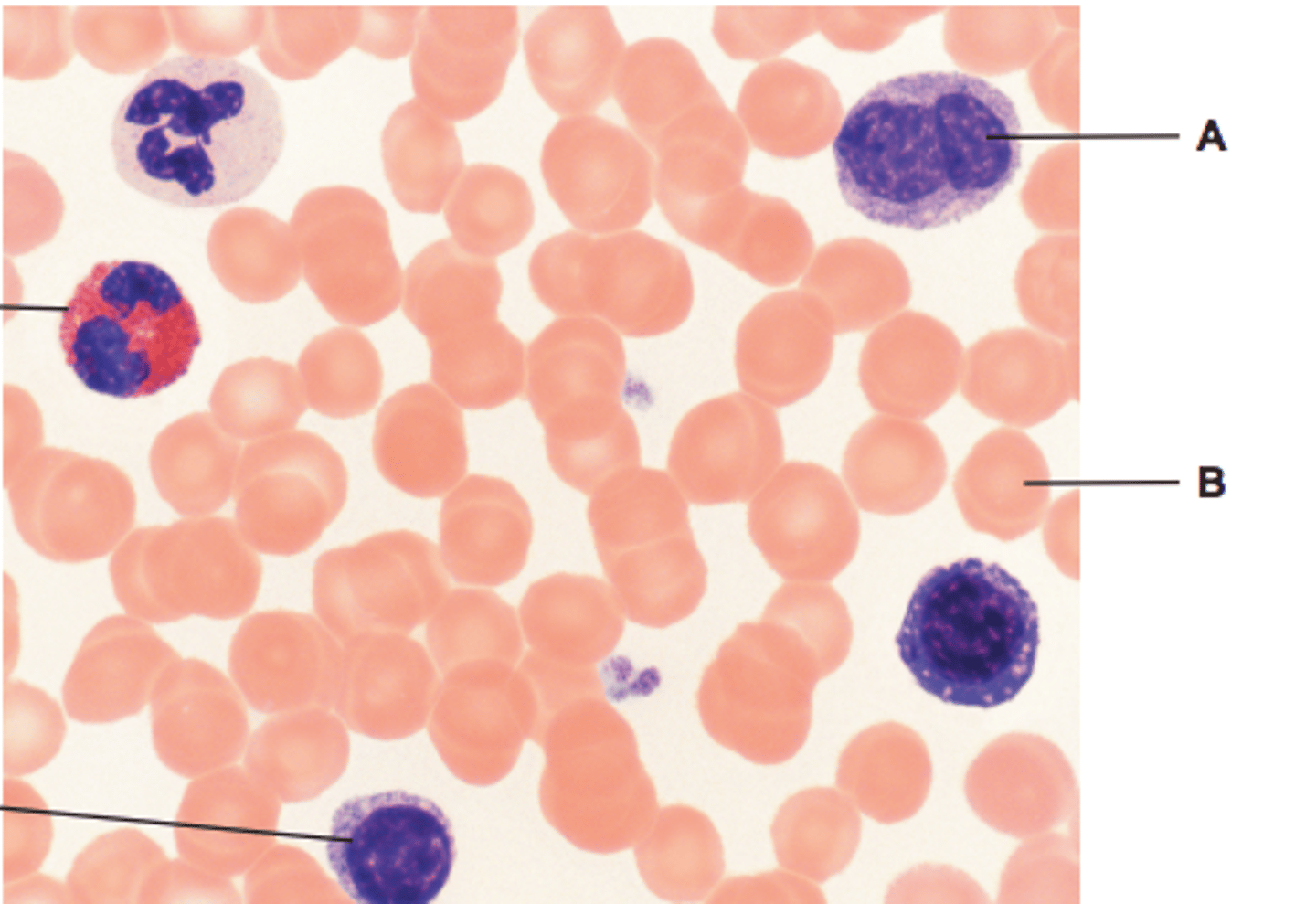
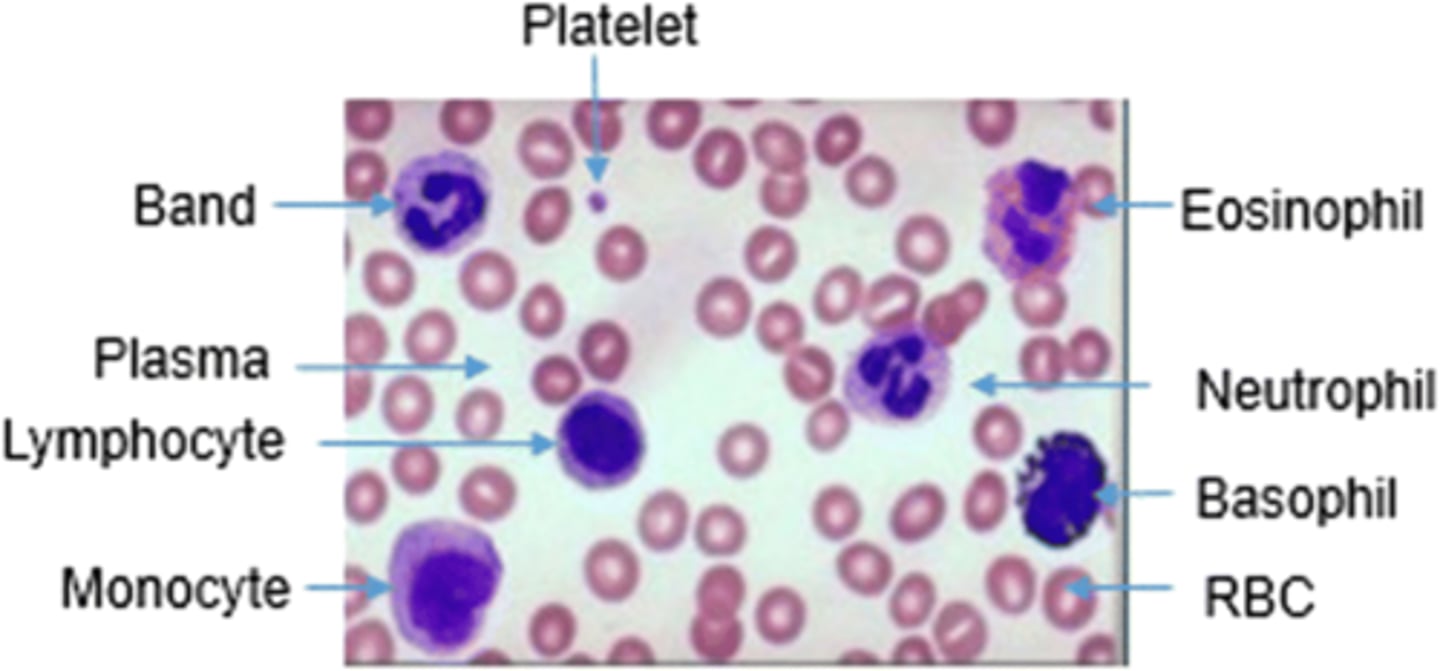
blood smear
identify A and B
lighter colour cells = erythrocytes
darker colour cells = neutrophils
turn
label A & B
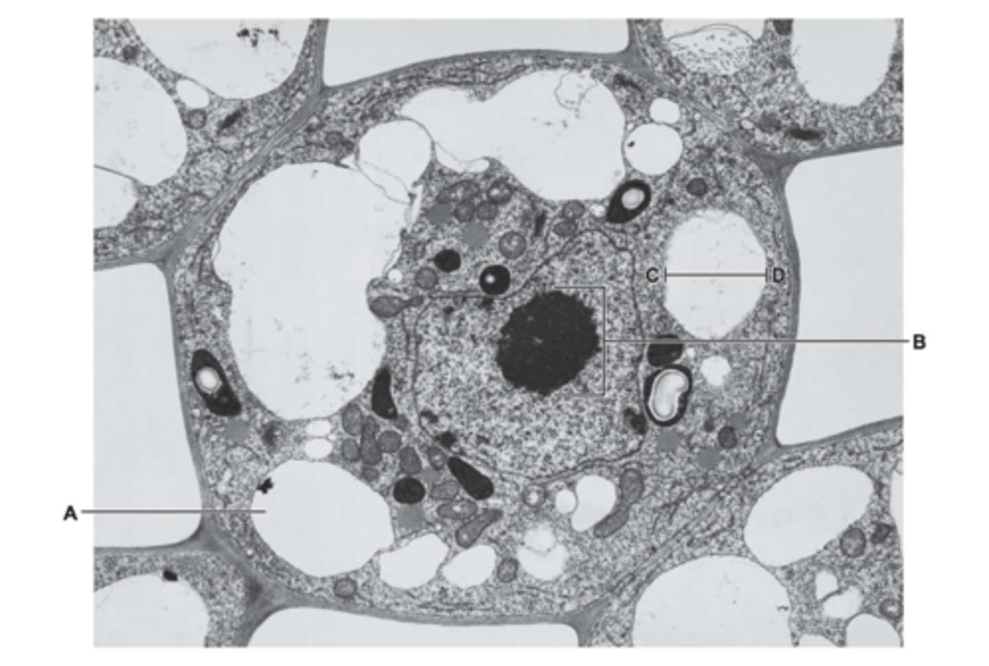
A - vacuole
B - nucleolus, not nucleus
turn
name A-F
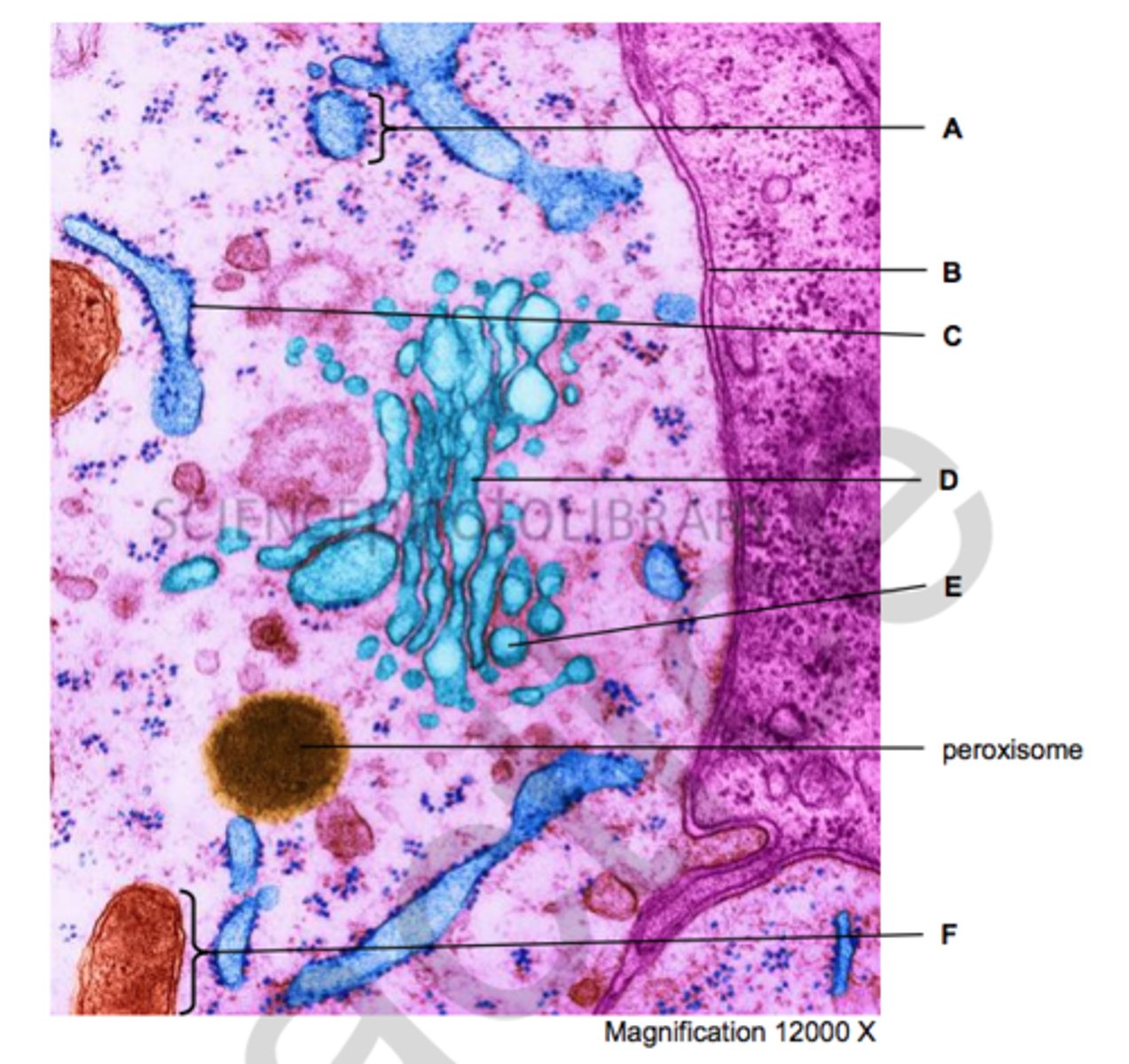
A - rough endoplasmic reticulum
B - plasma membrane
C - ribosome
D - golgi apparatus
E - secretory vesicle
F - mitochondria
turn
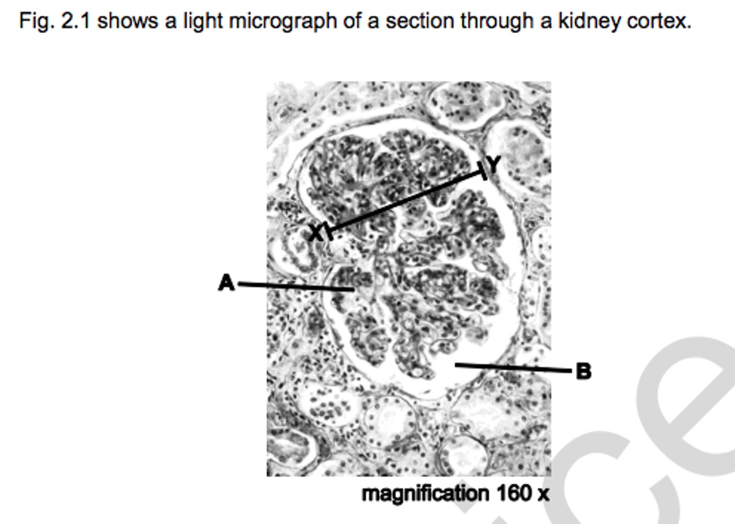
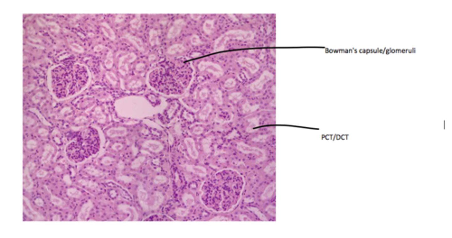
A - glomerulus
B - bowman's capsule
turn
name W, X, Y, Z
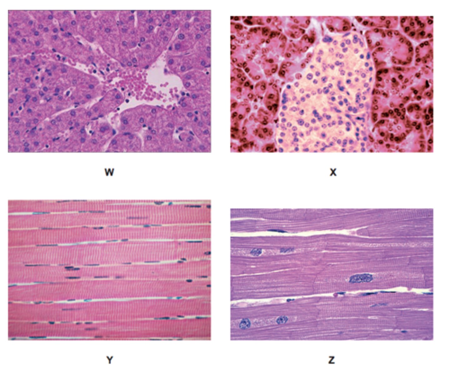
W liver / hepatic
X pancreas / pancreatic
Y skeletal / striated , muscle
Z cardiac muscle
turn
what stageisAin
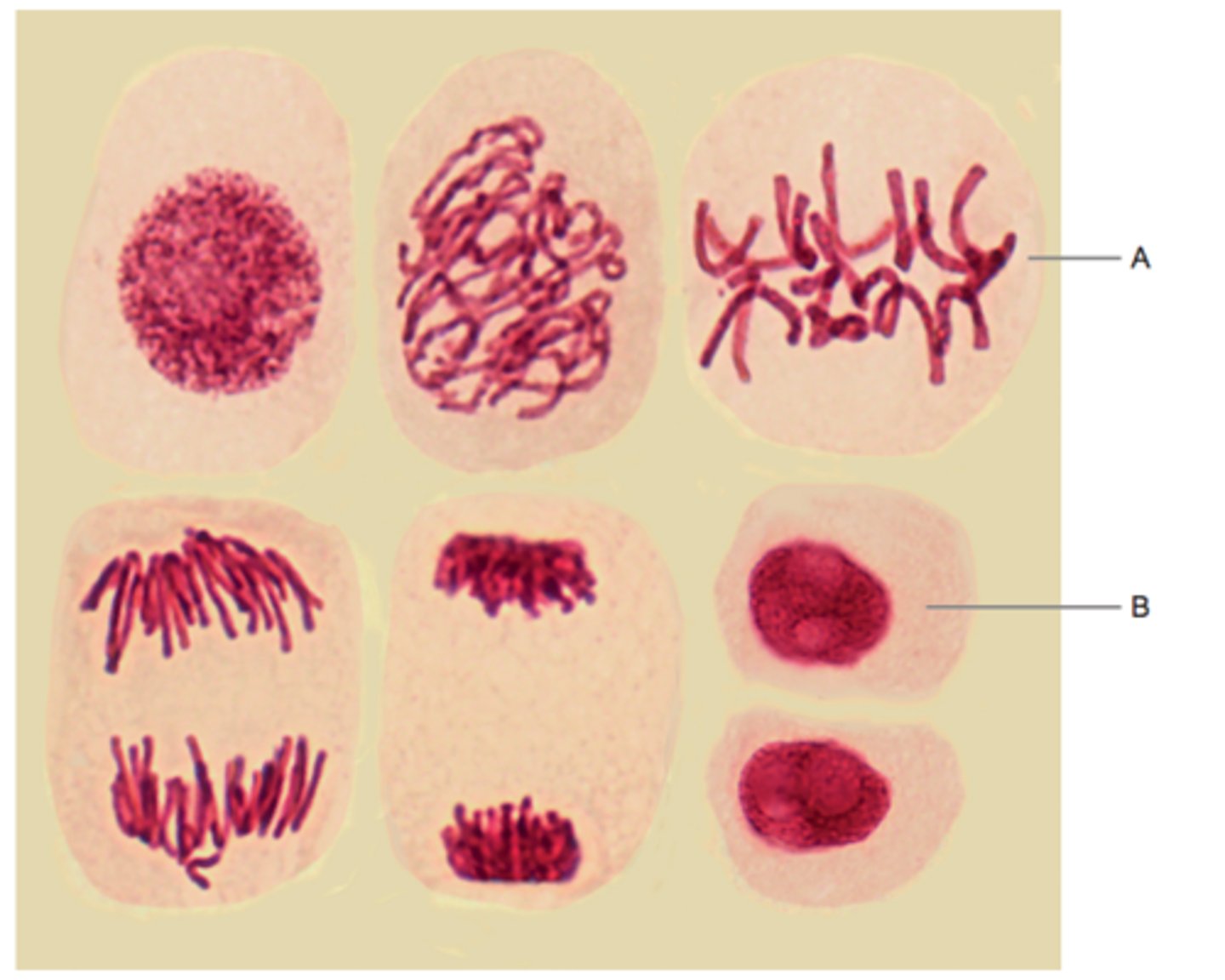
A - metaphase
turn
name A-D
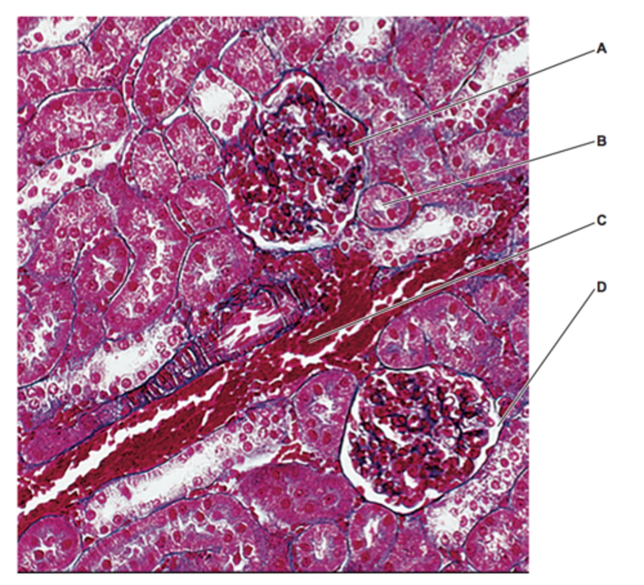
A - glomeruli
B - PCT
C - renal artery
D - bowman's capsule
(these are my guesses as the mark scheme don't have the actual answers)
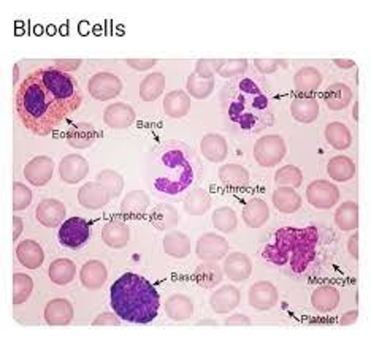
name A - E
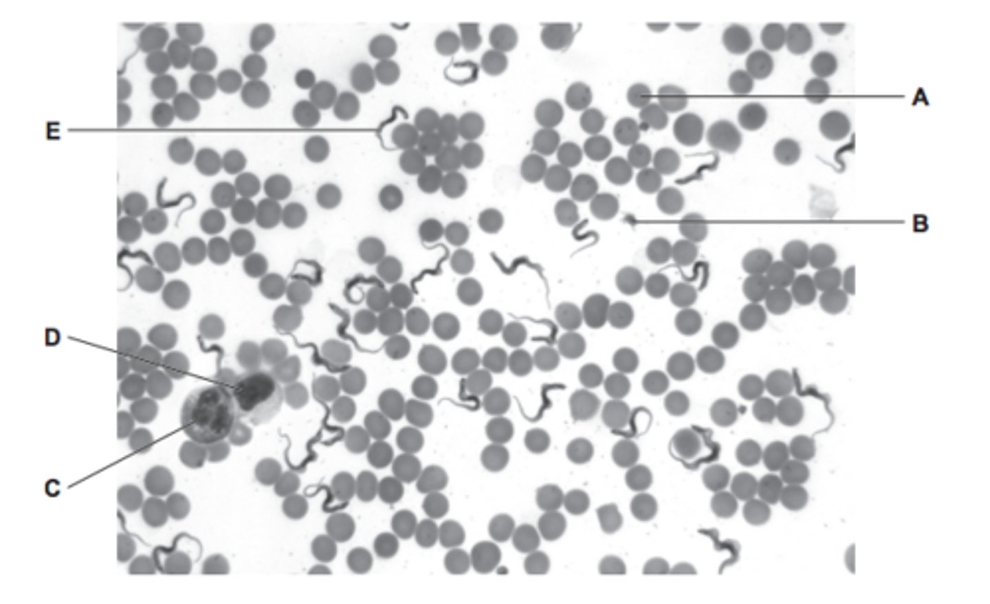
A - erythrocyte
B - platelet
C - neutrophil (lobed nucleus)
D - lymphocyte (large nucleus, takes up most of cell)
E - protist
how do you distinguish between a monocyte and lymphocyte under a microscope?
monocyte has a kidney bean nucleus, lymphocyte have a large nucleus that takes up most the cell
turn
name A-F
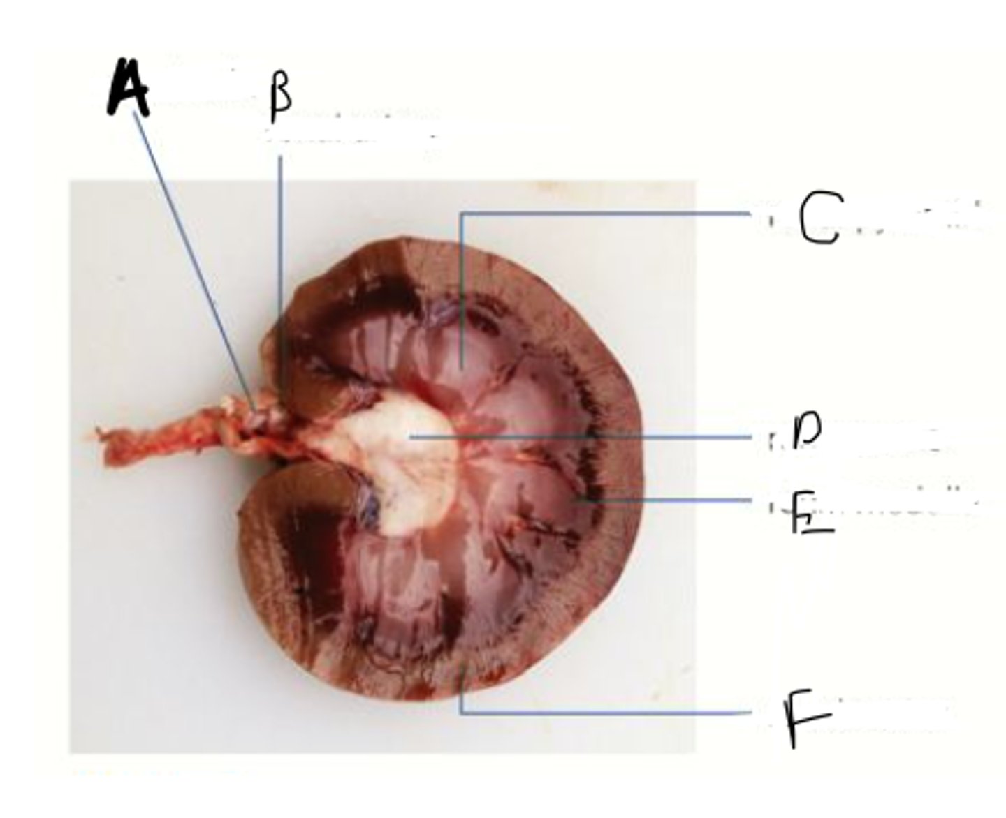
A - renal vein
B - renal artery
C - renal pyramid
D - renal pelvis
E - renal medulla
F - renal cortex
turn
name A & B
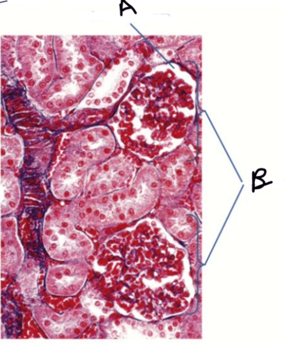
A - bowmans capsule
B - glomeruli
how do you distinguish between the PCT and DCT under a microscope?
PCT will have microvilli whereas DCT will not