Stomach, Sm Intestine
1/17
There's no tags or description
Looks like no tags are added yet.
Name | Mastery | Learn | Test | Matching | Spaced | Call with Kai |
|---|
No analytics yet
Send a link to your students to track their progress
18 Terms
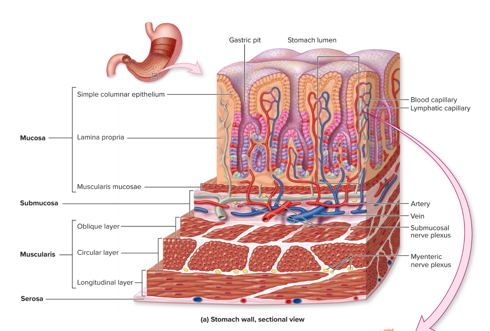
GI Tract histology: 4 layers of the gut wall
inner — outer
Mucosa → secrete mucus and fluid into the lumen
Submucosa → includes blood and lymphatic vessels that transport absorbed nutrients and a scattering of submucosal glands that release digestive secretions.
Muscularis → promote mechanical digestion, expose more of the food to digestive chemicals, and move the food along the canal.
Serosa → protective layer for internal organs, lubcrication
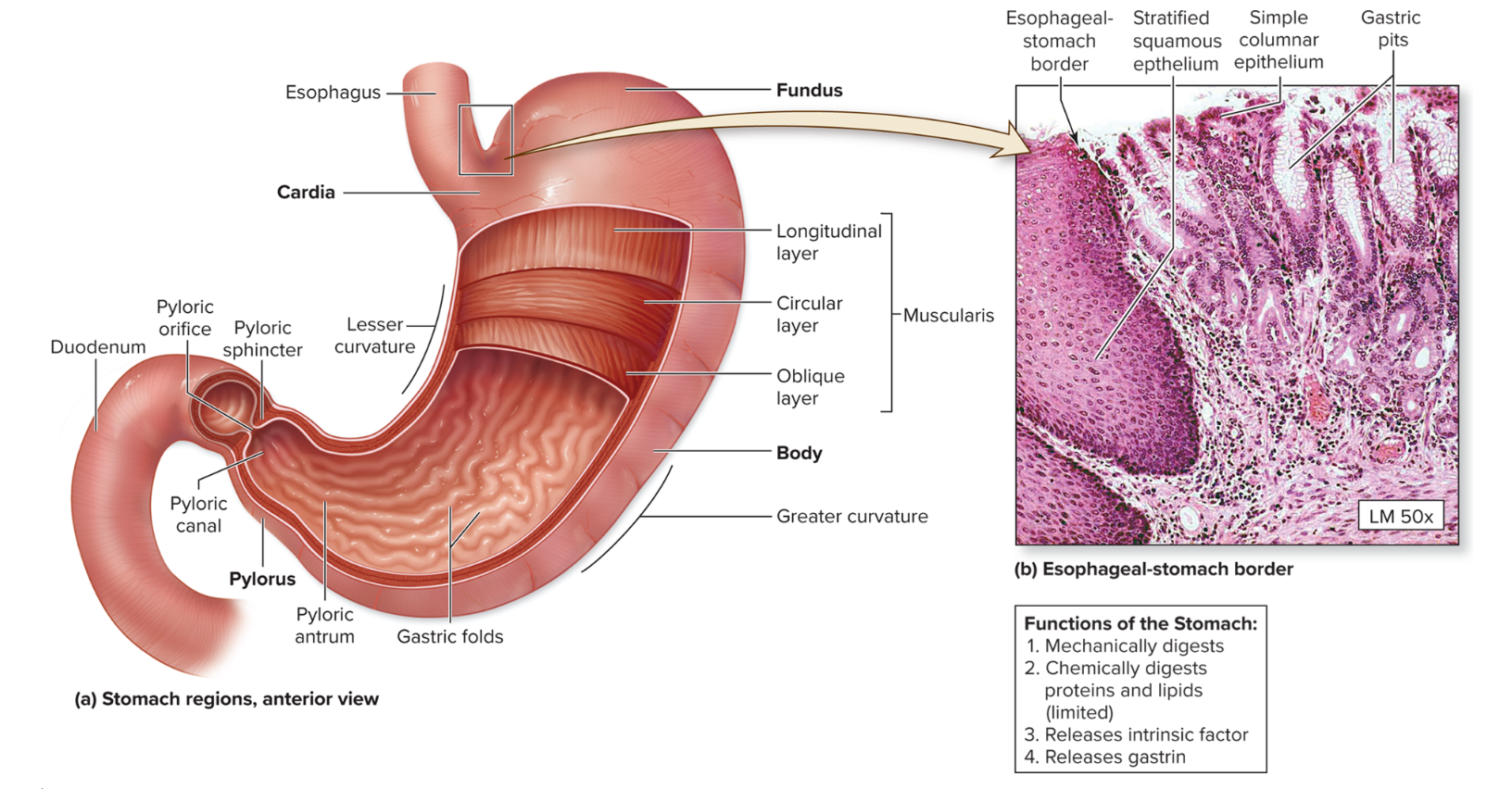
Stomach
lined with simple columnar: tunica muscularis had a 3rd layer (oblique) inside the usual circular and longitudinal muscle layers
watery contents = chyme
4 regions(in order): Cardia, fundus, body, and pyloris

Gastric glands
secretory cells within gastric pits
Parietal cells: HCI and intrinsic factor—B12 binding
Chief cells: pepsinogen
G cells: enteroendocrine cells
→ secrete gastrin which stimulates above cells

Small intestine
20 ft long!
90% of nutrients are absorbed here due to the high amounts of mucosal surface area

Small intestine: Plicae circularis
permanent internal ridges visible to the naked eye.
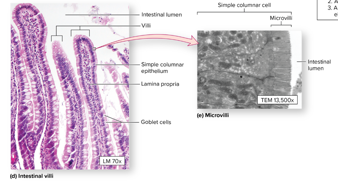
Villi
small, finger-like projections — lined with simple columnar epithelium
microvilli on apical surfaces

Lacteals
part of the lymph system that absorbs lipids

Intestinal glands (Crypts of Lieberkuhn)
found near the base of intestinal villi; secreting hormones and enzymes
Goblet cells: secrete protective mucus
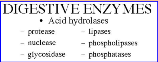
Digestive enzymes
from the pancreas and intestinal glands
digest all classes of molecules
What protects the lining from being digested?
Goblet cells (part of endothelium)
Brunner’s glands (deep into endothelium)
→ Both produce alkaline mucous
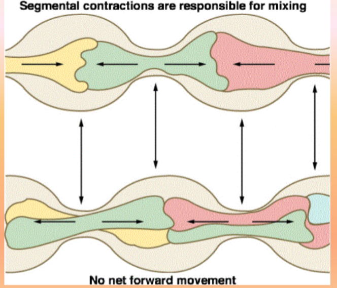
Segmentation
a movement characteristic of the small intestine
a churning motion that does NOT produce any net movement down the GI tract
mixes chyme with intestinal secretions
Peristalsis
starts in the esophagus, net movement down the GI tract
Peristaltic rush
diarrhea
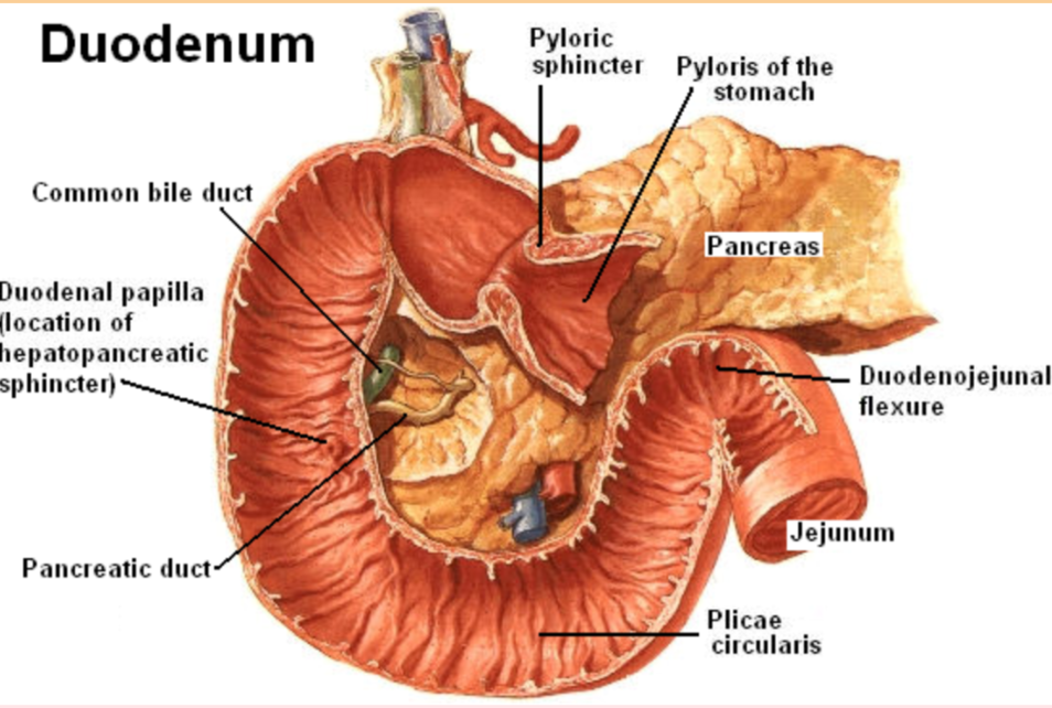
Duodenum
the first region begins after the pyloric sphincter and ends at the duodenojejunal flexure
is the shortest (~10in long) and widest segment of small intestine
forms a loop around the pancreas

Duodenum: Hepatopancreatic ampulla
where the common bile duct fuses w/ pancreatic duct
wide variation
Duodenal papilla w/ hepatopancreatic sphincter
opens to allow juices out, but keeps chyme from reversing back up
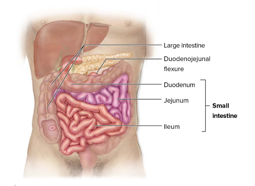
Jejunum
~8ft long; supported by mesentery
the bulk of chemical digestion and nutrient absorption occurs here

ileum
longest ~12ftl
continues absorption
ends at the ileocecal valve (controls rate of emptying)
large amount of MALT to counteract bacteria found in the large intestine → toward the end of small intestine
→ Mucosa Associated Lymphoid Tissue
MALT aggregates to form Peyer’s patches in the terminal region of the ileum