B3 gas exchange
1/50
There's no tags or description
Looks like no tags are added yet.
Name | Mastery | Learn | Test | Matching | Spaced |
|---|
No study sessions yet.
51 Terms
Organisms with ____ can rely on diffusion directly for gas exchange
high SA-V
Usually unicellular, like yeast, bacteria
What are 3 gas exchange surfaces
Alveoli in lungs of mammals, birds, reptiles
Gills in fish
Tracheal system in insects
Properties of gas exchange surfaces (5)
Thin (often 1 cell) → short diffusion distances
Moist → helps dissolve gases before they diffuse across the exchange surface (ex. Alveolar fluid)
Large SA → maximize diffusion as there is more membrane surface available
Permeable → must have pores allowing gases to be exchanged across the surface (ex. stomata)
Concentration gradient → diffusion involves difference in [ ] of the two gases
Describe how concentration gradients are used for gas exchange in gills and lungs
Gills
2 fluids involved: water passing over the gills, blood inside gill capillary
More O2 must be in water vs. blood → for diffusion into the blood
Animals
Concentration of O2 in lung capillaries is lower than air inspired into lungs
Concentration of CO2 in lung capillaries is higher than in air
Thus, diffusion gradients take a place
Adaptations at exchange surfaces to maintain steep concentration gradients? (3)
Ventilation → ensures the air or water rich in the gas is moved across exchange surface
Water (air) must be continuously passed over gills/ventilated in lungs
Continuous blood flow → ensuring that substances are immediately transported away when entering the blood
Ensures a low [ ] of that substance in the blood supply in relation to exchange surface
Dense network of blood vessels → many opportunities for substances to be exchanged between surface and blood
What is purpose of alveolar fluid/surfactant
Secreted by type II pneumocytes
Moistens the alveoli surface, allowing gases to dissolve before diffusing into the blood
Made of lipids + proteins
Reduces surface tension of moist inner surface and prevents alveolus from collapsing each time air is expired
What is the structure of bronchiole, why is it important
Each bronchiole branches into alveoli
Increases the SA for gas exchange = boosts the rate
Ensures air is distributed properly
Small diameter of bronchioles (compared to trachaea/bronchi) slows down rate of airflow → increases efficiency of gas exchange
What is the main purpose of capillary beds being around alveoli?
short diffusion distance
What are the main structures involved in lung ventilation
Diaphragm = sheet of muscle below ribs
Intercostal muscles = group of muscles between/anchored to ribs (internal and external, they are antagonistic)
Abdominal muscles
Tissue that makes up lungs is passive and not muscular (cannot purposefully move)
Breathing is based on _____ law
Boyle’s
An increase in V = decrease in pressure
Describe inspiration processes, 5 step
1. Diaphragm contracts to move downwards
2. External intercostal muscles and one set of abdominal muscles contract to raise rib cage, causing rib cage to move up
3. Since thoracic cavity has inc in V, pressure inside thoracic cavity decreases
4. Lung tissue responds to lower pressure by increasing its volume
5. Leads to a decrease in pressure inside the lungs (partial vacuum). Air comes in to counter this and fills alveoli
Describe the parts in lung volume
Measured using a spirometer
1. Tidal volume (air breathed in/out in one cycle/normal breath)
2. Inspiratory reserve volume (max volume of air that a person can breathe in)
3. Expiratory reserve volume (max volume of air that a person can breahte out)
4. Vital capacity (sum of inspiratory + tidal + expiratory)
*residual volume is the air in lungs that cannot be further exhaled, preventing lungs from collapsing
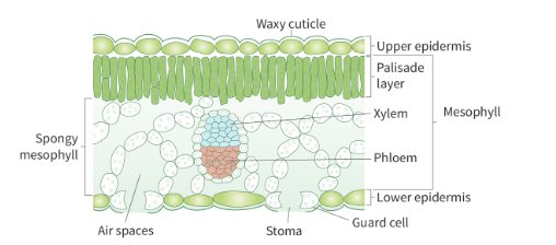
Leaf adaptations for efficient gas exchange (5)
1. Waxy cuticle (covers cells of upper epidermis, hydrophobic, prevents water loss and dehydration)
2. Epidermis (regulates exchange of gases between leaf and air using stomata)
Stomata: numerous microscopic openings on the lower surface of leaves. Each stoma = 2 guard cells, which can open or close.
When open = permit CO2 to enter, and H2O and O2 to exit (diffusion) → rate of gas exchange increases
Location on lower surface limits water loss due to transpiration
Stomata are also unprotected against physical factors (rain)
3. Palisade mesophyll: densely packed cylindrical cells in upper portion of leaf. Has many chloroplasts and located to get maximum sunlight for photosynthesis
4. Spongy mesophyll: loosely packed cells below palisade layer. Few chloroplasts and many air spaces → large SA for gas exchange
5. Thin and flat → large SA for gas diffusion and sunlight absorption
Define transpiration, how does it work, its function
Definition: the evaporation/loss of water through open stomata
Since there is more [ ] H2O in the leaf than outside, this water vapour diffuses out through stomata
Diffusion results in negative pressure (pulls on H2O in xylem due to capillary action, which is cohesive + adhesive properties of water)
A result of a leaf’s function to accomplish photosynthesis
Transpiration not only allows a plant to absorb water/nutrients from soil and transport it throughout
Also regulates temperature
Factors affecting transpiration (4)
Environmental factor | explanation |
Light intensity | When higher, guard cells cause stomata to open wider (for CO2 to enter for photosynthesis)
|
Temperature | More kinetic energy will be gained by H2O, so they can diffuse out of stomata Also increases saturation point of the atmospheric air, so it can hold more H2O vapour |
Wind intensity | The higher it is, the faster H2O are moved away from the leaf after transpiration
|
Humidity | Increased humidity, more H2O vapour in the air
|
Purpose of measuring stomata density
More stomata = higher rate of transpiration → indicate how efficiently a plant uses water
Changes in density in different environments understand effect of climate on plates
Similarities and differences indicate phylogenetic relationships
Density of past fossils indicate past environmental conditions
Describe haemologbin and its purpose
Haemoglobin = protein molecule in erythrocytes responsible for carrying most of oxygen
Purpose: transport O2 from lungs to tissues, transport CO2 from respiring tissues to lungs
Can reversibly bind to CO2 or O2
Composed of 4 polypeptides with a quaternary structure
Each polypeptide has a haem group with an iron atom
When binding to O2, it’s the iron that does it
Can bind to 4 O2 (saturated)
Describe structure and adaptations of foetal haemoglobin
Quaternary, 2 alpha + 2 gamma polypeptide chains, each has a haem group
Presence of gamma allows it to higher affinity for O2 (compared to mother haemoglobin)
Increases efficiency in which foetus gets O2 from mother blood across placenta
Important since O2 [ ] in foetal blood is lower than in the mother
Describe structure of adult haemoglobin
quaternary structure, 2 alpha + 2 beta polypeptide chains, each has a prosthetic group (haem) containing an iron
Has lower affinity to O2 than foetal
Since it has a higher affinity for (2,3 BPG) (organic phosphate) which competes with O2 for binding
What is myoglobin
*another type of haemoglobin is myoglobin, found in muscle tissue. Consists of 1 polypeptide chain and has much higher affinity for O2 than HbF or HbA
What is cooperative binding?
Any O2 bonded to haemoglobin subunit increases its affinity for more O2 (conformational change)
Called cooperative binding since oxygen molecules interact with each other to increase its affinity
haemoglobin with ____ O2 has the greatest affinity for O2
3
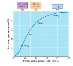
Describe shape of oxygen dissociation curve
Y axis = % of haemoglobin saturation with O2
X axis = partial pressure (concentration) of oxygen
This varies depending on where the blood is
Cooperative binding of haemoglobin to O2 results in a sigmoidal shape (otherwise would be linear)
Rate of O2 intake by haemoglobin increases rapidly
However, it eventually levels off as it becomes fully saturated with o2 in areas of higher O2 partial pressure (ex. lungs)
Define partial pressure
Definition = the pressure exerted by a single gas within a mixture of gases
This partial pressure of O2 decreases in the body and blood as it’s used for aerobic cellular respiration
How is CO2 transported in body?
Most is dissolved in plasma (carbonic acid, low pH), small amount binds to haemoglobin
What molecule can undergo allosteric binding to haemoglobin? describe. Why is this important?
CO2 binds to allosteric region of haemoglobin (polypeptide regions) whereas O2 binds to haem group
Allostery → when binding of CO2 to polypeptide chains of haemoglobin results in a conformational change, decreasing haemoglobin's affinity for O2
Important to ensure that haemoglobin unloads O2 in areas of low partial pressure of O2 (respiring tissues)
Define bohr shift, why is it important
Definition: shifting the oxygen dissociation curve to the right with higher partial pressures of O2 in the blood
The change in affinity of haemoglobin for O2 in the presence of CO2
Essentially, when CO2 is present, haemoglobin has a greater tendency to give up O2
why important: Due to decreased affinity for O2, haemoglobin releases more O2 into tissues with higher partial pressures of CO2 (respiring muscles) → important while exercising
Describe the 2 types of alveolar cells
Type I pneumocytes | Type II pneumocytes |
Cover 95% alveolar surface | Cover 5% alveolar surface, found between type I |
Function: allow gas exchange between alveoli and capillaries | Function: produce pulmonary surfactant (Reduces surface tension to prevent alveoli from collapsing and sticking while breathing) |
Adaptations
| Adaptions
|
Capillary structure
small, thin walled blood vessels for material exchange between blood and itnernal/external environment
Highly branched (more SA-V, and slows flow of blood for more time exchanging materials)
Wall is 1 cell thick (endothelial cells) → short diffusion distance
Has a basement membrane (thin layer of ECM providing structural support to endothelium to regulate material exchange)
2 types of capillaries
Most capillaries are continuous (complete endothelial lining)
Allows selective exchange of small molecules/ions
Other capillaries are fenestrated
Definition = gaps in endothelial cells allow for more rapid exchange of materials
Found in organs with high metabolic demands (kidney, small intestine)
Capillary adaptations
Small inside diameter
Thin walled
Permeable
Large SA
Fenestrations
Describe capillary beds and their functions
All chemical exchanges in lungs and body tissues occurs in capillary beds
Structure
Capillaries receive blood from arterioles (smallest artery)
Arteriole branches into capillary bed within body tissues (network of capillaries receiving blood from same arteriole)
Capillary bed drains blood into venules (smallest veins)
Some metabolically active tissues in the body have more capillary beds (highly vascular tissue)
Some tissues have capillary beds that are more permeable to substances (fenestrated)
Small slits allowing large molecules to pass, and faster movement
Examples: capillaries in kidneys and intestine
When blood enters a capillary bed → much of pressure and velocity is lost
Blood cells line in a single file because the lumen of capillary is small
Capillary bed is extensively branched leading to large SA, so it can reach body cells
What is the artery’s purpose and adaptations
Transports high pressure blood away from the heart
Main one is the aorta → branches into smaller arteries → branches into arterioles → connects to capillaries for substance exchange
Adaptations
Wall is made of smooth muscle
Regulates diameter of artery/blood flow by contracting or relaxing based on signals (ex. Hormones, blood pressure)
Can contract → diameter of lumen decreases (vasoconstriction) to increase blood pressure
Can relax → diameter of lumen increases (vasodilation), decreasing blood pressure
Controlled by autonomic nervous system (ANS) → unconscious
Walls contain proteins (elastin and collagen)
Reduces fluctuations in blood pressure (prevents damage to smaller blood vessels)
When blood is pumped in → these fibres are stretched and allow blood vessel to accommodate more pressure
Once blood passed, the fibres recoil and provide pressure to propel the blood forwards
Thus, blood in arteries maintains high pressure during pumping
Muscle + elastic tissues permit withstanding of high pressure (ensure blood is moving)
small lumen
Describe veins and their adaptations
Return low pressure blood to the heart
Also has 3 layers (same as artery)
Blood vessels that return blood to heart after passing through a capillary bed
Adaptations
To account for the loss of pressure and velocity in blood in capillary beds
Thin walls → easily compressed by muscles to make blood pushed along
Ex. in the limbs during vigorous exercise
Large lumen (more blood flows at lower pressure)
Has one way valves → unidirectional flow to the heart → prevents backflow
Important in lower parts of body, like feet, when force of gravity is difficult for blood to flow back
Describe process of transpiration and its purpose
To transport water and minerals unidirectionally against gravity using xylem
Rely on tension force
From roots → other plant parts
Possible due to transpiration (loss of water from plants through stomata)
When water evaporates through stomata, a negative pressure potential is created/tension is created (lower water potential outside stomata vs. inside of leaf) at upper end of xylem tube
Results in water moving up xylem → entire water column due to cohesion moves up
Cohesion-tension theory
Describe structure of xylem
Structure: thin, continuous columns running from the roots through the stems
Formed from specialized cells (which lose their cell contents and membrane as they mature and die)
Dead → leave behind the cell wall
Have lignin for strength → binds to cellulose → provides strength and rigidity to withstand tension (due to tension in transpiration)
Also waterproofs the xylem
Even the end walls (where cells were joined to each other in the time) → degenerate
partial/total lack of these allows unobstructed water to flow upwards
The specialized cells that make up xylem walls contain pit areas
Where cell wall is thinner, no lignin
Allow water to move between adjacent cells
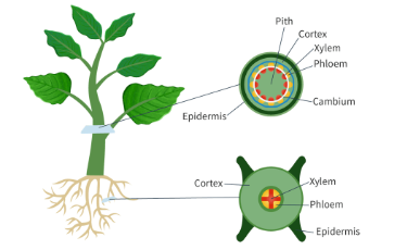
Describe the 5 main parts of stems and roots
Stem
Tissue | Function |
epidermis | Prevents water loss, protection from microorganisms |
cortex | Unspecialized cell layer, can store food |
xylem | Transport tubes that bring up water from roots |
Vascular bundle | Contains vessels of xylem and phloem |
phloem | Transports carbohydrates, usually leaves to other parts of the plant |
Root
Tissue | Function |
Epidermis | Grows root hairs that increase SA for water uptake |
cortex | Unspecialized cell layer that stores food reserves |
xylem | Transport tubes for water and minerals, starting in roots |
Phloem | Transport tubes that receive sugars from leaves |
Vascular bundle | Center of root, contains xylem and phloem |
What is tissue fluid?
Tissue fluid = made from blood plasma that is pushed through capillary walls into surrounding tissues
Contains water and small solutes (ions, hormones)
It bathes cells and facilitates exchange of substances between blood and cells
How is tissue fluid created
Amount of fluid/solutes pushed out of capillary wall is due to the hydrostatic pressure of blood on the wall
Arteriole end → hydrostatic pressure is high (fluid is pushed out of wall into tissue) due to ultrafiltration (pressure filtration)
This pressure is high enough to open gaps between cells making up capillary walls
Venule end → fluid has already been lost inside capillary, decreasing hydrostatic pressure → majority of tissue fluid draws back into capillaries (some amount doesn’t and enters lymph ducts)
Exchange between cells and tissue fluid can occur in 2 ways
Can occur through diffusion or active transport
What can leave capillaries and enter tissue fluid? What can’t?
RBC and large proteins don’t leave capillaries (remain in blood) since they are too large
Some WBC can squeeze through capillaries into tissue fluid (fight infection)
How much tissue fluid reenters venous side of capillary bed vs. entering lymphatic capillaries?
90% vs 10%
What is lymph? Lymph vessels? Lymph nodes?
Lymph = the fluid that enters lymphatic capillaries
Prevents fluid build up around body cells
Helps transport immune cells and remove toxins from body
Lymph vessels
Low pressure → relies on contracting muscles to squeeze liquid
Valves
Join into larger lymph ducts
*Fluid entering lymphatic vessels is routed through lymph nodes before returning to a vein
Lymph nodes = filter bacteria, viruses, cancer cells = part of immune system
What is single circulation
Single (to complete 1 circulation, blood passes through heart once)
Bony fish
2 chambered heart (one receives blood, another pumps out)
When pumped out, sent to gills for gas exchange
Reoxygenated blood collected from gills and sent to capillary beds in body tissues
Deoxygenated blood returns to heart
Limitation: loss of blood pressure when blood is in gill capillaries
What is double circulation, give some benefits
Double (to complete 1 circulation, blood passes through heart twice through 2 types of circulation)
Mammals
Heart has 4 chambers
One side pumps to capillaries in lungs (pulmonary circulation)
Heart → lungs → heart
Blood returns to other side of heart to pump to body tissue capillaries (systemic circulation)
Heart → rest of organism → heart
Benefits
Physical separation of oxygenated + deoxygenated blood helps maintain high [ ] gradients (efficient gas exchange)
This additional trip to the heart restores blood pressure
Describe left and right side of the heart, and adaptations (Structural features)
Right side
Sends blood to and from lung capillaries (pulmonary circulation)
Left side
Sends blood to and from body tissues (systemic circulation)
Advantage of this 2 compartments → both lung and body capillaries receive blood (allows pressure filtration to occur in all capillaries)
Adaptations for efficient blood flow
Heart is a double sided pump
Cardiac muscle
Highly vascular
Thick in the ventricles (left is thickest)
Pacemaker
Also called SA (sinoatrial mode) → an area of specialized cells in right atrium create a spontaneous electrical impulse to start a heartbeat
Atria
Thin muscular chambers
Receive low pressure blood through veins
Sends blood to ventricles
Ventricles
Thick muscular chambers
Pump pressurized blood
Atrioventricular valves
Between atria + ventricles
Closes each heart cycle to prevent backflow of blood into atria
Semilunar valves
Close after blood enters aorta to prevent backflow of blood into ventricles
Septum
Wall of muscular/fibrous tissue
Separates right from left side of heart
Coronary vessels
Blood vessels providing oxygenated blood to heart muscle
*heart valves open/close due to different blood pressure on either side
most water transport in plants is due to ____ but it can also occur using ____
transpiration (negative pressure pulling water through xylem using capillary action)
Root pressure
Define root pressure and how it works
Generated by active transport of mineral ions (K+, Mg2+) from soil → root hair
This reduces water potential inside root hair
Water moves through osmosis from soil → root
Movement of water → xylem generates hydrostatic pressure that pushes it upwards
Define purpose of phloem and the direction of movement
Phloem = the vascular tissue in plants that transports organic molecules from 1 location to another (called sap, basically sugar)
Direction of movement based on = movement from a source to a sink
Source = plant organ that is a net producer of sugar (photosynthesis or hydrolysis of starch) → like leaves
Sink = plant organ that uses or stores sugar (buds, stems, seeds) → parts actively growing or metabolically active
However, some sources are sources and sinks
Roots can store sugar or hydrolyze starch to provide it depending on the season
How are carbon compounds transported in phloem? what is the name of this process? give a 5 step method
source —> sink
called translocation (movement of sap within sieve tube elements)
requires active transport
nslocation: the movement of sap within sieve tube elements
1. Occurs as sugars/carbon compounds are actively transported into phloem (sieve tube elements) (from companion cells through plasmodesmata) at the source → decreases water potential
2. Water is drawn into phloem from xylem due to osmosis
3. Increases hydrostatic pressure exerted on sap
4. Creates a pushing effect, moving sap through sieve plates to the sink (mass flow) (where there is lowest pressure)
5. At the sink, solutes are unloaded (passively, active transport) → decreases solute concentration, causing water to return to xylem by osmosis to lower hydrostatic pressure
Describe phloem structure (2 cell types)
both living
Phloem sieve tubes
Connected to each other by porous sieve plates to form sieve tube elements
Cannot remain alive without companion cells (providing metabolic activities)
No nucleus/organelles since they must be empty to carry fluid as their function
Companion cells
Contain many mitochondria → produces ATP for active transport and loading nutrients into phloem at the source
Can also unload nutrients from phloem at the sink
Both have connections called plasmodesmata (pores)
For transporting nutrients + communication between cells
Sap doesn’t travel through cytoplasm of these cells, but the tube like area of sieve tube elements
Which has reduced plasma membrane and cytoplasm