MIP 260 Exam 3
1/28
There's no tags or description
Looks like no tags are added yet.
Name | Mastery | Learn | Test | Matching | Spaced | Call with Kai |
|---|
No analytics yet
Send a link to your students to track their progress
29 Terms
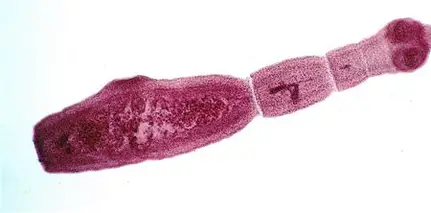
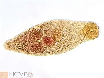
Dipylidium caninum
“double-pored dog tapeworm”
Cestode
2 sets of reproductive organs and a genital pore on each side
armed rostellum
found in cats (sporadically seen in humans)
infects flea then animal → gravid proglottids passed in feces
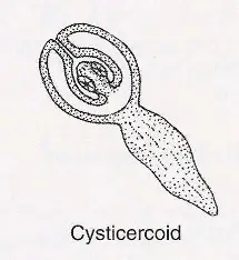
egg packets → oncosphere → cystercoid (infective)
Diphyllobothrium latum
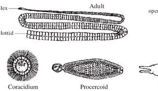
cestode
“broad fish tapeworm”
Host: fish-eating mammal
Order: Pseudophyllidea
Bothria on scolex
broad proglottids
eggs embryonate in water → coracidia infect copepod/crustation → develops into procercoid which fish eats → becomes plerocercoid → big fish (paratenic host) eats small, infected fish → human/fish eater eats fish
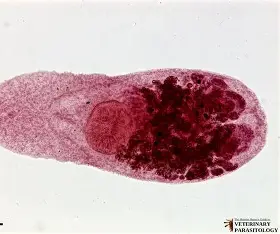
Taenia solum

“pork tapeworm”
Cestode
Order: Cyclophyllidea
Scolex has 4 acetabula suckers and rostellum with 2 rows of hooks
cysticercus in pork muscle →Human eats pork and hexacanth embryo attaches to small intestine wall → eggs or gravid proglottids shed into feces → pigs eat eggs and become infected
cysticercosis larvae
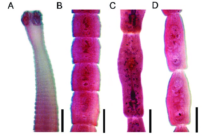
Moniezia
DH: hooved animals, IH: mite in orbatidae family
up to 6 meters, large
wide proglottids
unarmed scolex
two sets of reproductive organs in each proglottid
eggs released → eggs ingested by mites → animal eats mite → worm matures in small intestine
Mesocestoids
medioventral genital pores
widespread in carnivores
4 simple suckers and no rostellum
complete life cycle unknown
many infect rodent or reptile as IH
May undergo Asexual reproduction inside a IH by longitudinal fission at the scolex
Larval form - tetrathyridium (cysticercoid type)
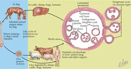
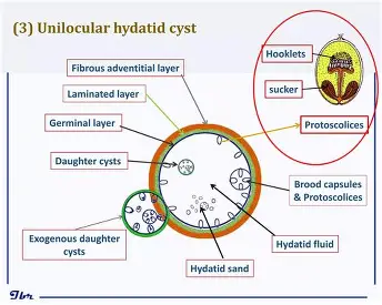
Echinococcus
Hyatid disease
common in sheep raising areas
small, only 3-6 segments
eggs are taeniid-like (meaning similar to taenia and echinococcus, they are in the same family)
Larval tapeworm form - hyatid cyst
DH: canids
IH: sheep, cattle, pigs, moose, elk, humans, deer, rabbits, goats, horses
Trematodes
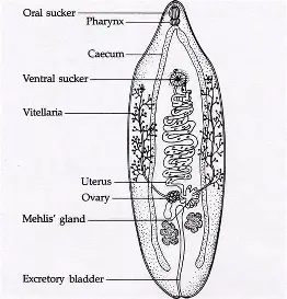
“Flukes”
Platyhelminth
Digenea
At least 1 IH, usually a snail
Found in most all bodily organs
Leaf life
Trematode Life Cycle
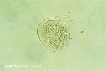
Egg → Miracidium → Sporocyst → Daughter sporocyst (+/- Rediae) → Cercaria → Metacercaria (INFECTIVE) → Adult
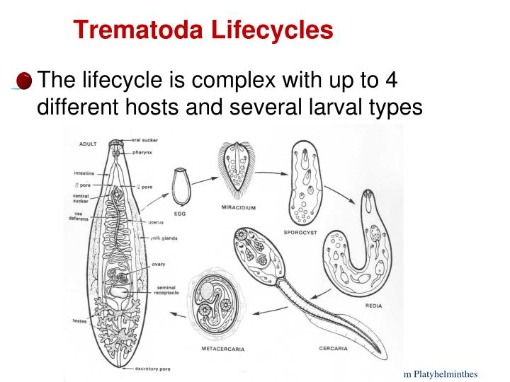
ENTER SNAIL: Miracidium
EXIT SNAIL: Cercaria
ENTER DH: Metacercaria
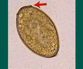
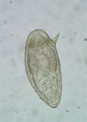
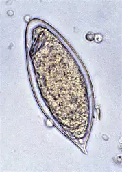
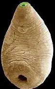
Fluke Egg
Heavier than other eggs
Operculum
Schistosoma sp.
“Blood flukes”
Trematode
Found in domestic animals and birds
Diecious (2 sexes)
Cercaria infective (no metacercaria)
Males have gynecophorical canal in which females lie
Eggs in bladder and kidneys cause granulomas and fibrosis
Schistosoma mansoni
eggs released in feces
lives in intestine
lateral spine on egg
Signs of Schistosoma
Cercarial dermatitis
Fever, malaise
Abdominal pain
Liver/spleen/intestinal tenderness, enlargement
Diarrhea
Hematuria (blood in urine); dysuria (painful urination)
Eosinophilia
Schistosoma haematobium
eggs released in urine
found in kidney and bladder
egg has spine at top
Schistosoma japonicum
eggs released in feces
found in GI and liver
eggs have small bump
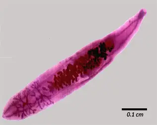
Fasciola spp.

“Liver Fluke”
Trematode
common in herbivores
ventral acetabulum close to oral sucker
Live in liver then bile ducts
cercaria leave snail and become metacercaria on vegetation (watercress)
Fasciolosis Signs
Can be found with fluke finder
Fever
Abdominal pain, malaise
Weight loss
Anemia
Hepatomegaly
Dicrocoelium dendriticum
“Lancet fluke” due to bladelike shape
Trematode
Lives in bile duct of sheep, cattle, goats, pigs, and cervid
1st IH: land snail → 2nd IH: ant
Land snail covers cercaria in slime balls which ants eat
Metacercaria develop in ant and move to nervous system, causing the ant to climb to the top of vegetation to be eaten
Clonorchis sinesis
“Chinese Liver Fluke”
Trematode
Leaf like with no spines and weak musculature
eggs released into water
1st IH: snail → 2nd IH: freshwater fish → DH: fish-eating mammal
IN SNAIL: miracidia→ sporocyst → rediae → cercariae
Adults live in bile ducts
Clonorchis sinesis Signs
Eggs found in stool samples
Upper abdominal pain, nausea; anorexia
Diarrhea
Gallbladder inflammation, granulomas
Hepatomegaly, jaundice
Pancreatitis
Heterophyes heterophyes
Trematode
Lives in small intestine of fish-eating birds and mammals
Small, tear shaped flukes
cercaria swim towards surface of water and penetrate fish musculature
1st IH: aquatic snail → 2nd IH: fish → DH: fish-eater → egg passed in feces
Nanophyetus salmincola
Trematode
causes rickettsial disease: hyperparasitism
large oral sucker
Lives in small intestine of birds and bird eating mammals
Snail host inhibit fast moving stream
unembryonated eggs pass down stream and hatch
rediae inhabit all areas of snail
metacercariae common in fins, muscle, and kidneys
1st IH: snail → 2nd IH: fish (often salmanoid) → DH: fish eater (commonly skunks and racoons)
Paramphistomum cervi
“rumen fluke”
Trematode
amphistome
large, pigmented cercariae with eye spots
NO 2nd IH, metacercariae develop on vegetation
penetrates gut, migrates to lumen, migrates to rumen
often kills host, no adequate treatment
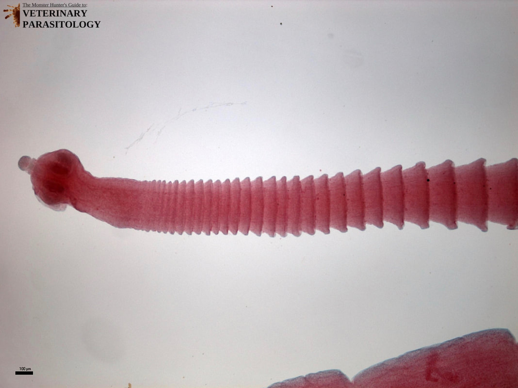
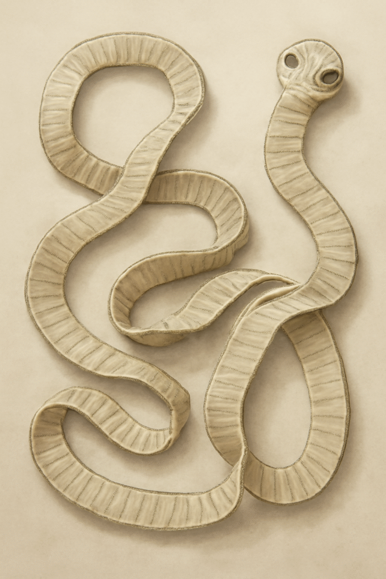
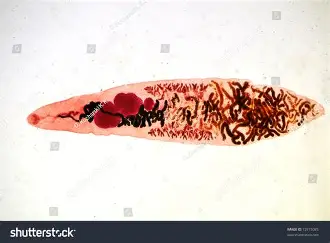
Cestodes
“Tapeworms”
platyhelminths
segmented worms
flattened dorsoventrally (top to bottom)
Scolex with 3 types of suckers:
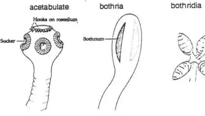
Scolex may bear a rostellum with hooks
Monenzia benedeni
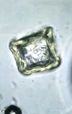
square egg
Monezia exoansa
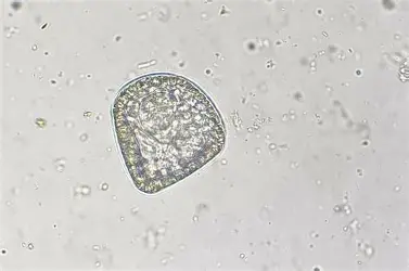
triangle egg
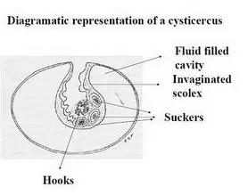
Cysticercus
Procercoid
Hydatid Cyst
Cysticercoid