Final exam diagrams
1/123
There's no tags or description
Looks like no tags are added yet.
Name | Mastery | Learn | Test | Matching | Spaced | Call with Kai |
|---|
No analytics yet
Send a link to your students to track their progress
124 Terms
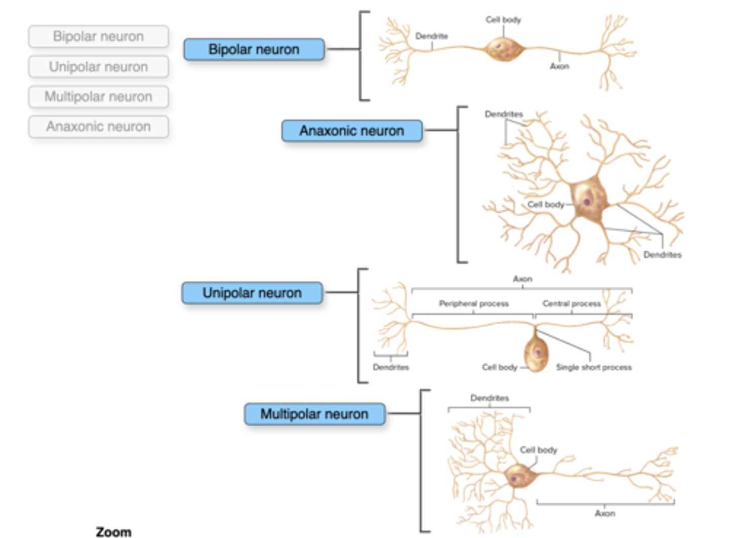
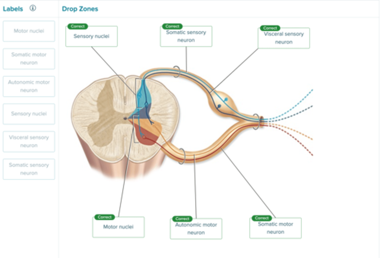
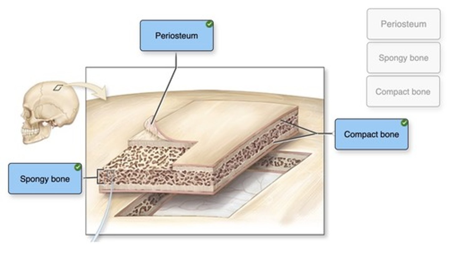
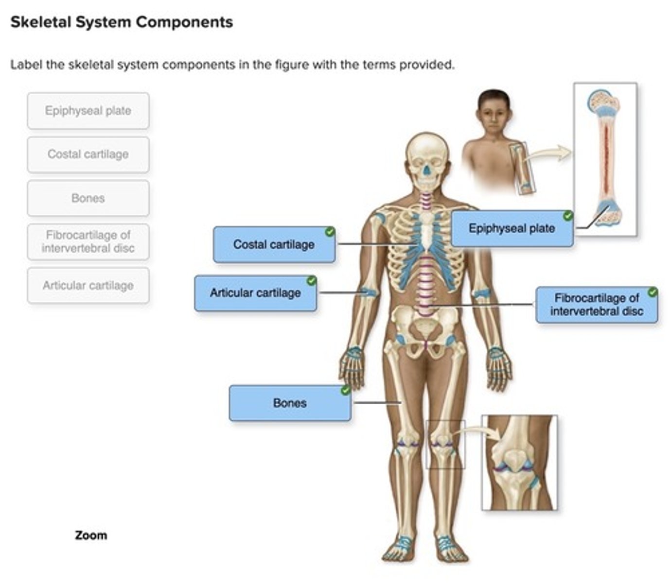
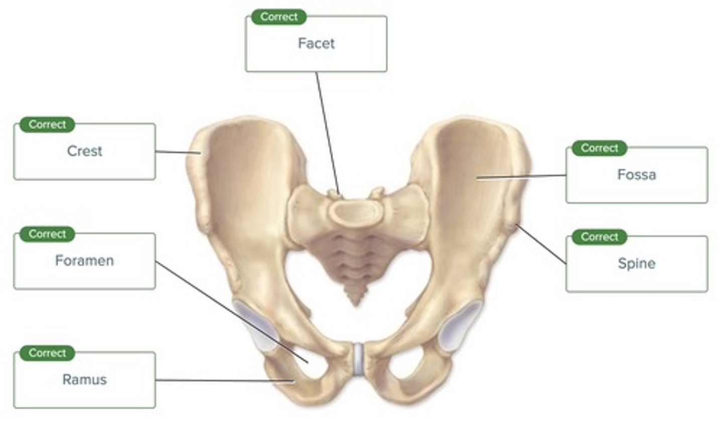
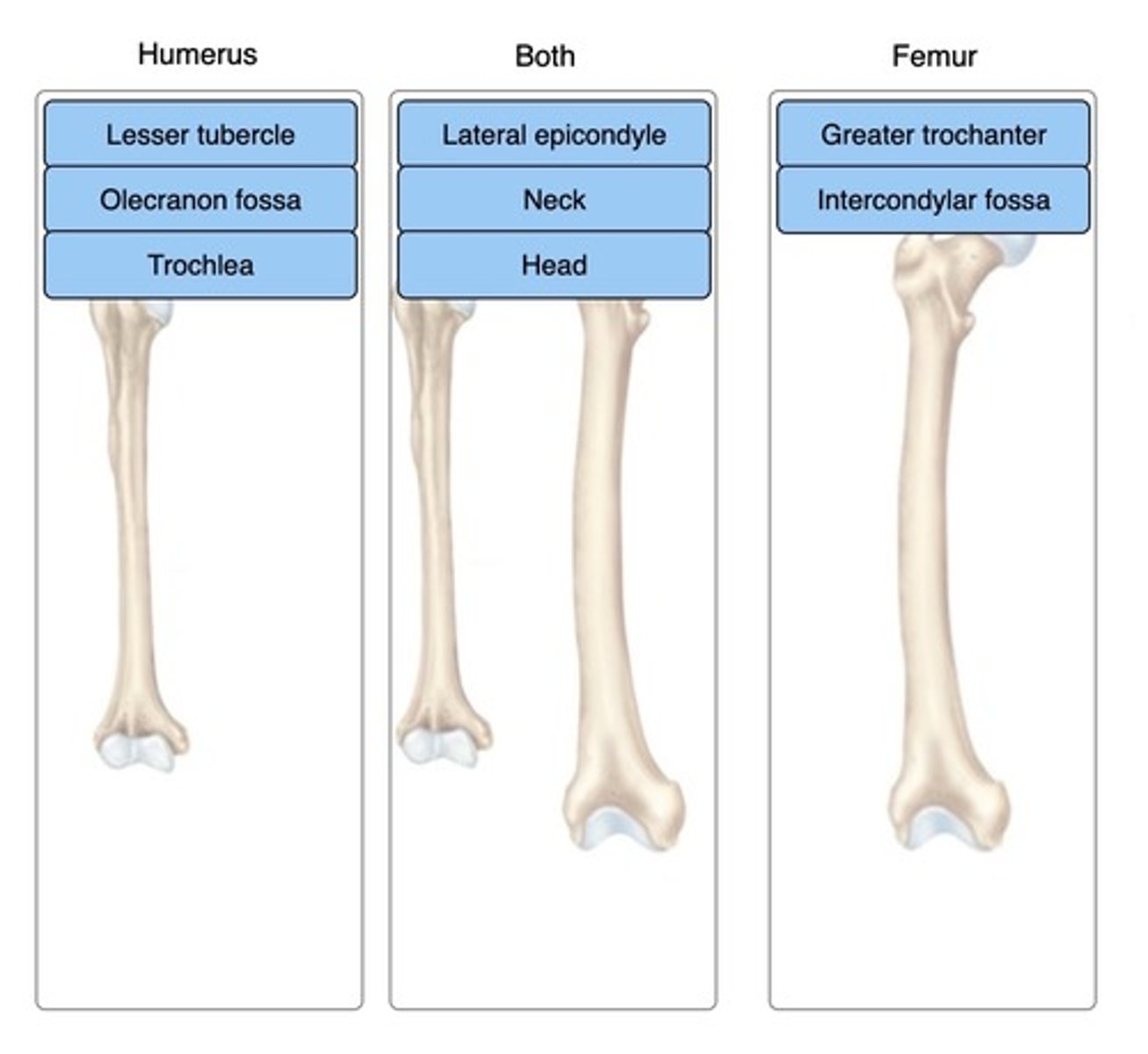
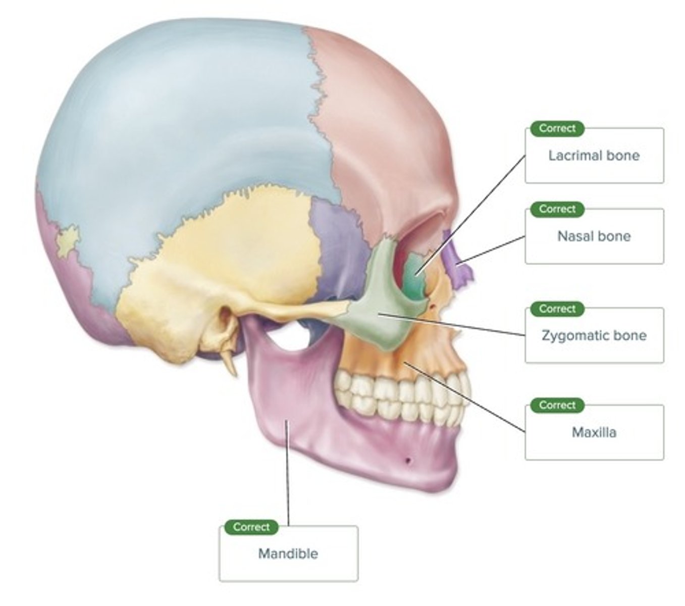
Label the figure with the items provided.
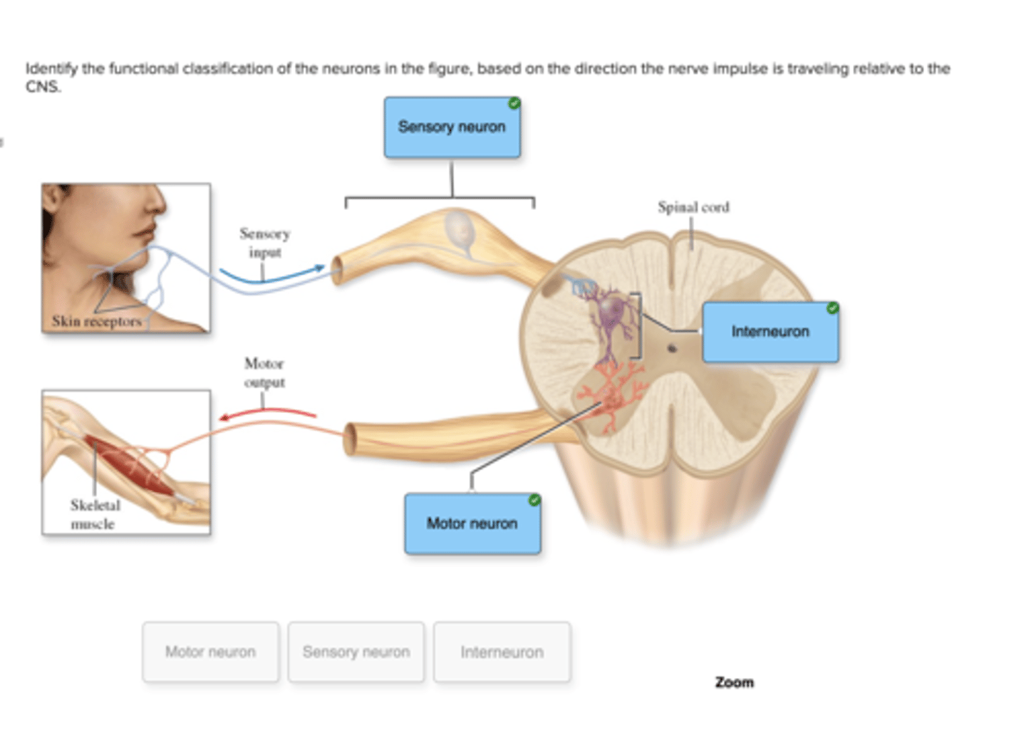
Identify the functional classification of the neurons in the figure, based on the direction the nerve impulse is traveling relative to the CNS.
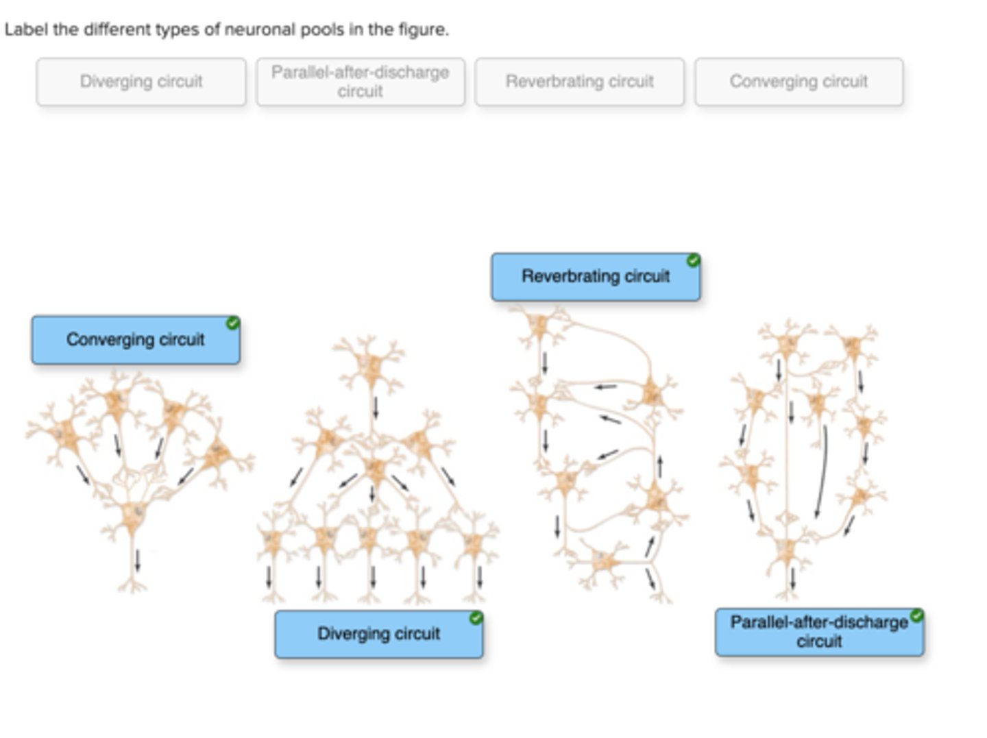
Label the different types of neuronal pools in the figure.
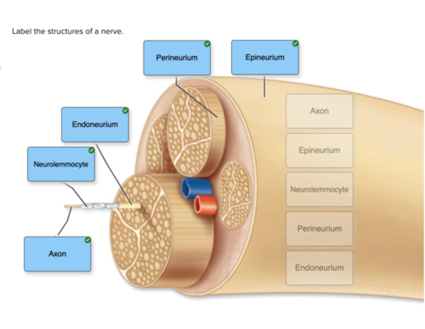
Label the structures of a nerve.
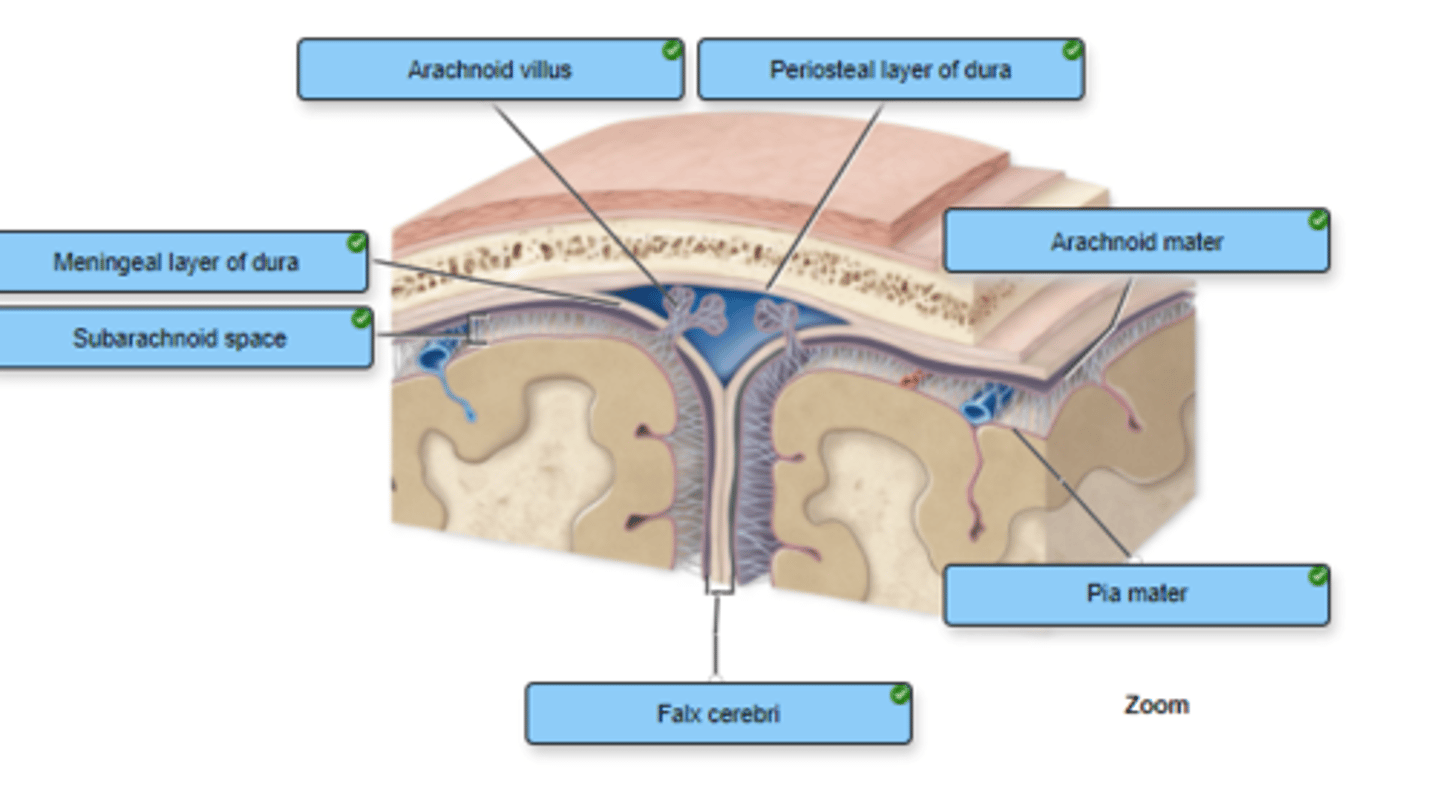
Correctly label the following meninges and associated structures.
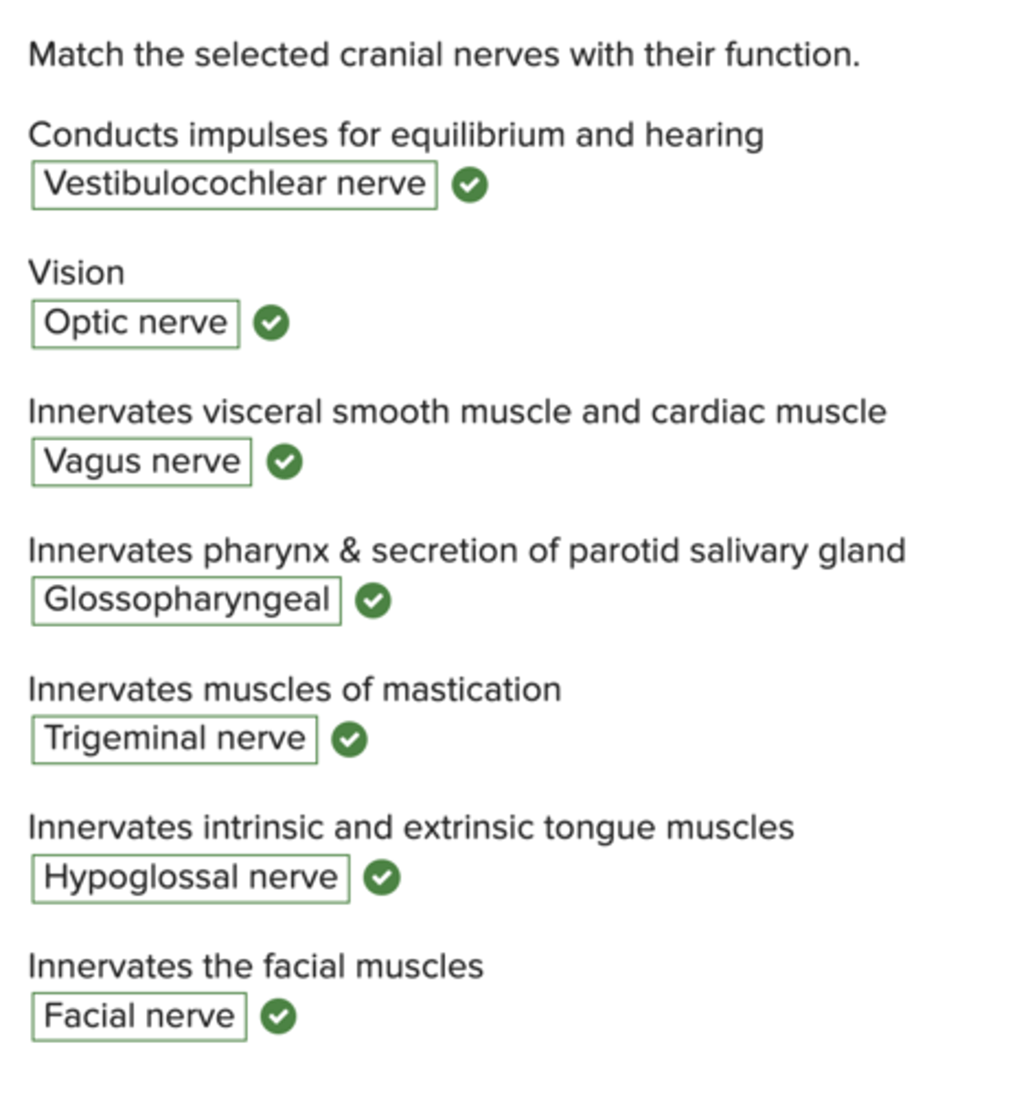
Match the selected cranial nerves with their function.
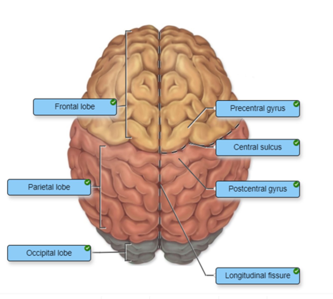
Identify the cerebral lobes on the left side of the figure. Label the additional cerebral structures on the right side of the figure.
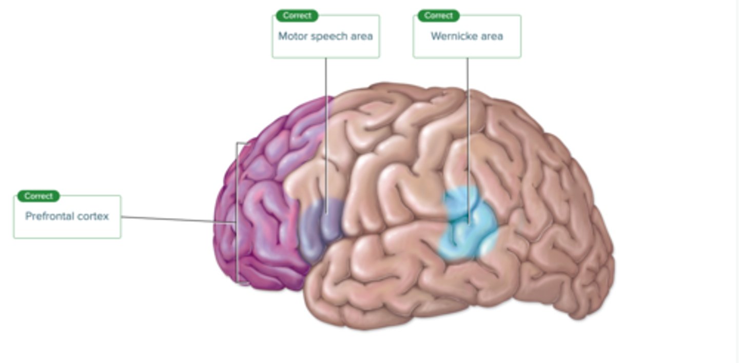
Label the regions involved in interpreting and carrying out speech information.
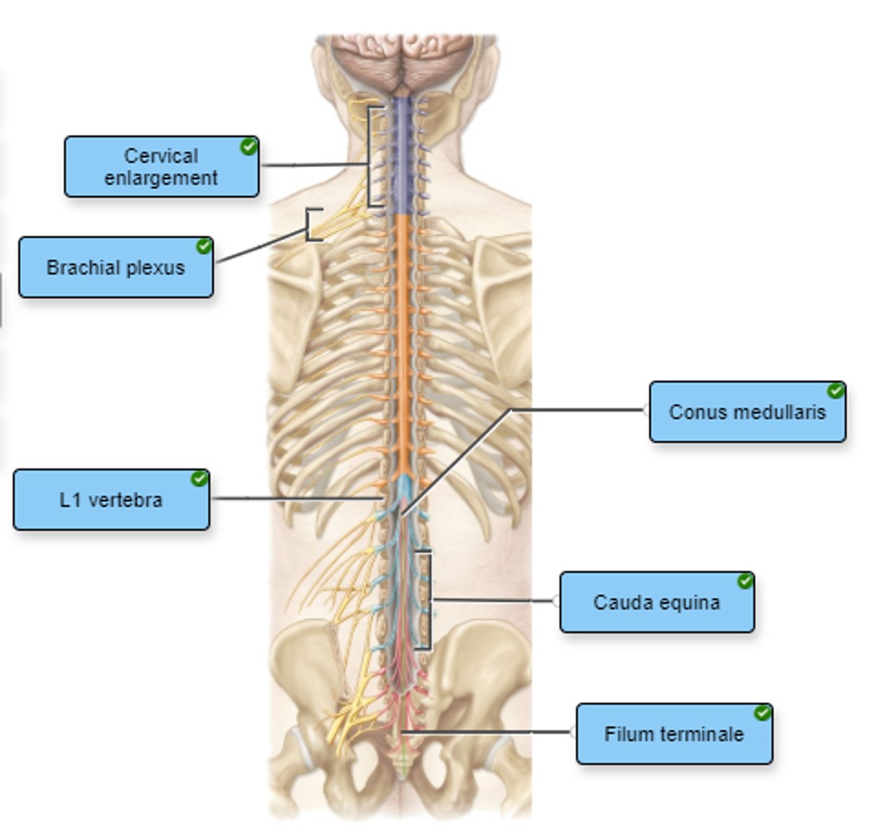
Label the structures of the spinal cord.
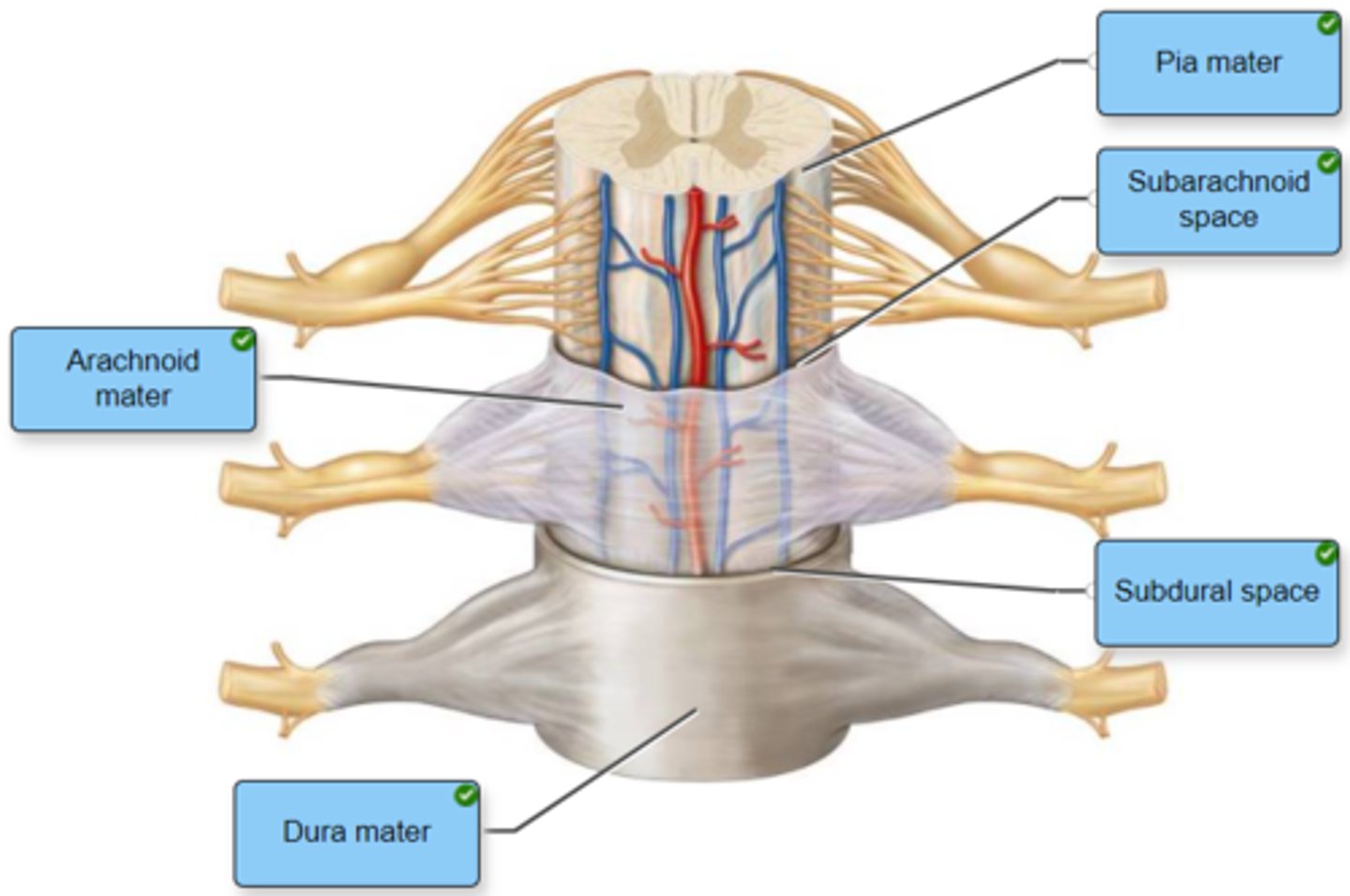
Label the spinal cord meninges and spaces.
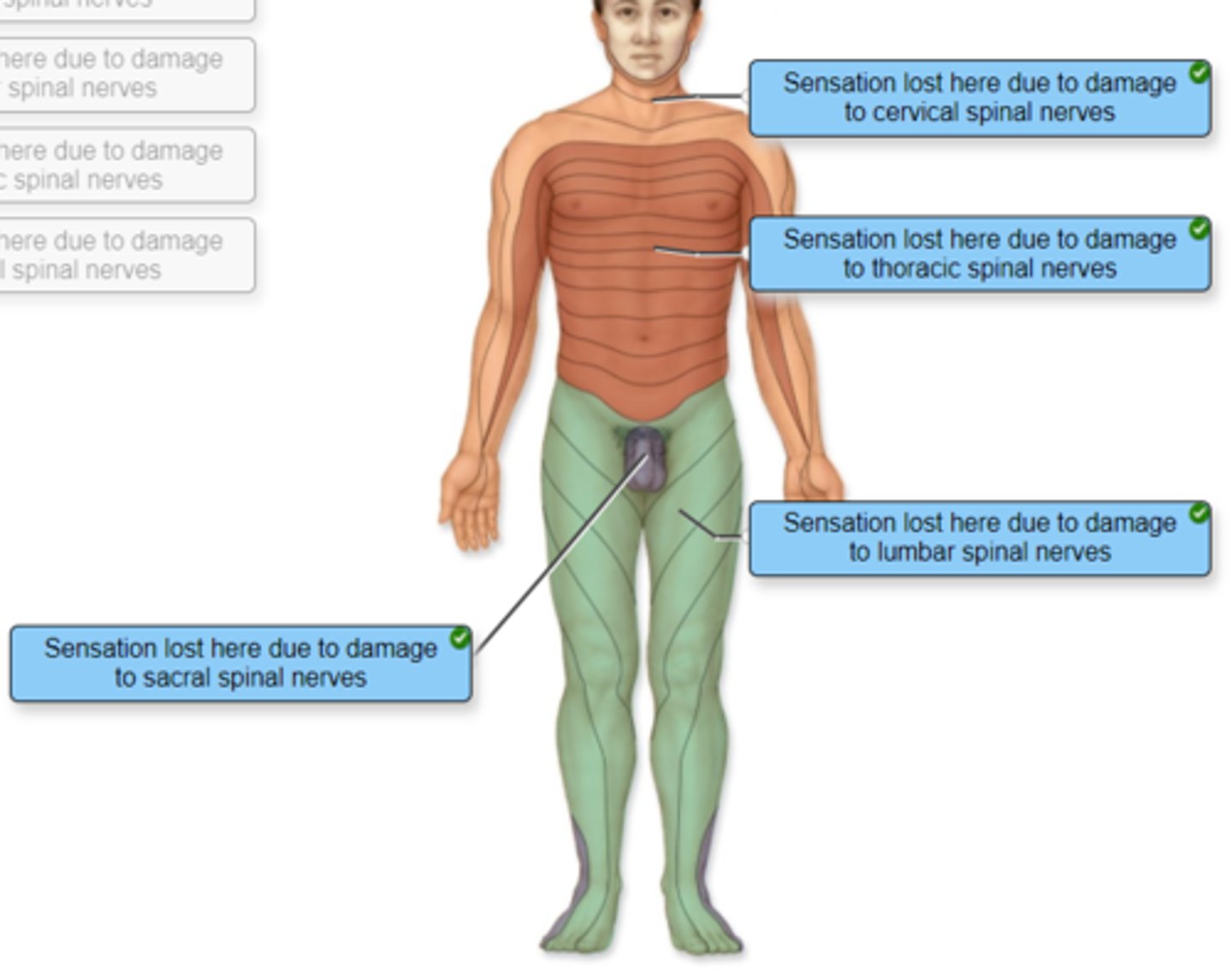
Correctly identify and label the dermatome(s) represented by the statement(s) associated with them.
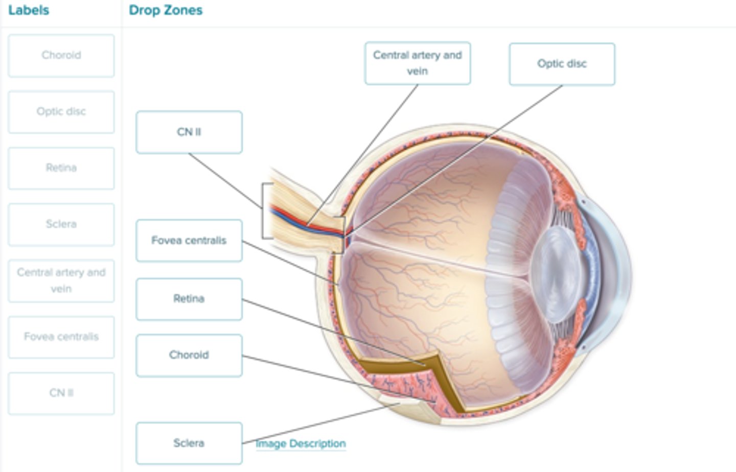
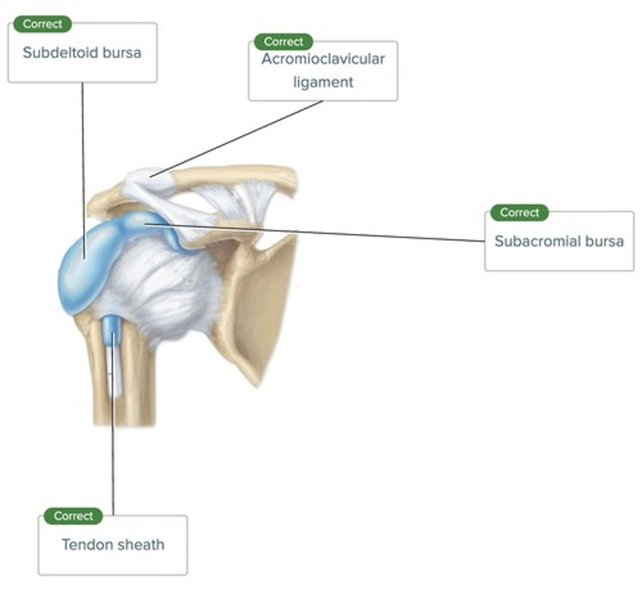
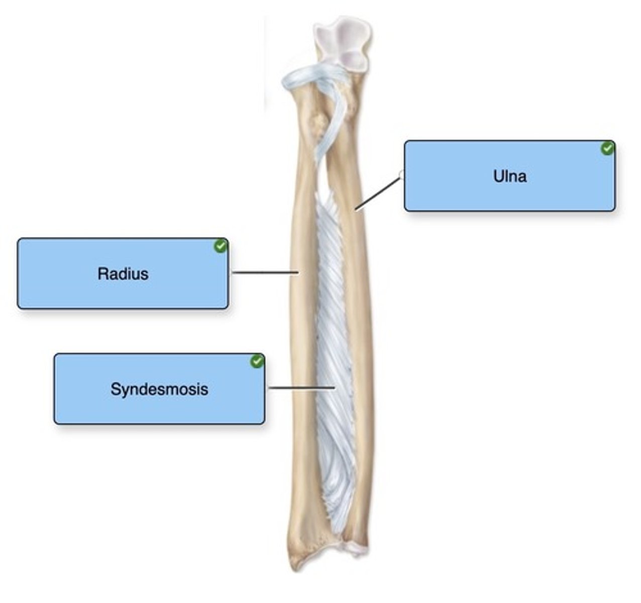
Label the figure with the items provided.
Drag and drop the labels into the appropriate location on the figure.
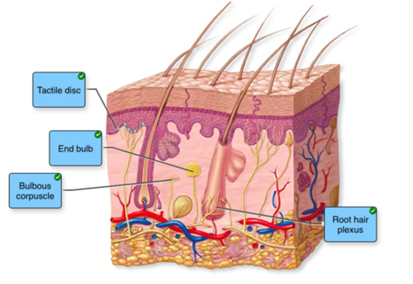
Label the type of tactile receptors in the image.
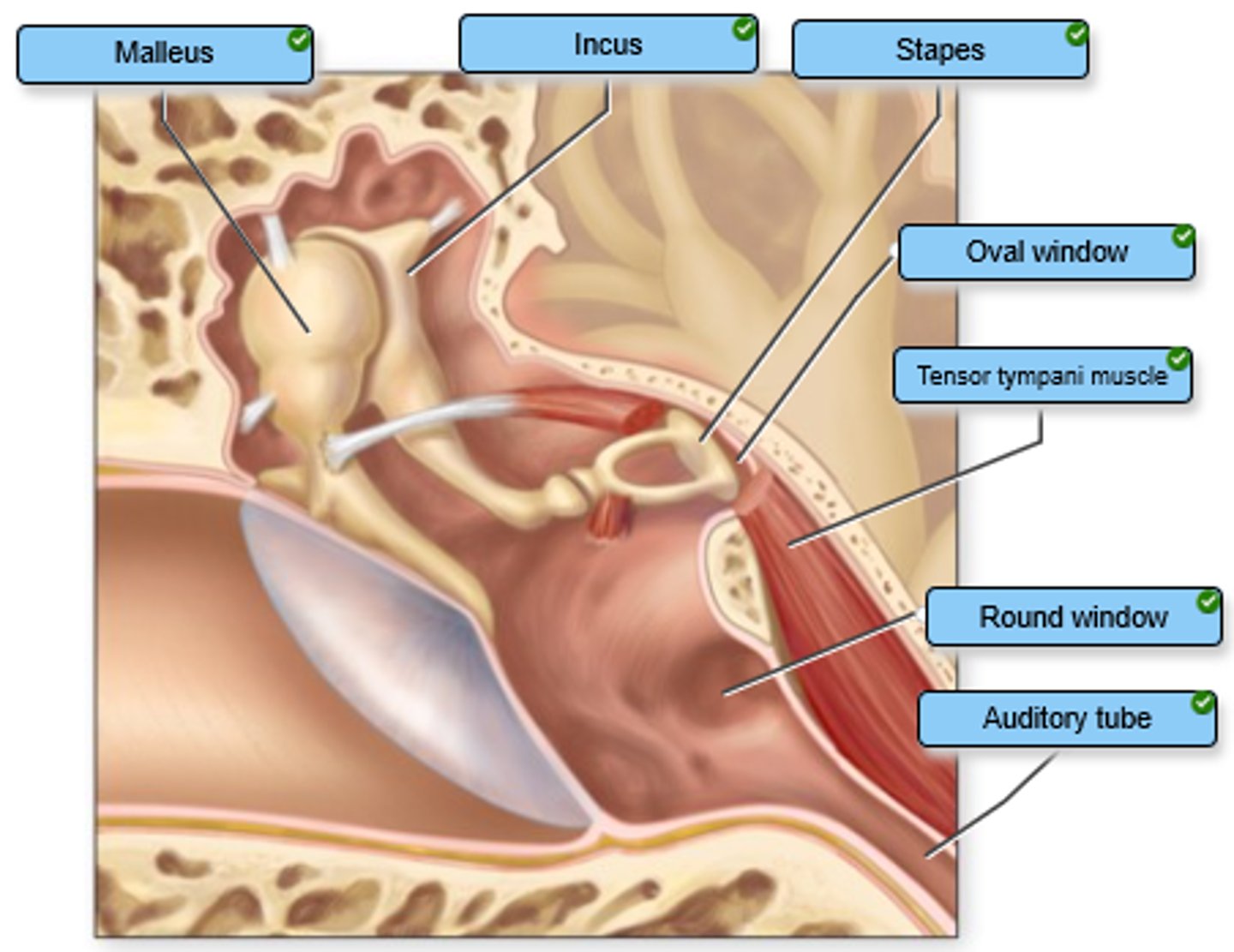
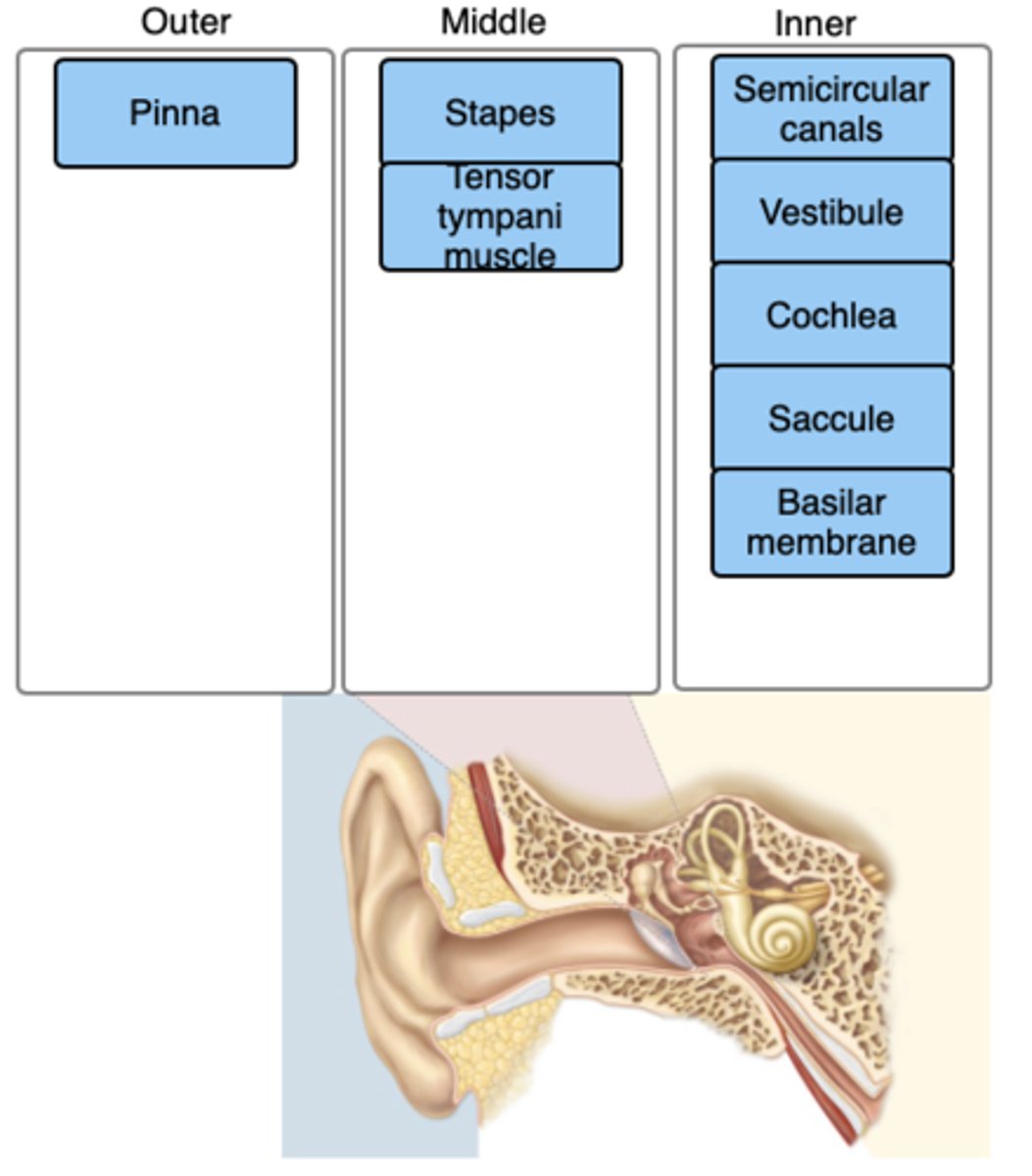
Label the structures of the middle ear.
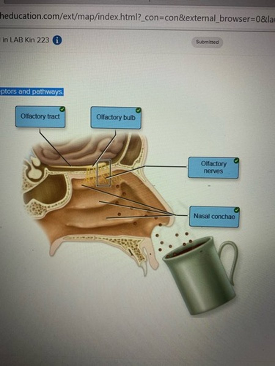
Label the olfactory receptors and pathways.
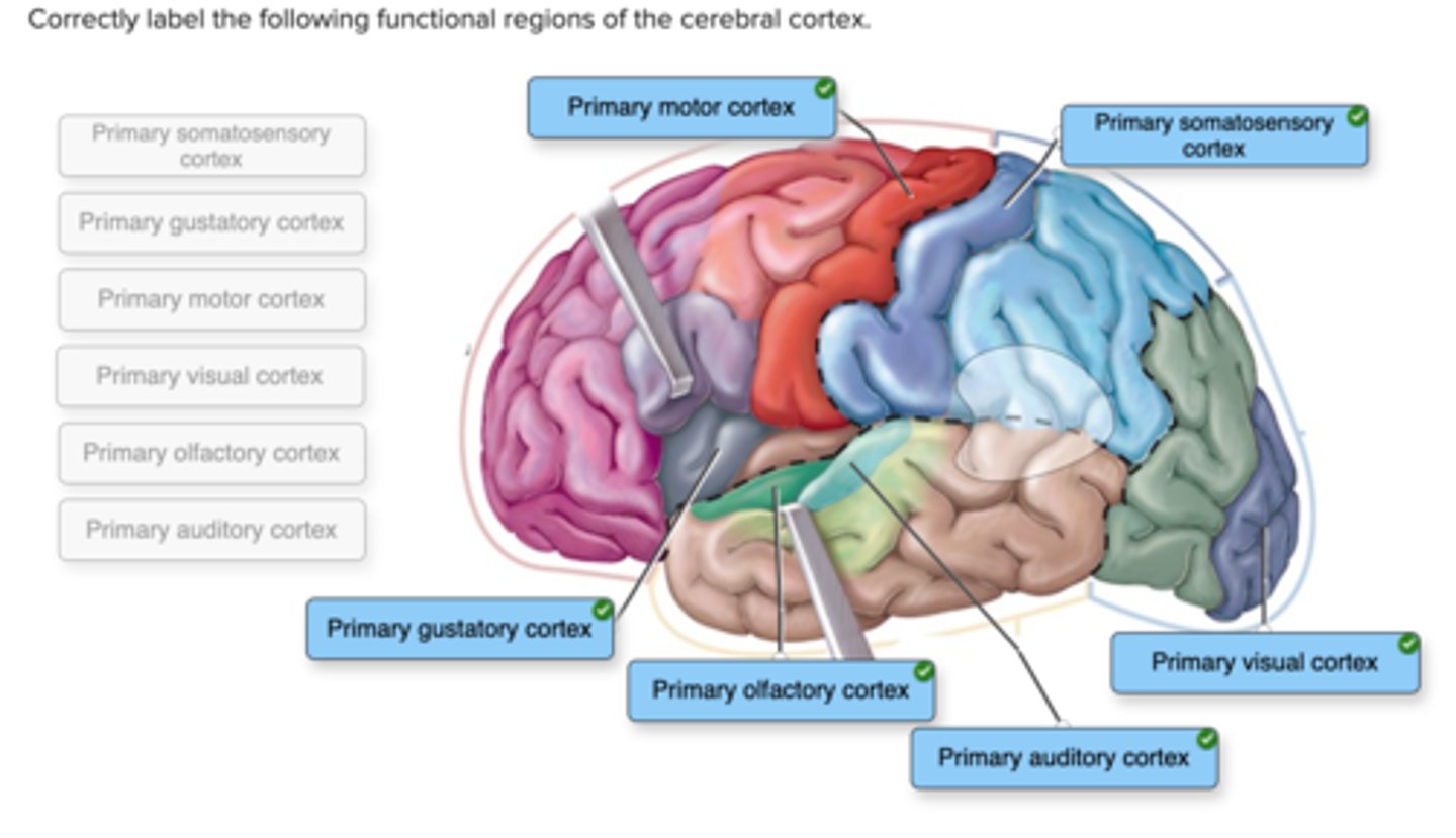
Correctly label the following functional regions of the cerebral cortex.
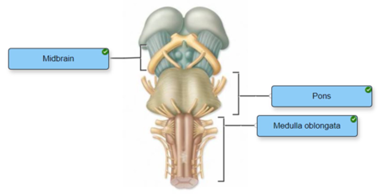
Identify the components of the brainstem.
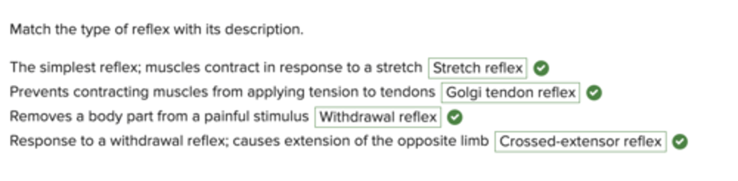
Match the type of reflex with its description.
Indicate whether the given structure is located in the outer, middle, or inner ear.
Check all of the statements that are true regarding the changes that occur in the muscular system as a result of aging.

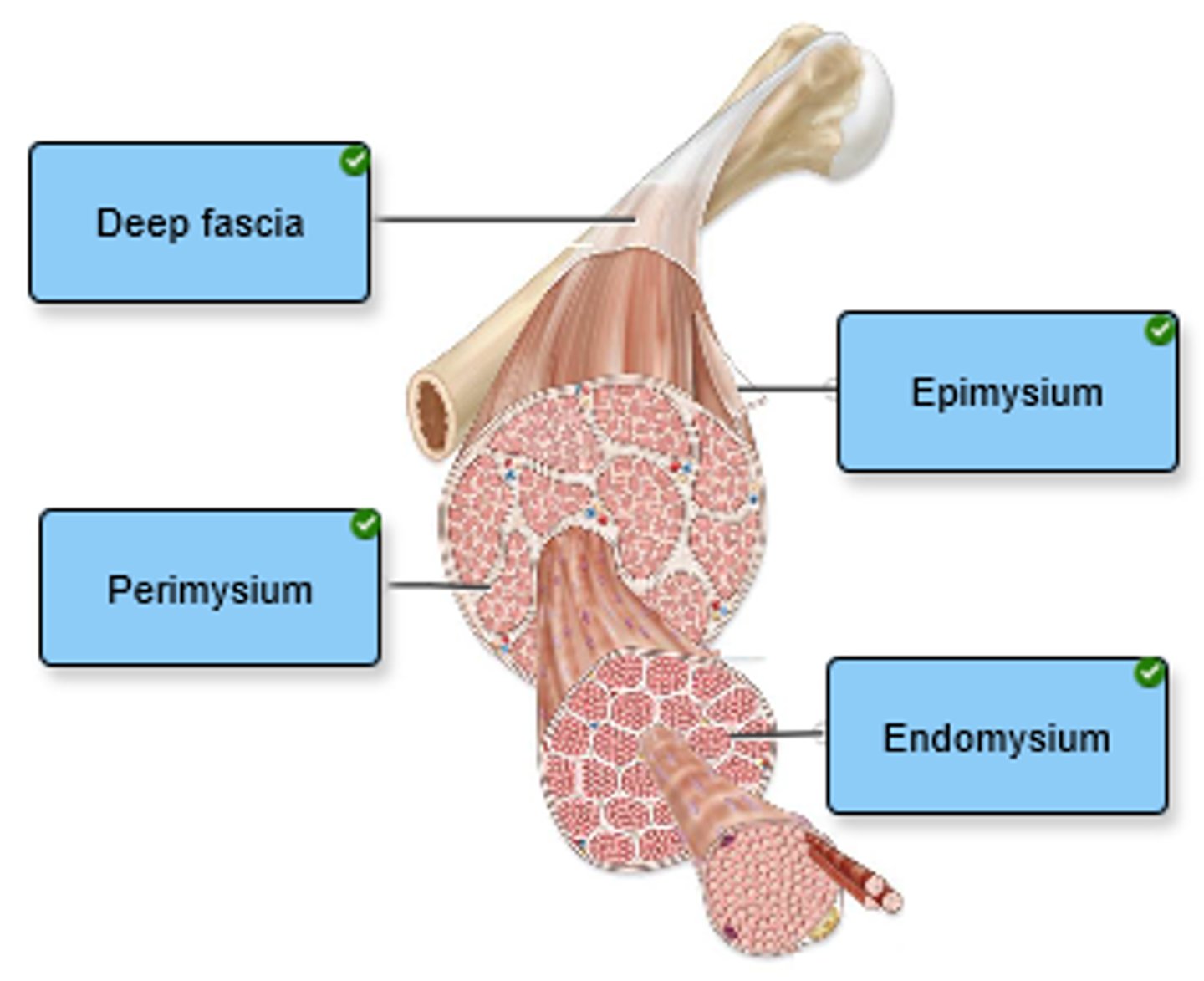
Label the connective tissue in the figure.
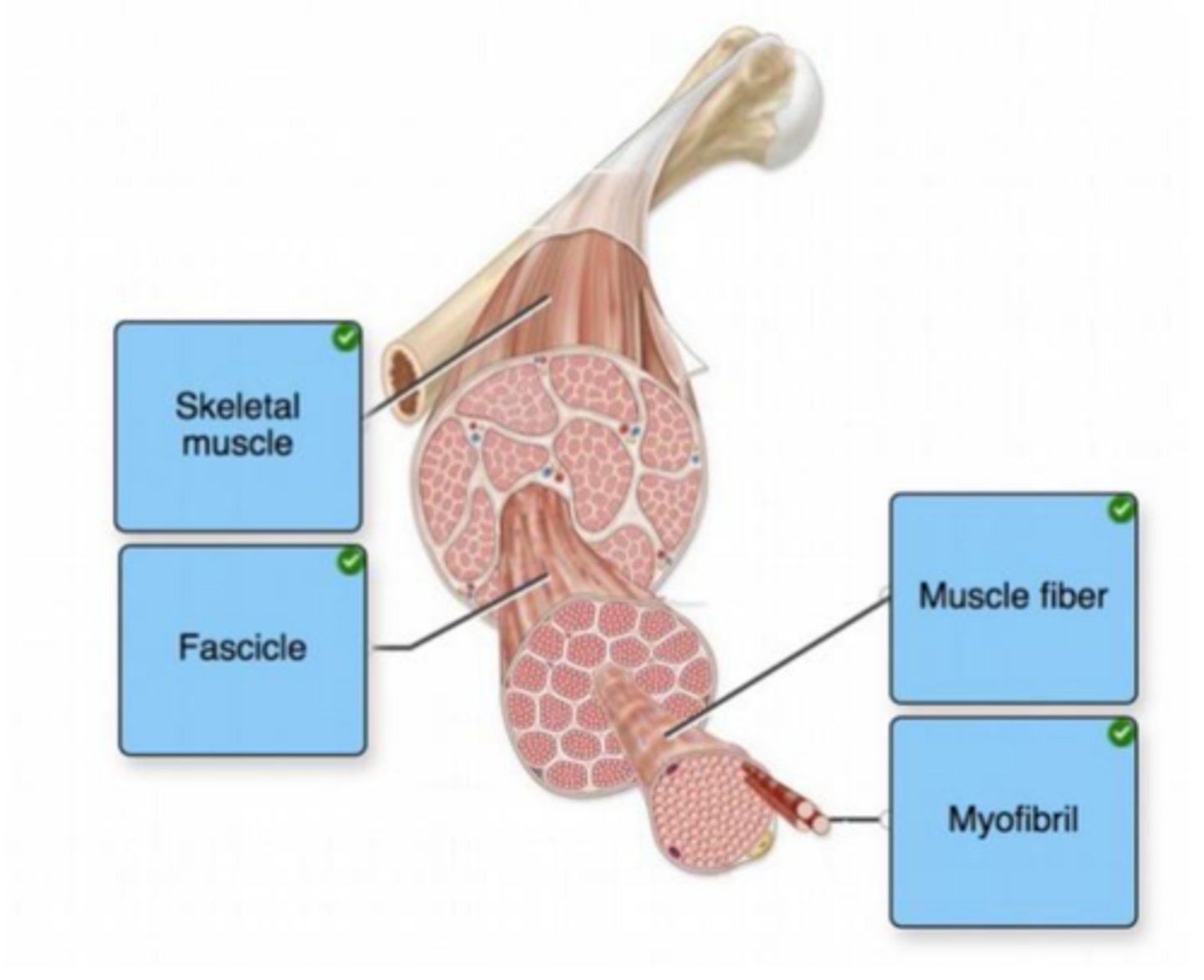
Label the components of skeletal muscle.
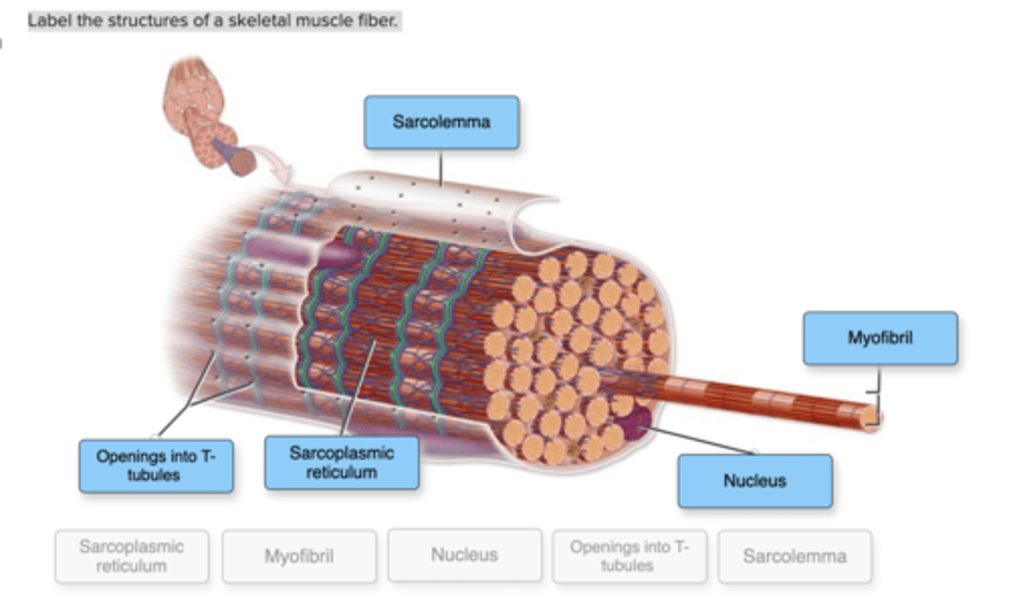
Label the structures of a skeletal muscle fiber.
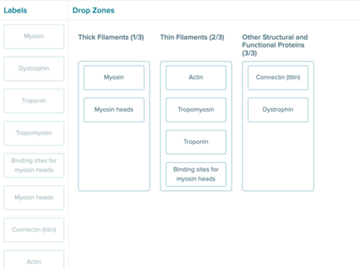
Determine whether each structure is part of a thick filament, part of a thin filament, or is a different structural/functional protein.
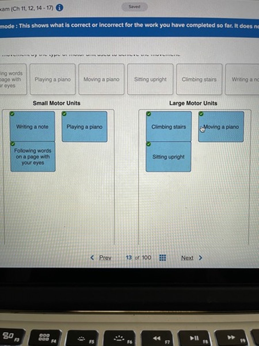
Sort each movement by the type of motor unit used to achieve the movement.
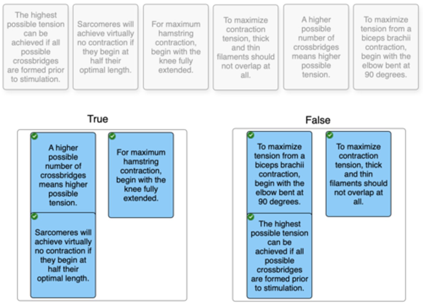
Click and drag each statement into the correct box to identify whether it is true or false regarding the length-tension relationship.
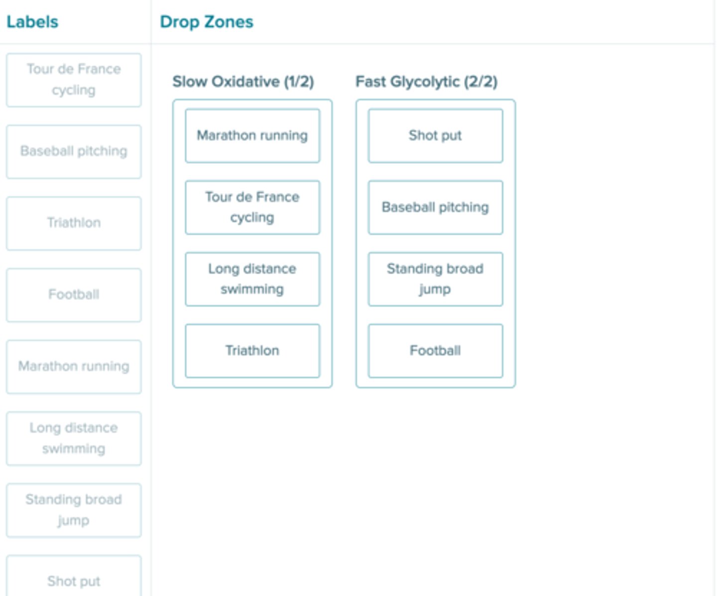
If gene therapy in the future would allow one to artificially alter the expression of muscle fibers for athletic success, which increase in fiber types would make the most sense for the following activities?
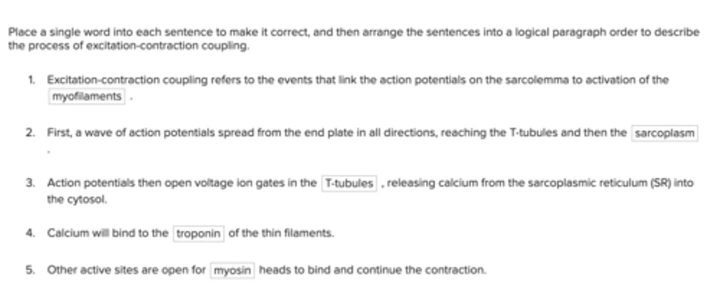
Place a single word into each sentence to make it correct, and then arrange the sentences into a logical paragraph order to describe the process of excitation-contraction coupling.
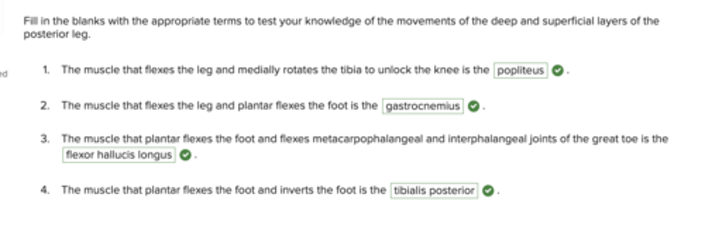
Fill in the blanks with the appropriate terms to test your knowledge of the movements of the deep and superficial layers of the posterior leg.
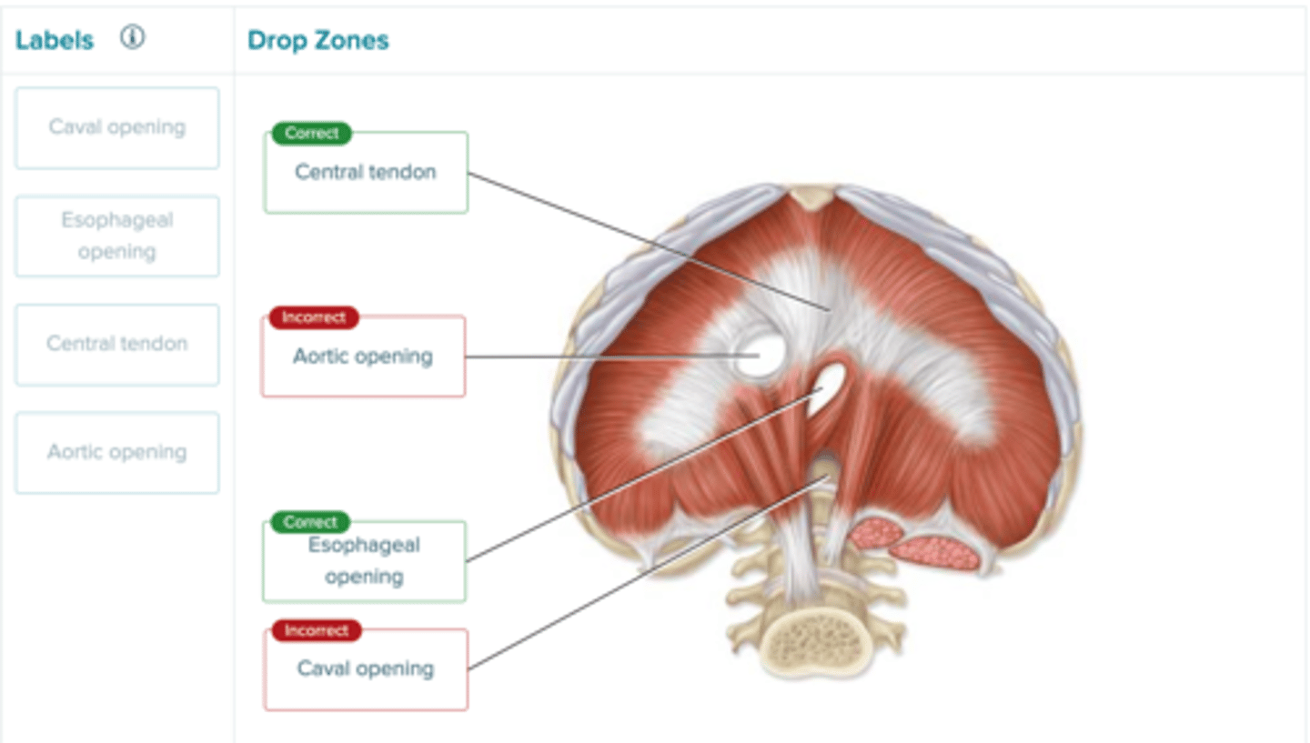
Label the parts of the diaphragm in the image.
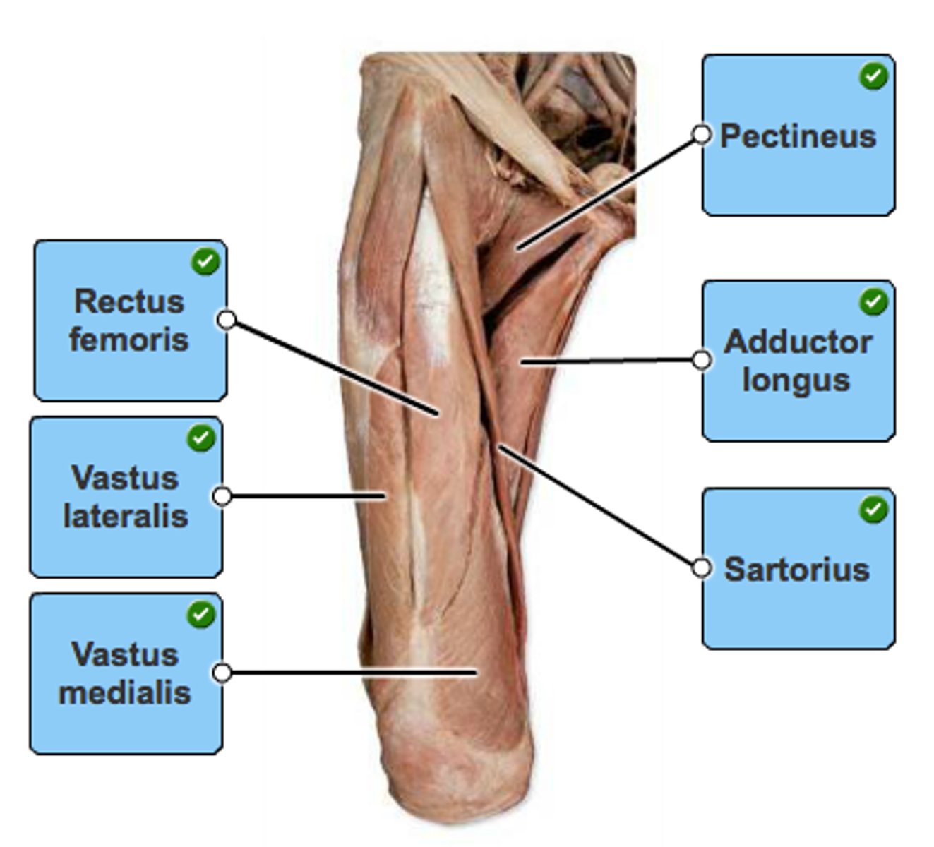
Label the muscles of the anterior thigh.
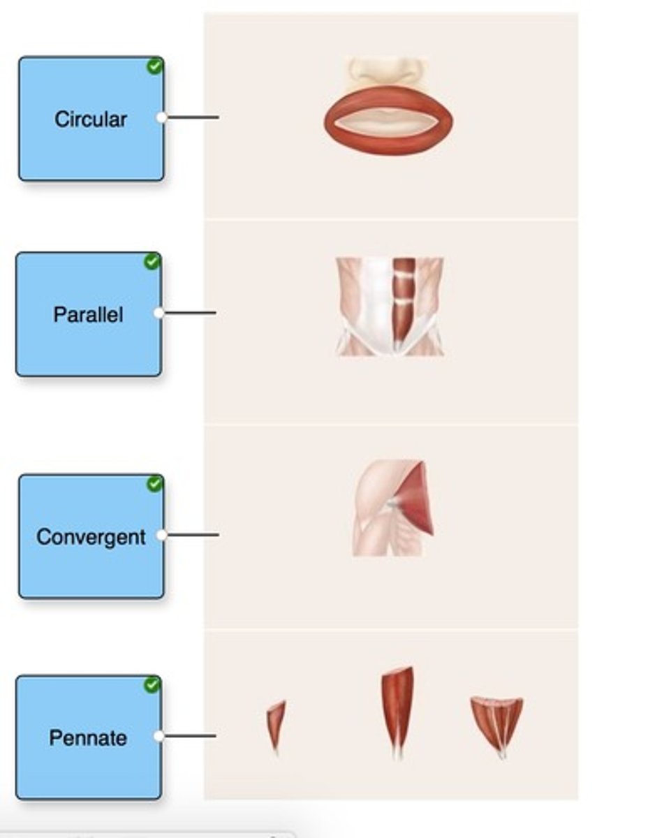
Identify the organizational pattern of the fascicles in each muscle.
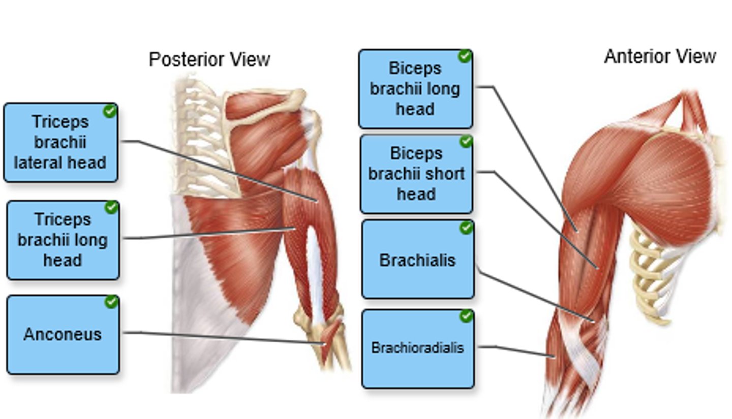
Label the anterior and posterior arm muscles in the figure.
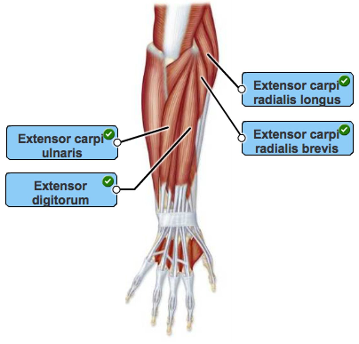
Label the muscles of the right posterior forearm in the figure. Not all labels will be used.
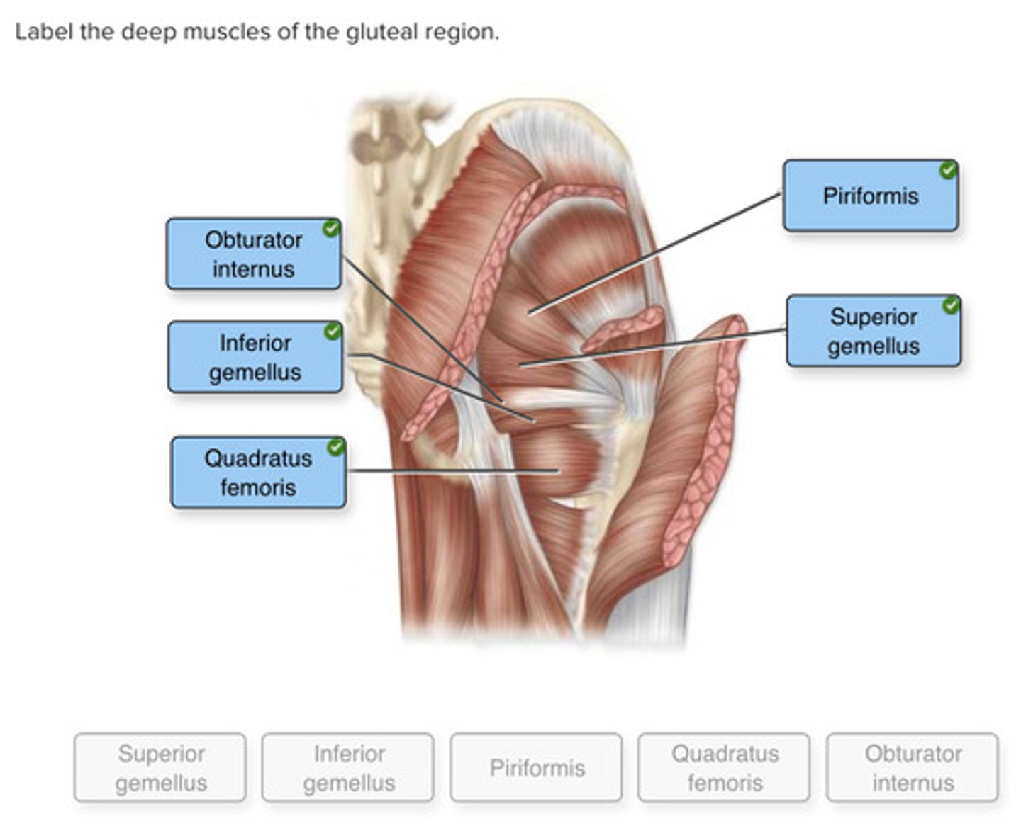
Label the deep muscles of the gluteal region.
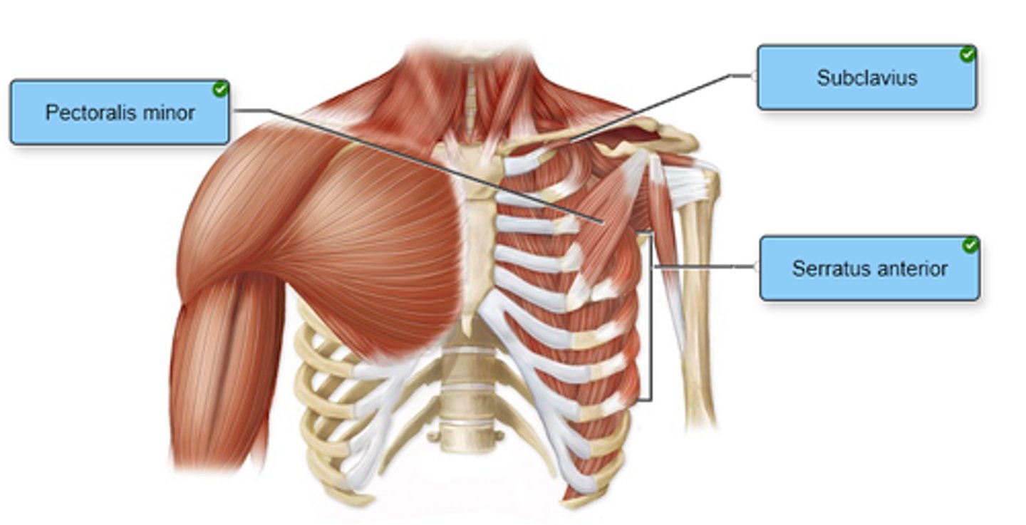
Label the anterior thoracic muscles in the figure.
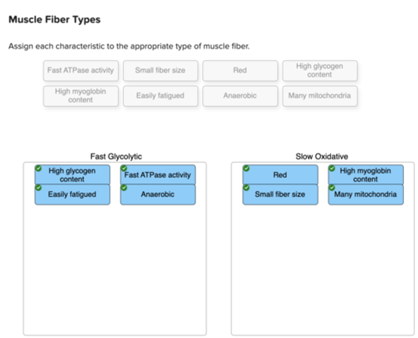
Assign each characteristic to the appropriate type of muscle fiber.
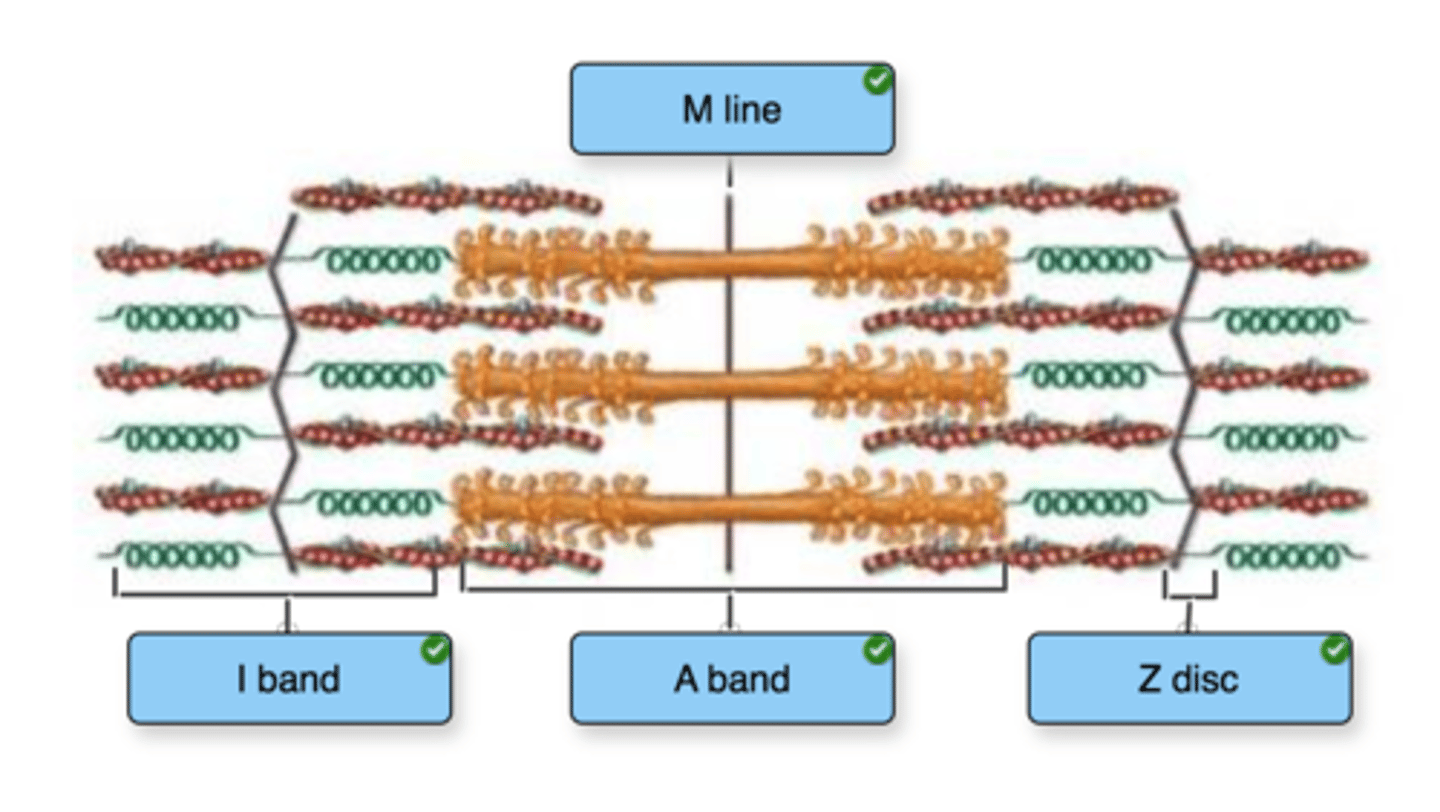
Label the structures of a sarcomere.
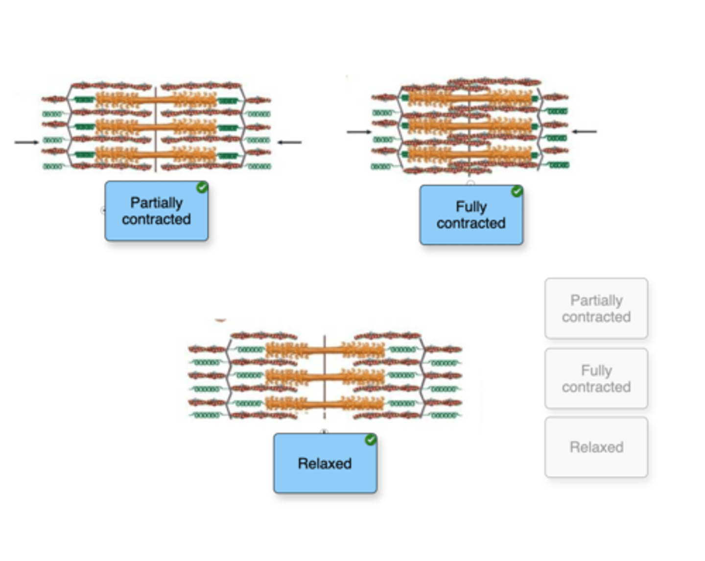
Based on your understanding of the process of skeletal muscle contraction, indicate whether each figure represents a relaxed, partially contracted, or fully contracted skeletal muscle.
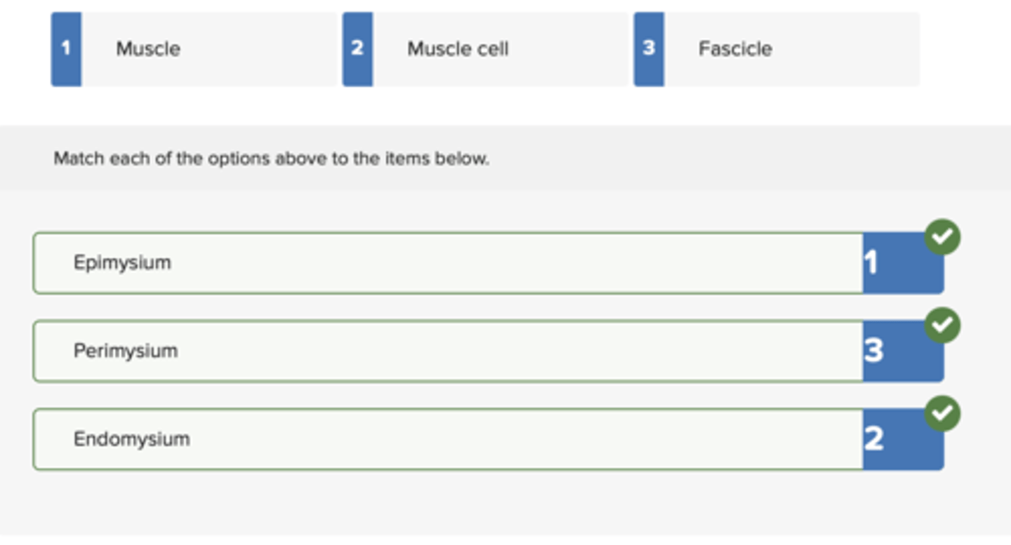
Match each structure with the connective tissue layer that surrounds it.
Epi
Peri
Endo
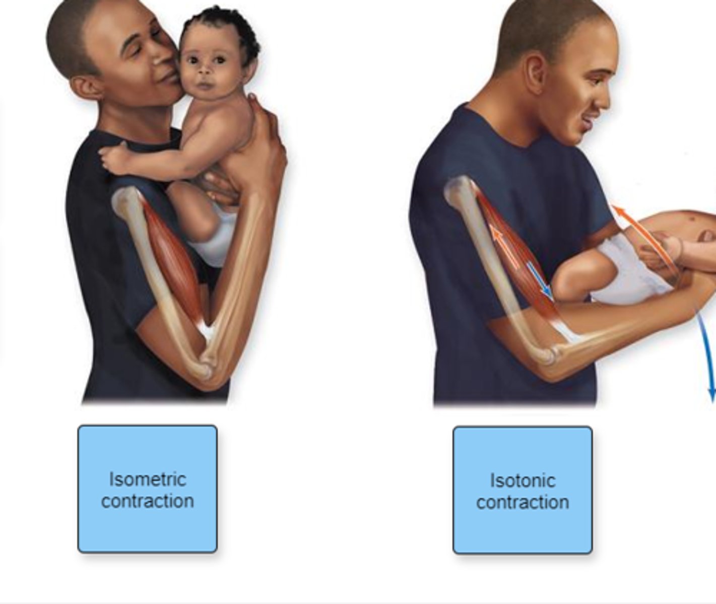
Indicate which type of contraction each figure represents.
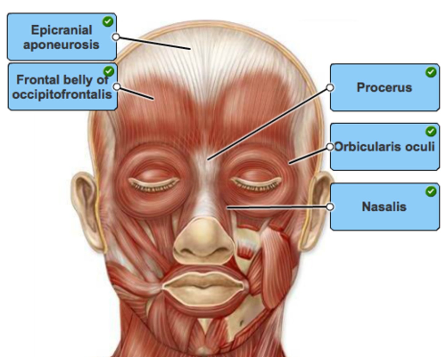
Label the muscles of the forehead and muscles around the eyes in the figure.
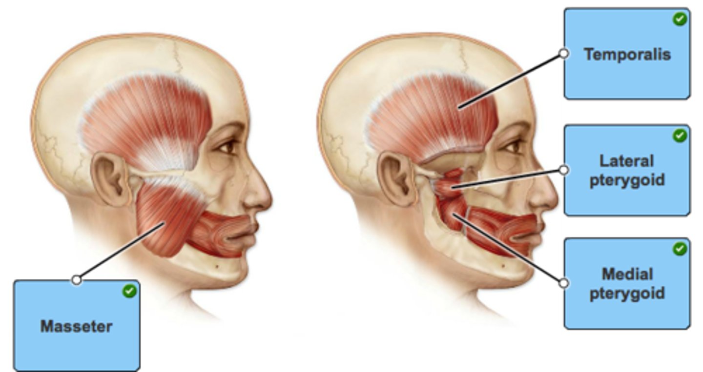
Label the muscles of mastication in the figure.
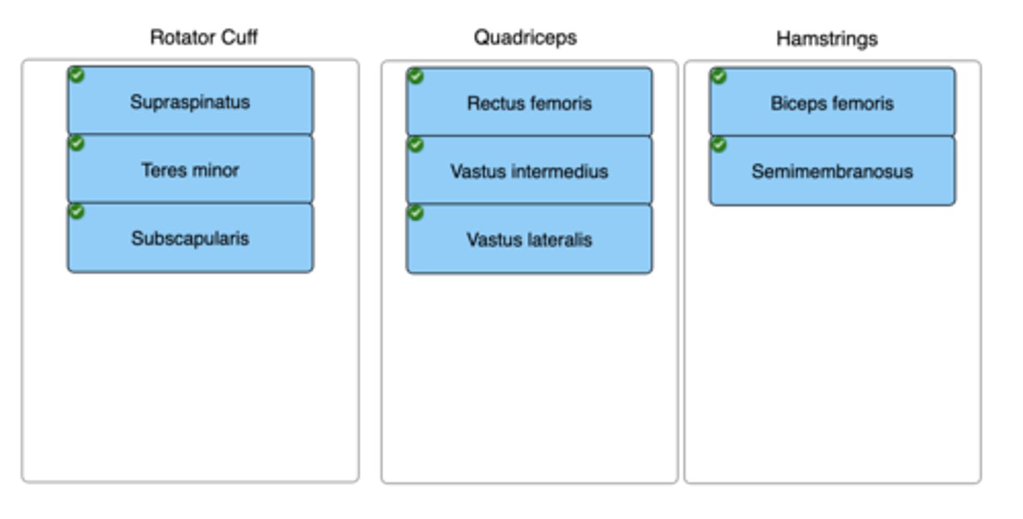
Drag each muscle to the muscle group in which it belongs.
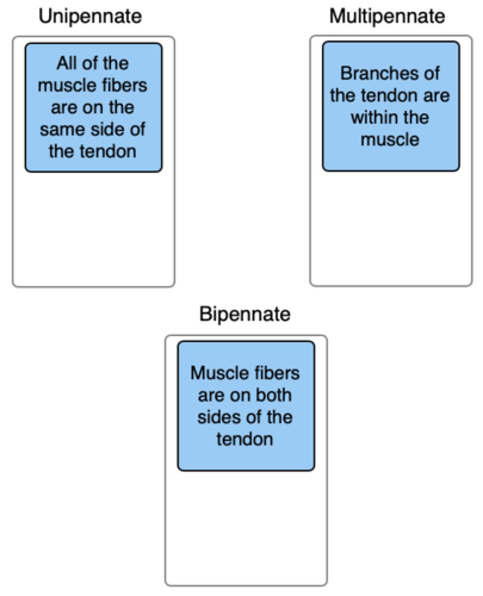
Classify the type of pennate muscle with its description. Not all labels will be used.
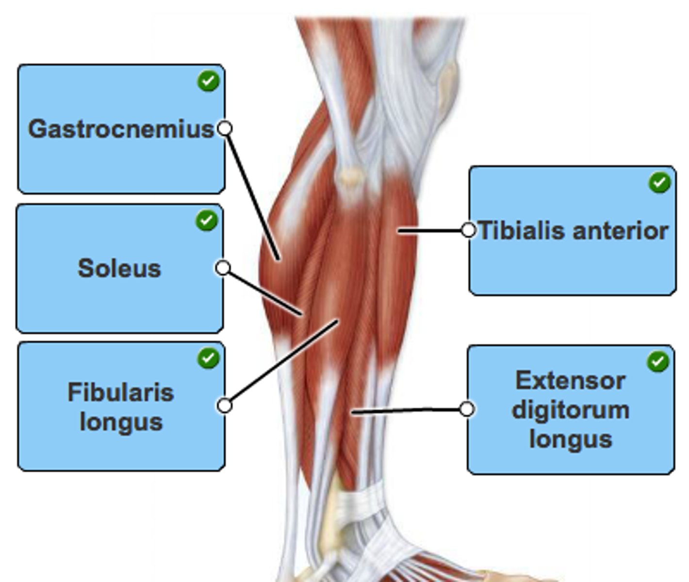
Label the muscles in a lateral view of the leg.
Label the anterior and posterior arm muscles in the figure.


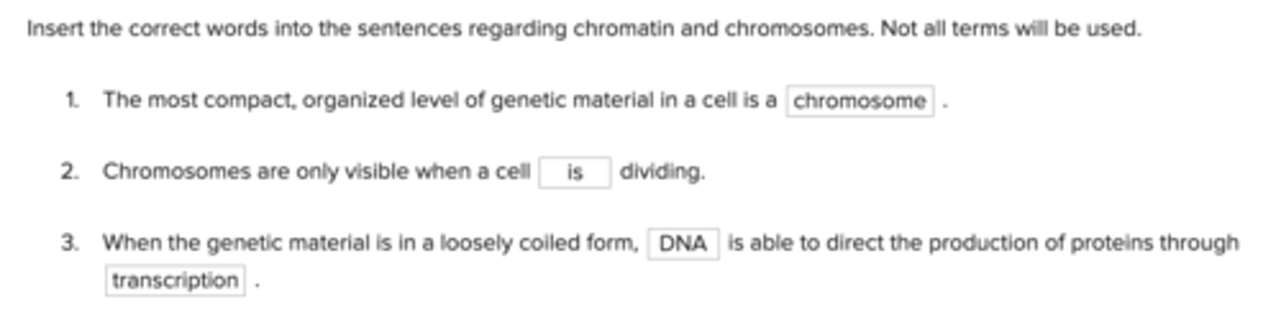
Insert the correct words into the sentences regarding chromatin and chromosomes. Not all terms will be used.
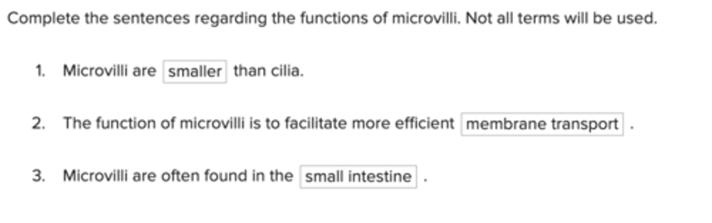
Complete the sentences regarding the functions of microvilli. Not all terms will be used.
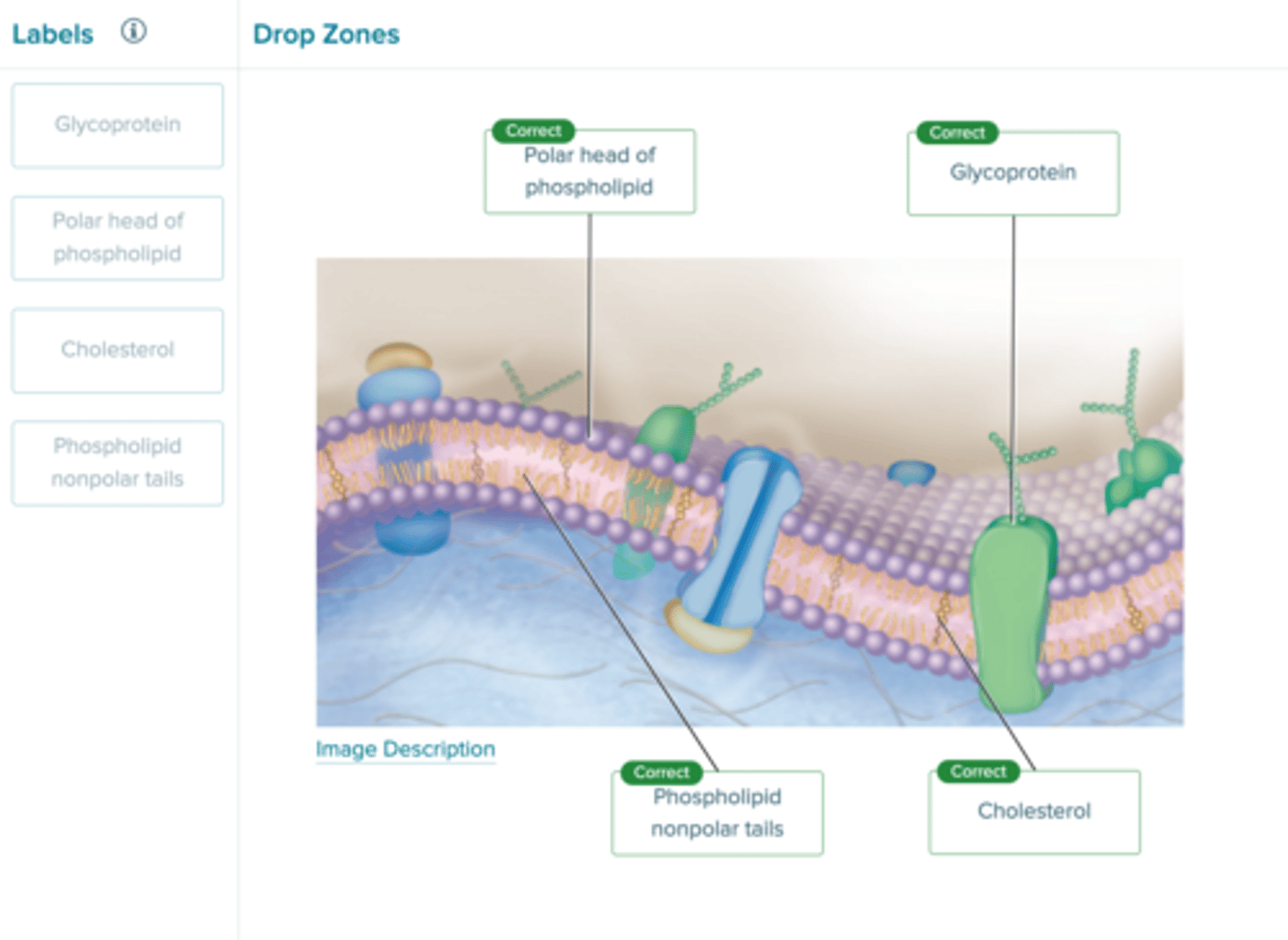
Identify the components of the plasma membrane.
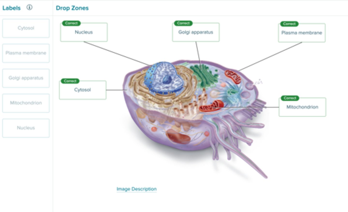
Identify the structures of a prototypical human cell.
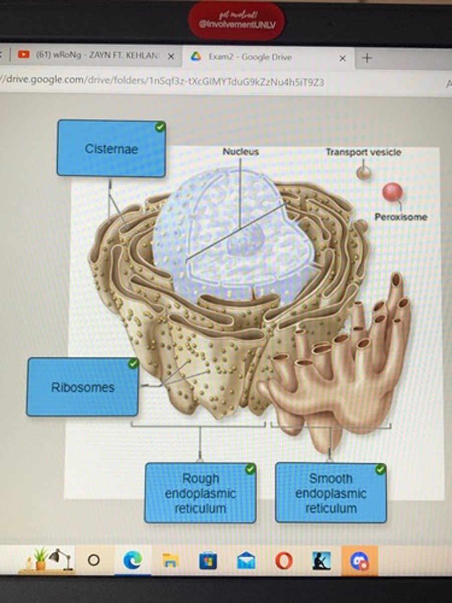
Correctly identify the parts of the endoplasmic reticulum.
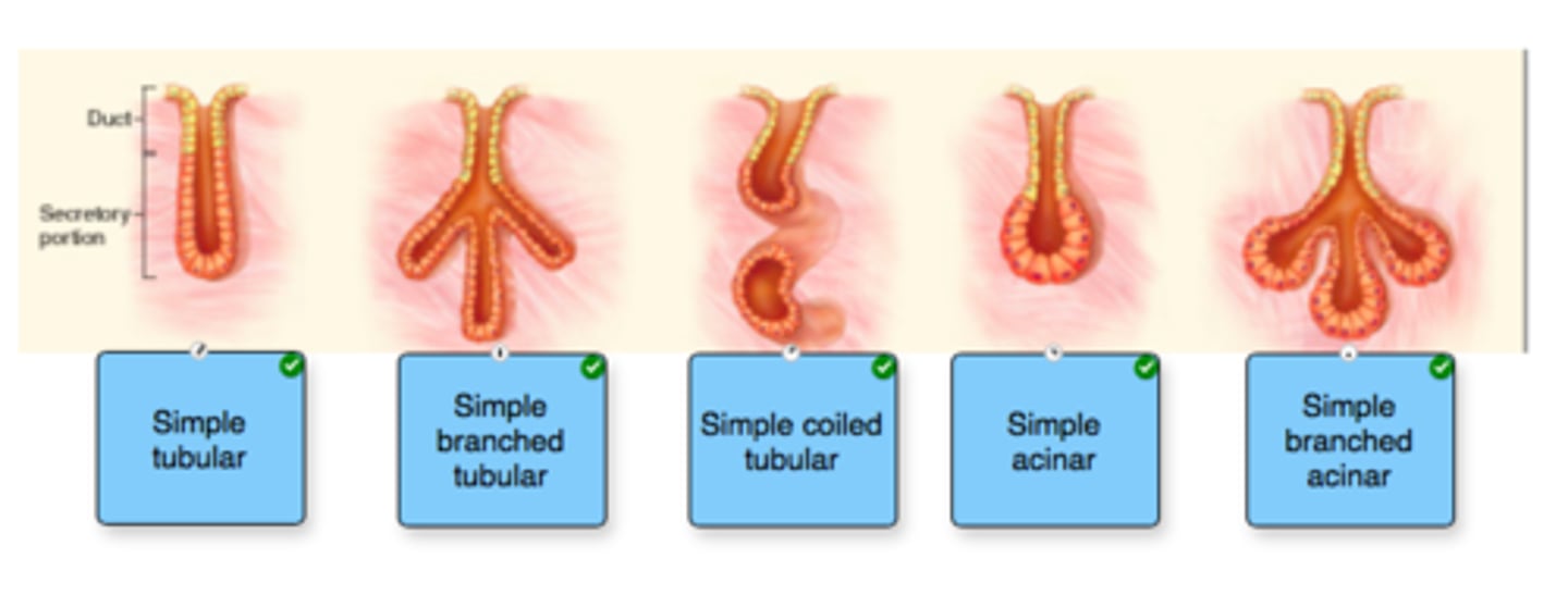
Label the types of simple exocrine glands based on their structural classification.
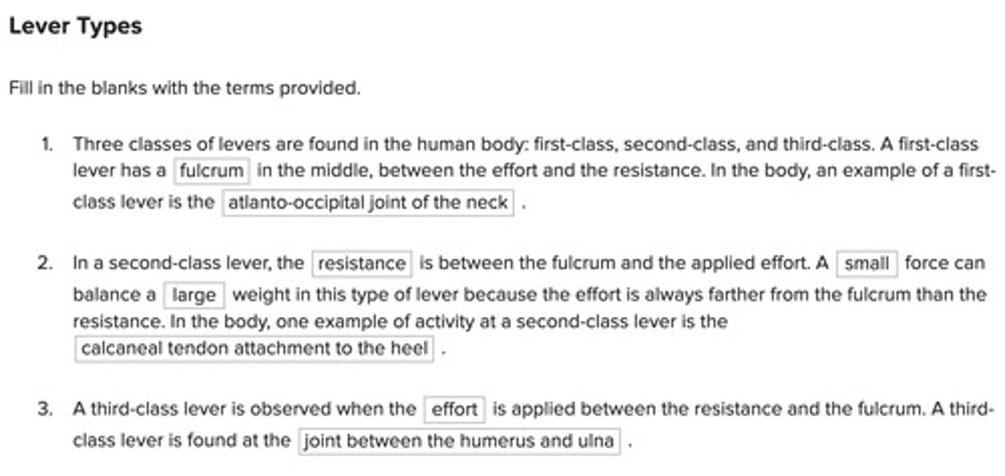
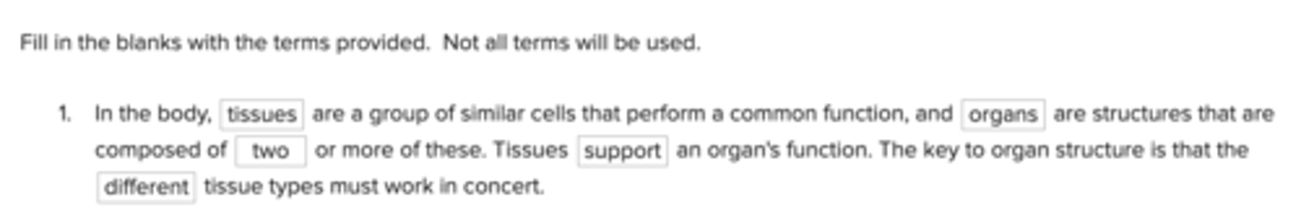
Fill in the blanks with the terms provided. Not all terms will be used.
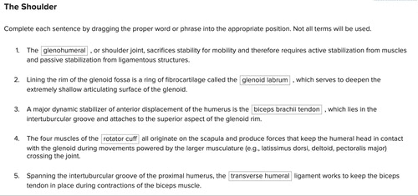
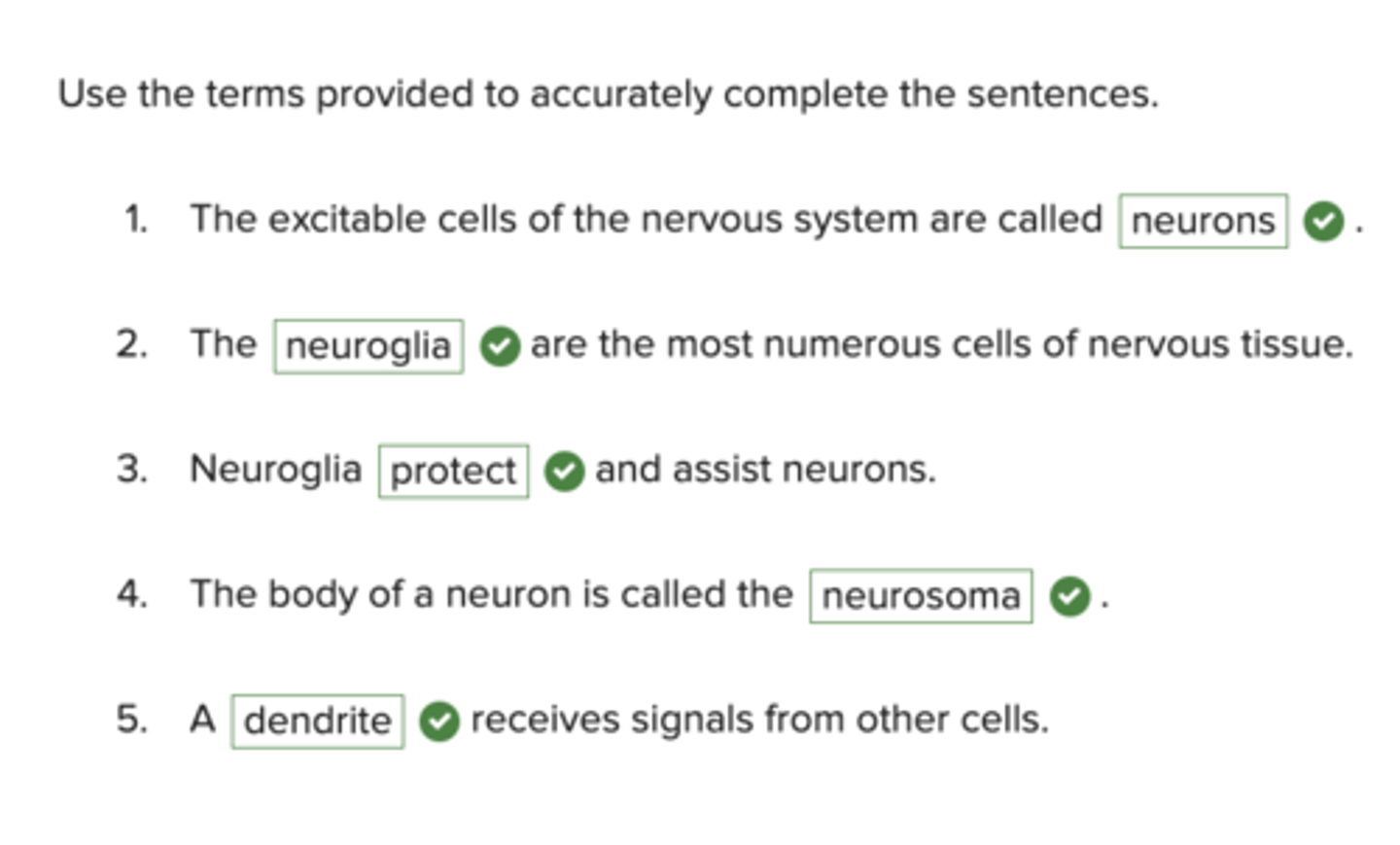
Use the terms provided to accurately complete the sentences.
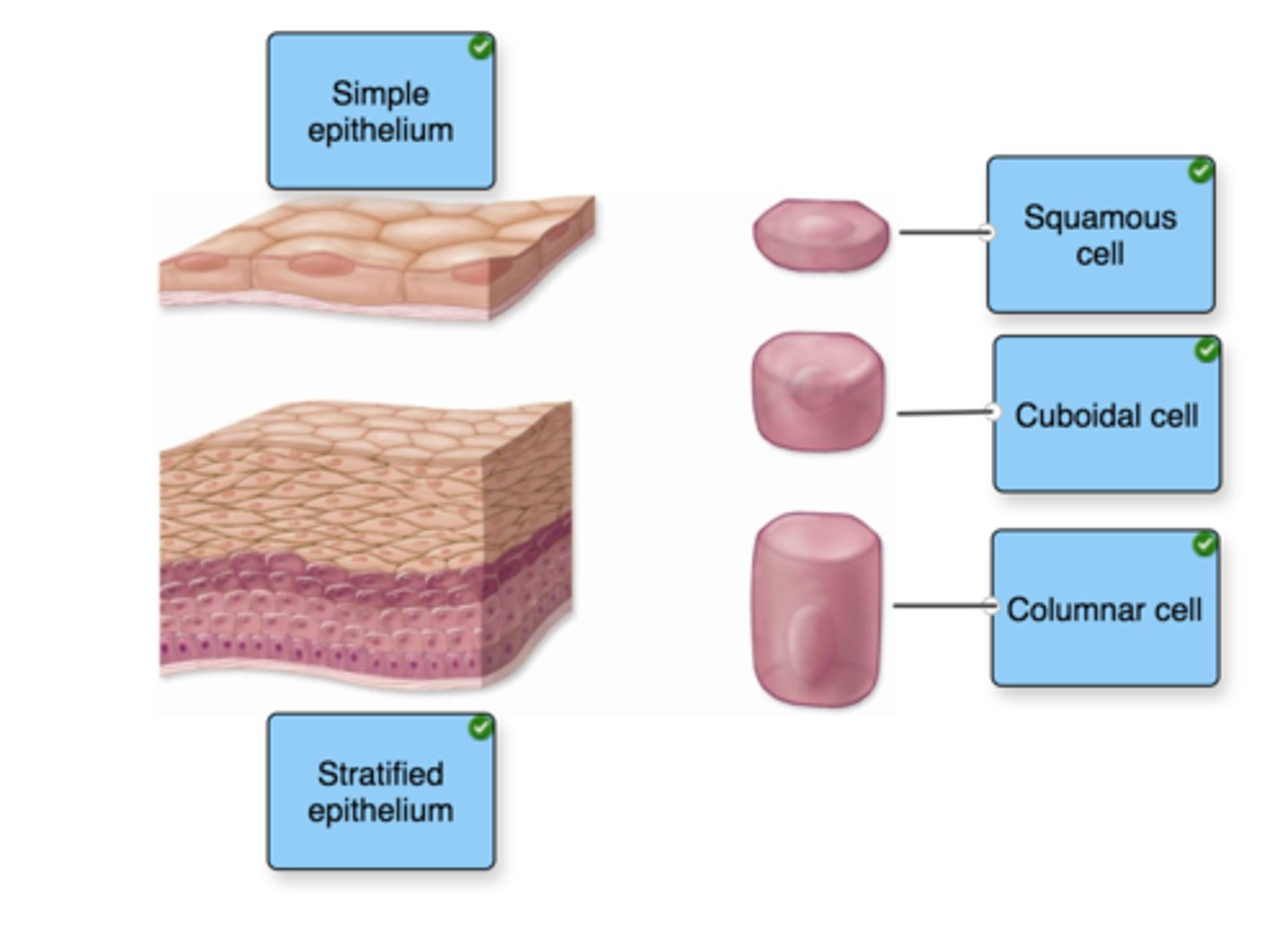
Label the types of epithelium based on their number of layers. Label cell types by shape. Not all terms will be used.
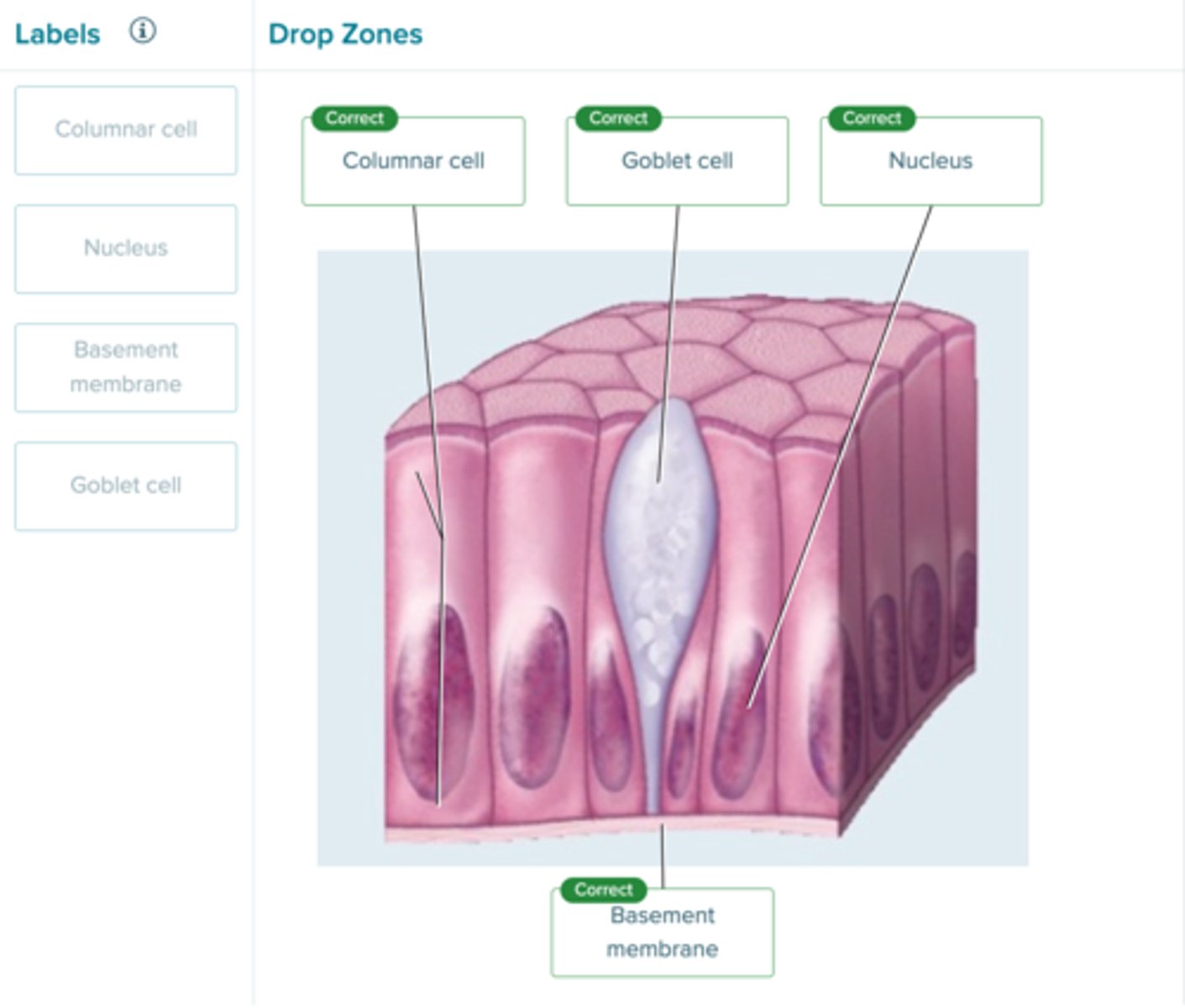
Label the components of a simple columnar epithelium.
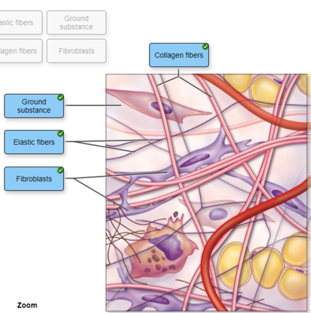
Correctly label the following areas on a slide of areolar connective tissue.
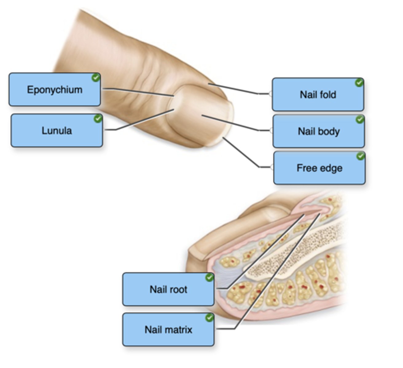
Correctly label the following structures of a nail.
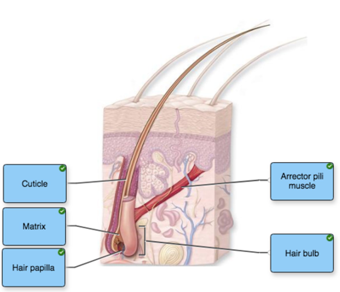
Label the structures of hair in the figure.
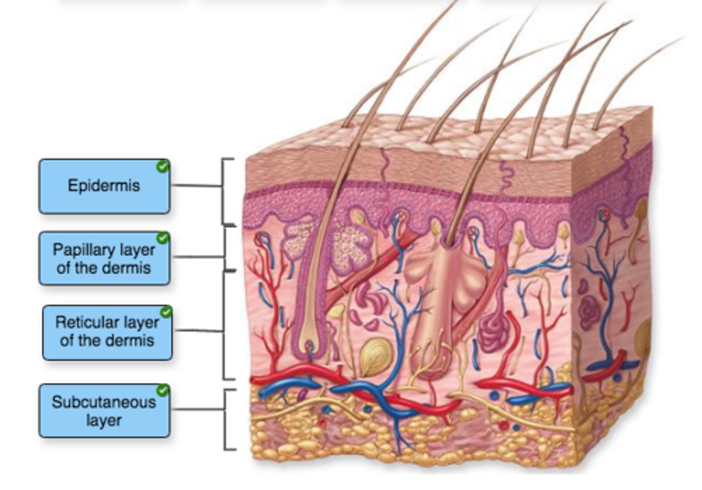
Label the structures of the integument.
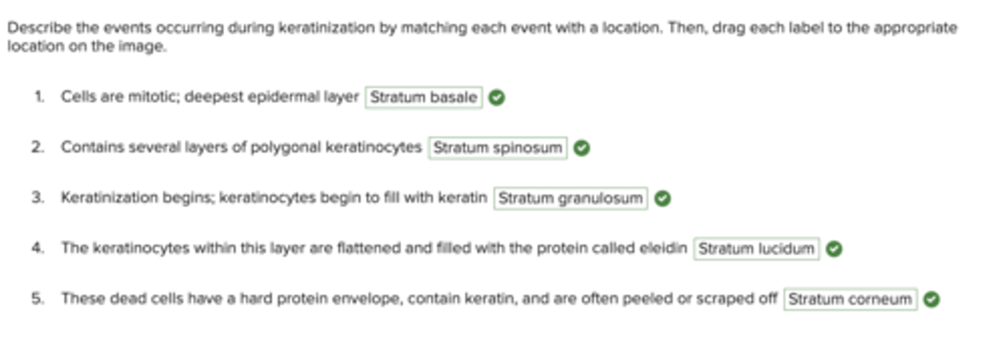
Describe the events occurring during keratinization by matching each event with a location. Then, drag each label to the appropriate location on the image.
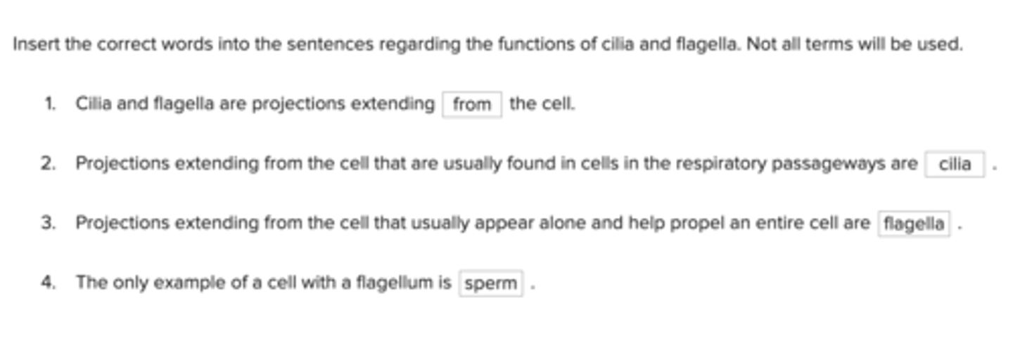
Insert the correct words into the sentences regarding the functions of cilia and flagella. Not all terms will be used.
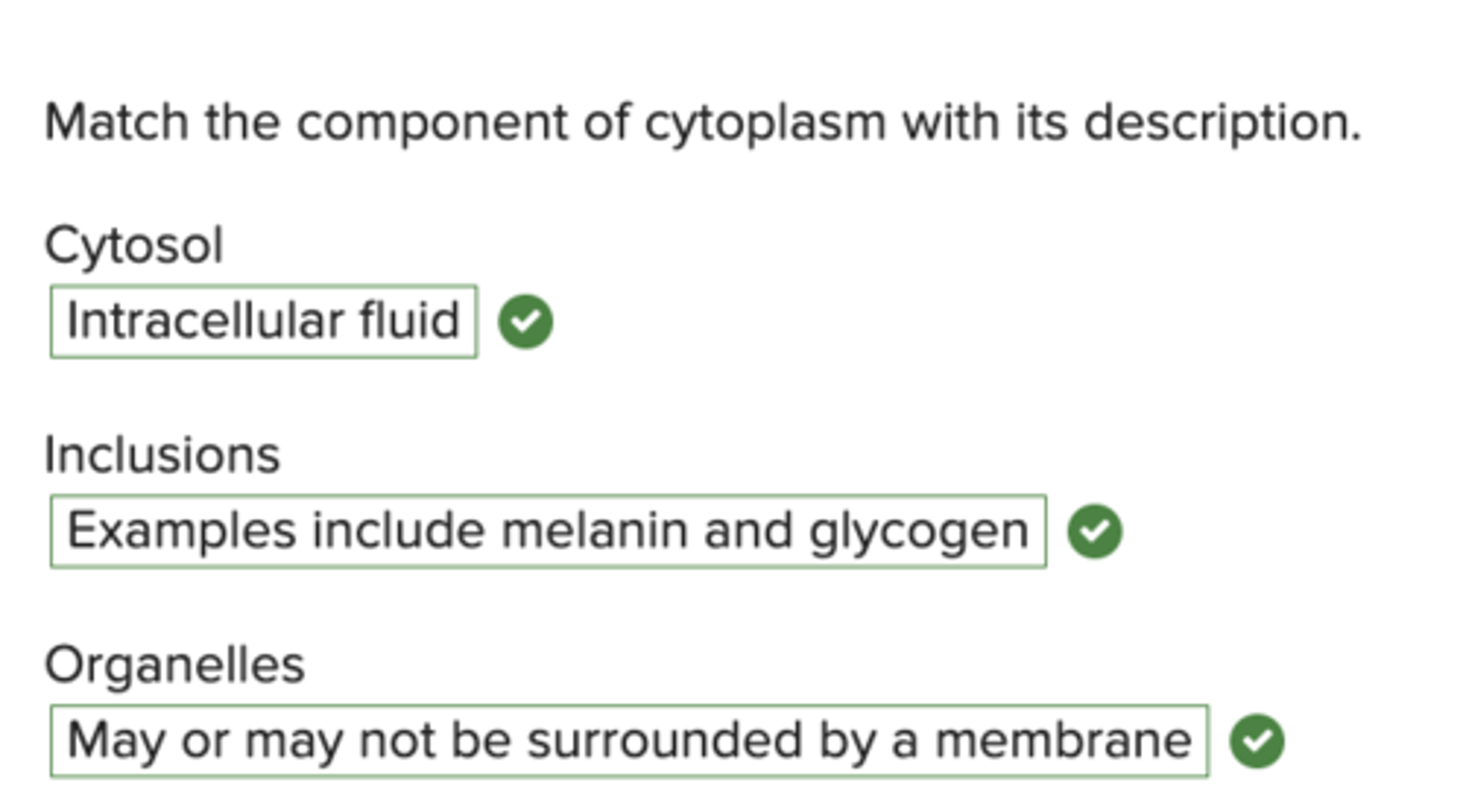
Match the component of cytoplasm with its description.
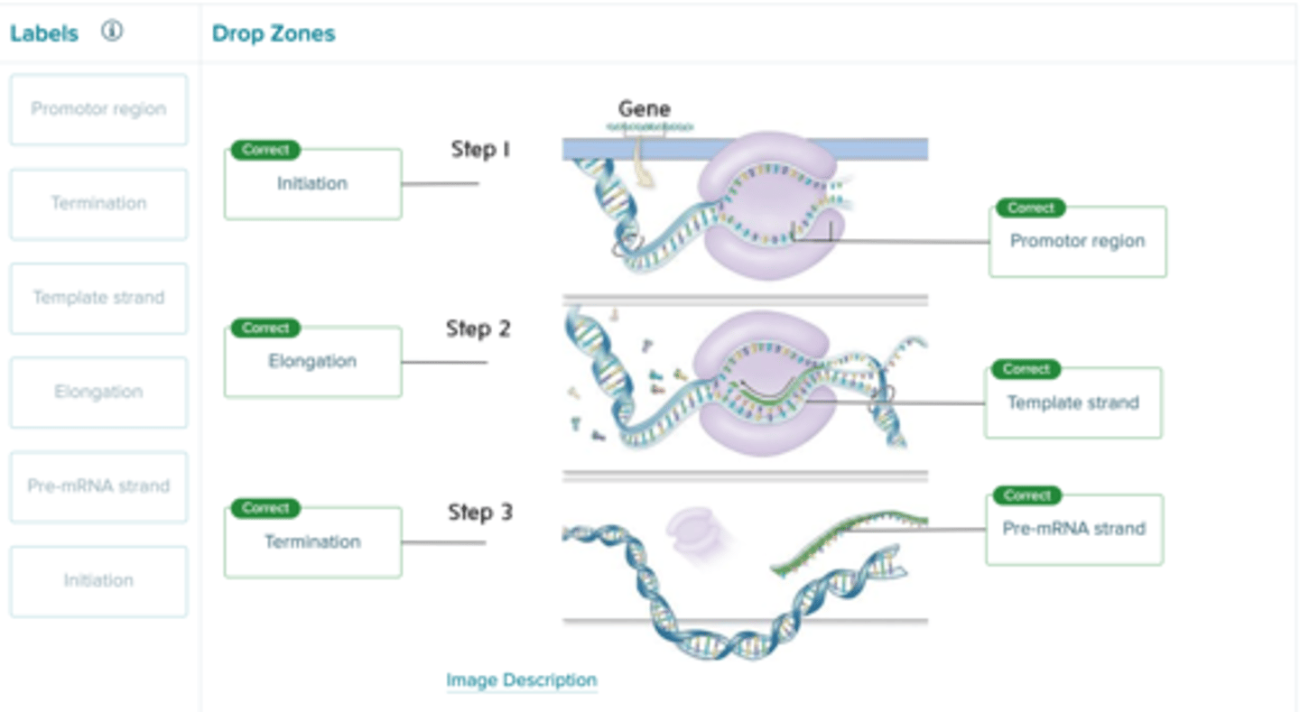
Identify the events that are occurring in the process of transcription in the figure.
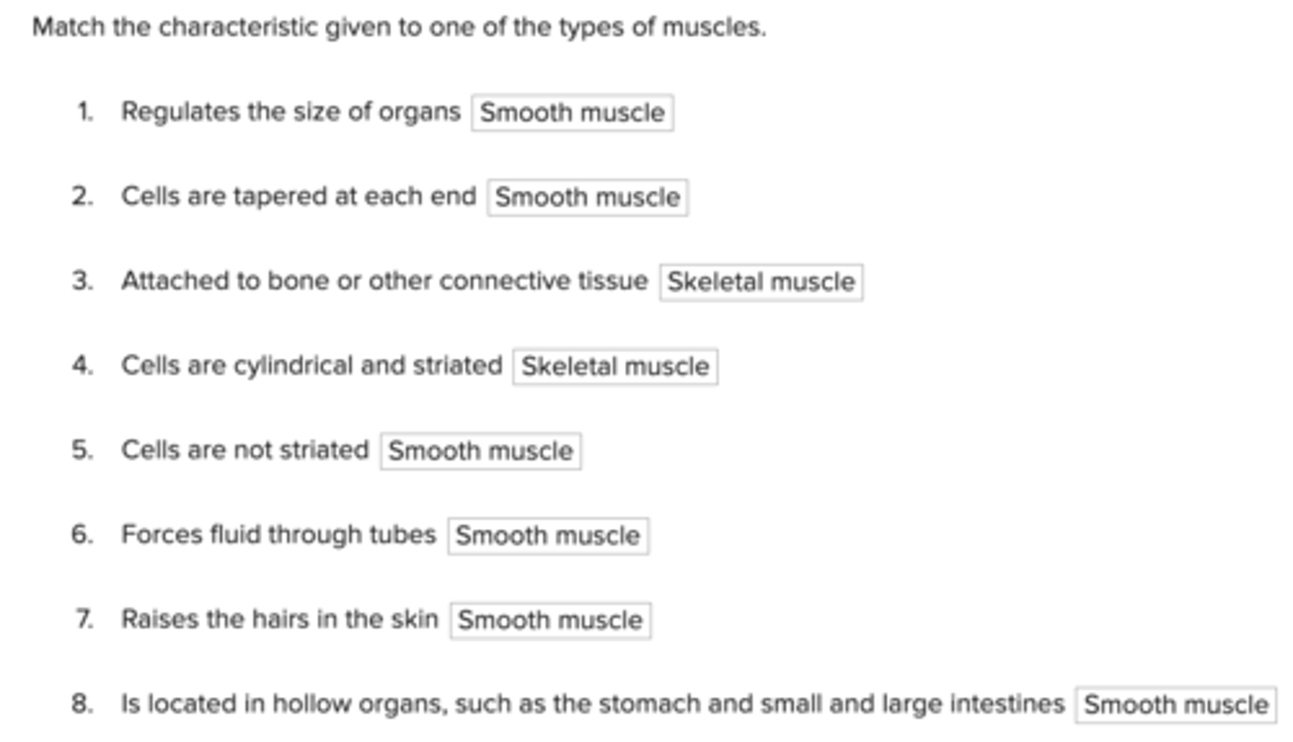
Match the characteristic given to one of the types of muscles.
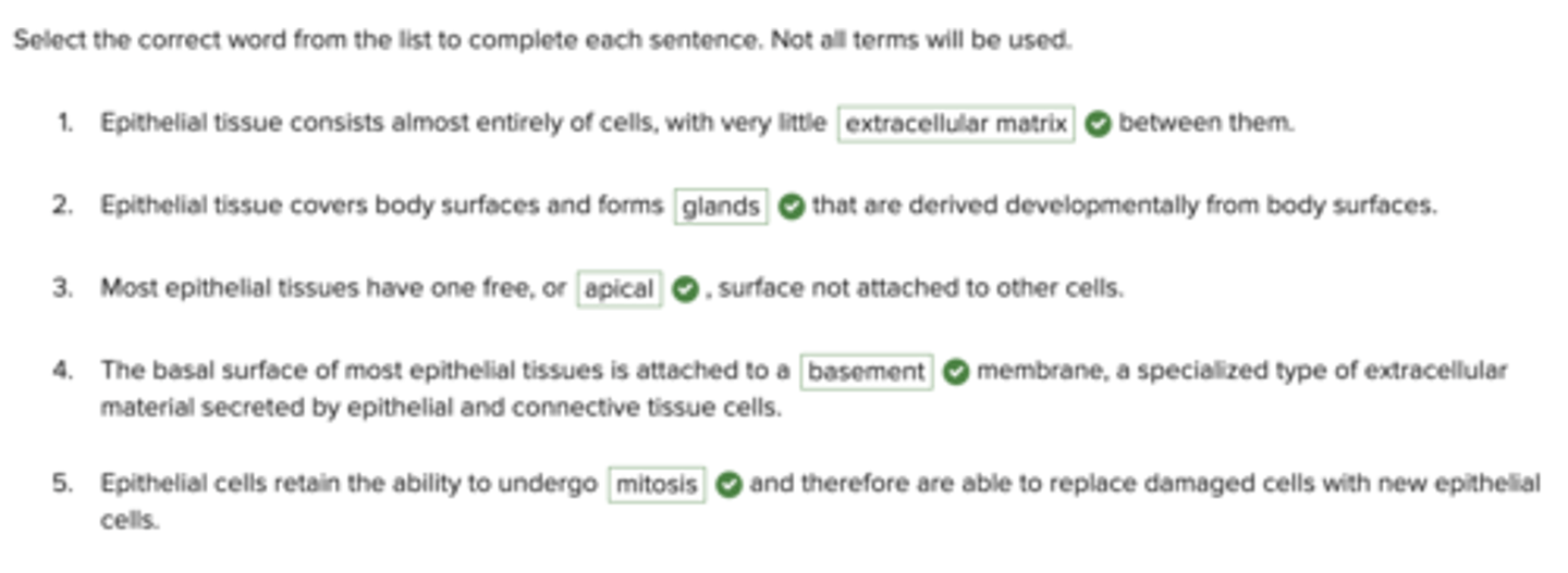
Select the correct word from the list to complete each sentence. Not all terms will be used.
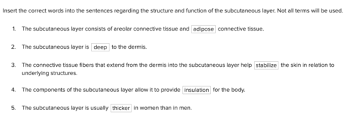
Insert the correct words into the sentences regarding the structure and function of the subcutaneous layer. Not all terms will be used.
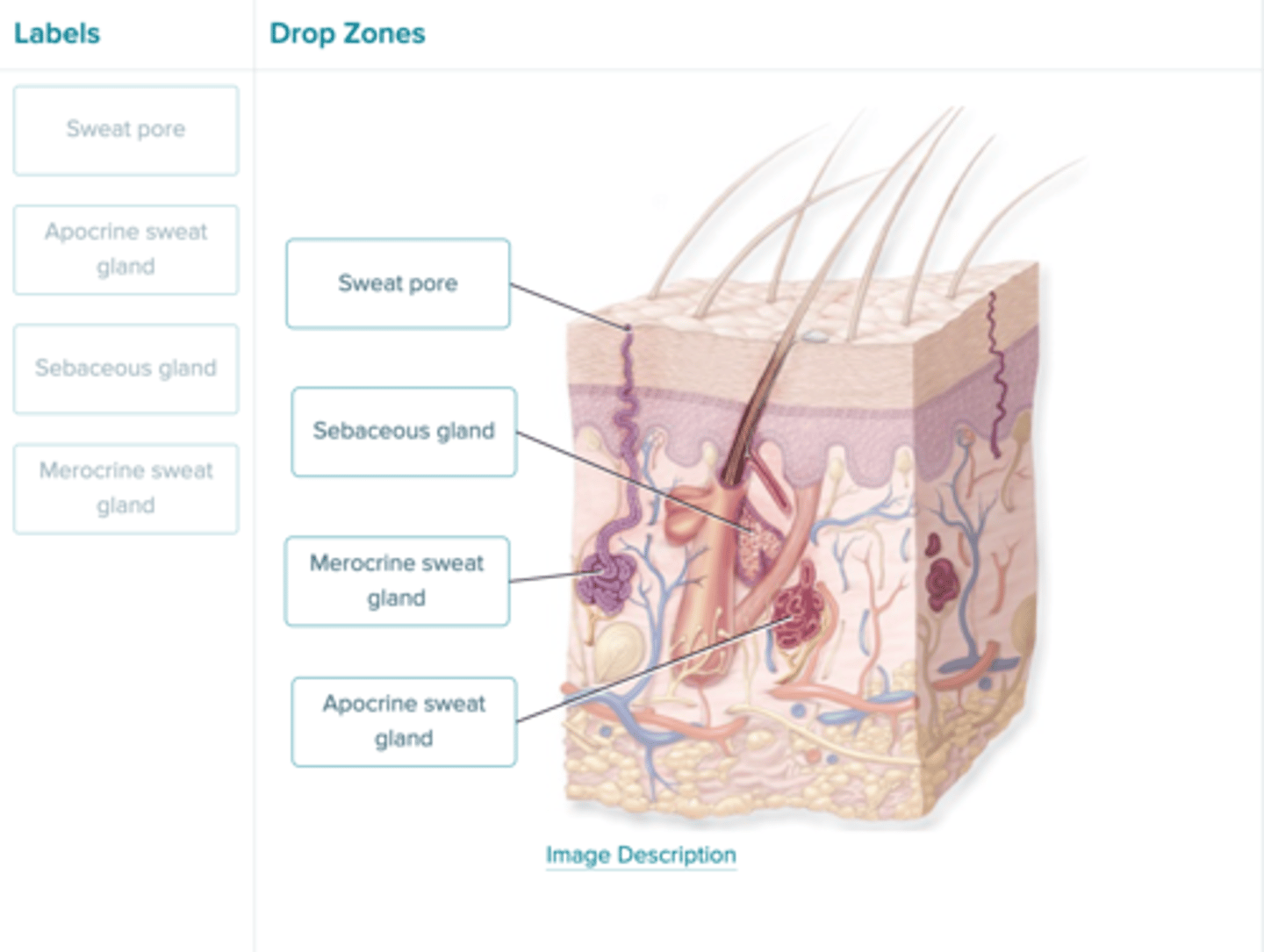
Identify the structures associated with the exocrine glands of the skin.
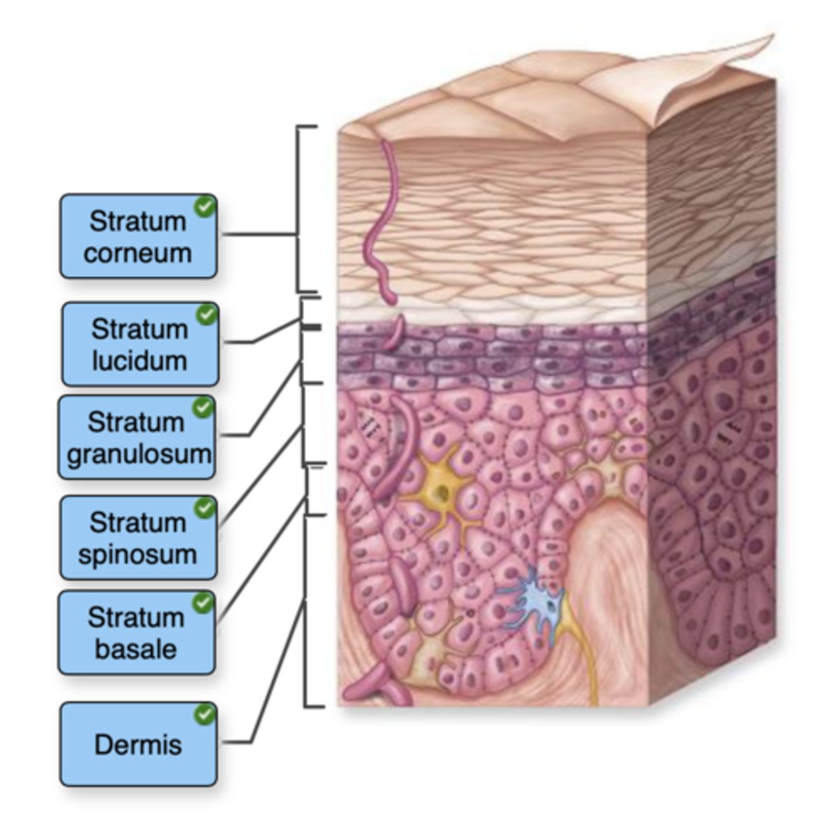
Label the layers of the epidermis.
come
lets
get
sun
burnt
Dermis
Fill in the blanks with the terms provided.
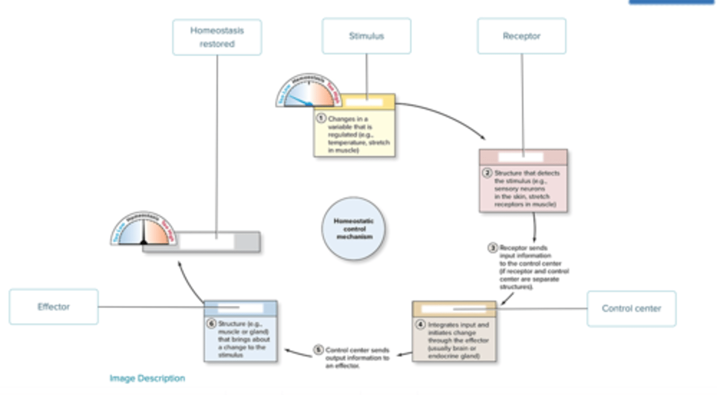
Label the components of a homeostatic control mechanism using the terms provided.
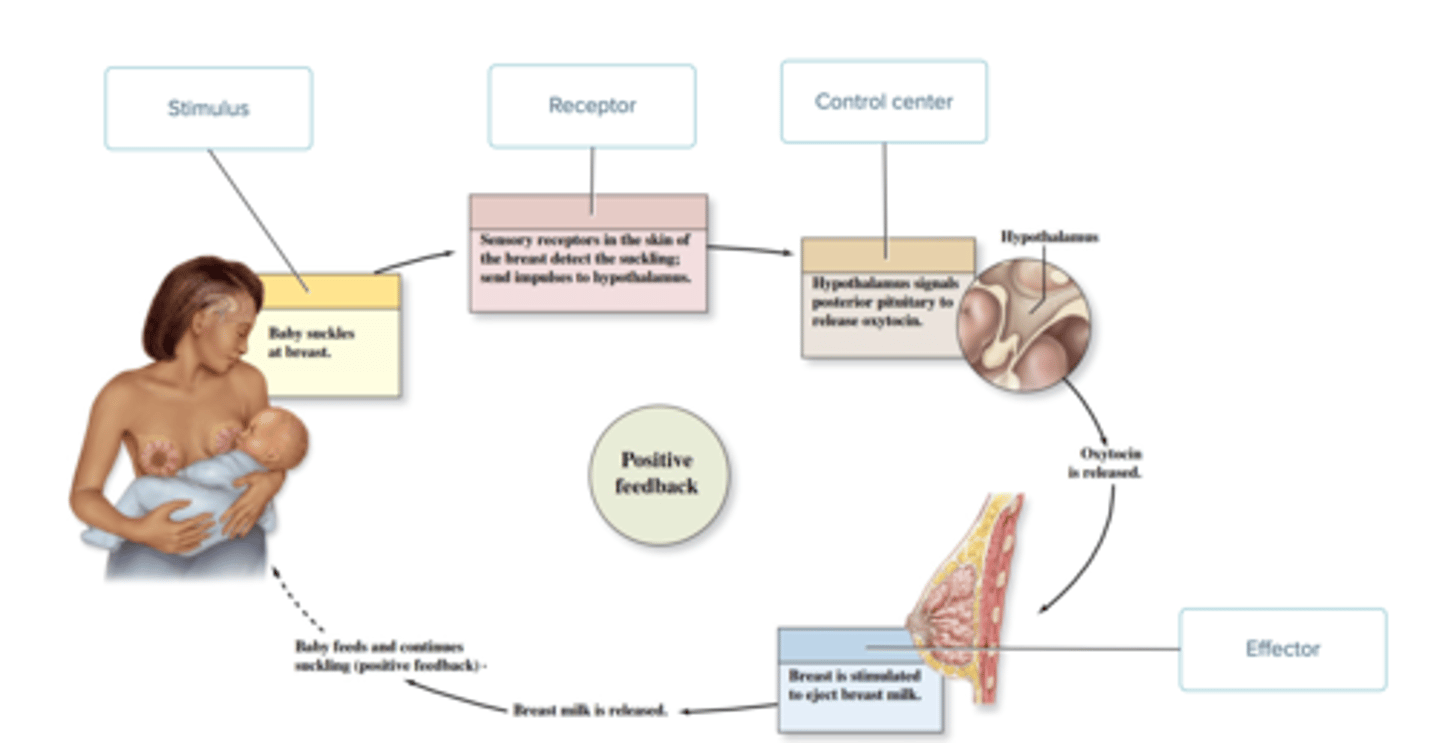
Label the positive feedback figure with the correct terms.
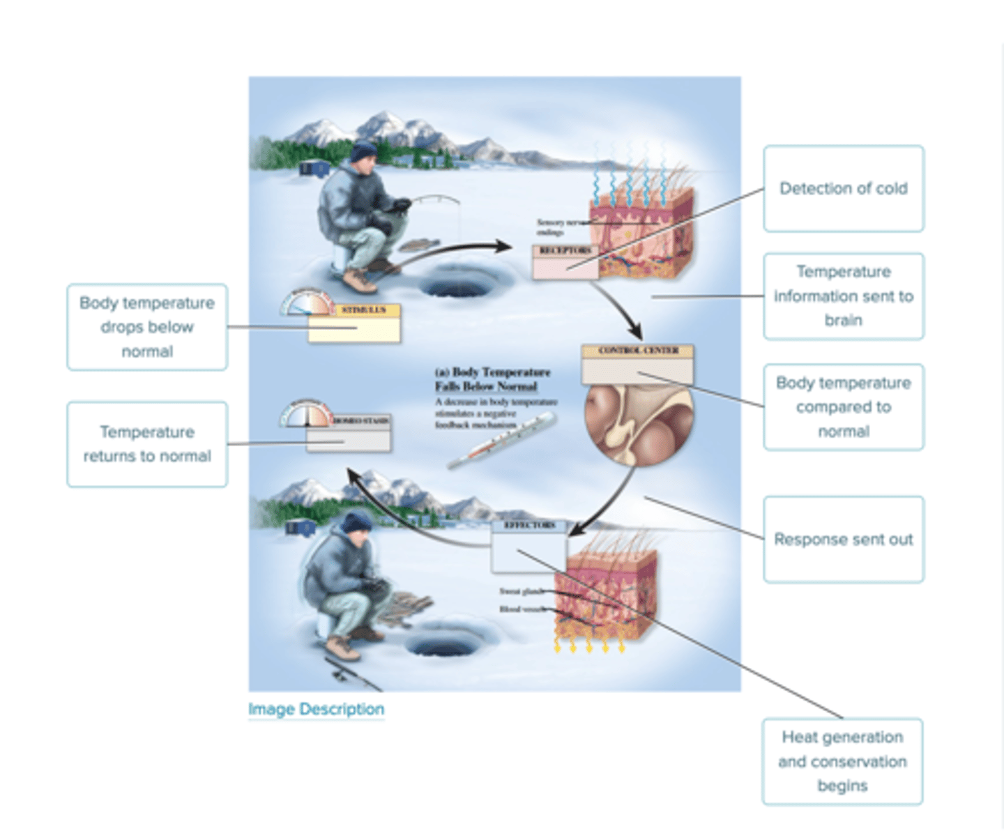
Label the figure for negative feedback regulation when the body temperature falls below normal.
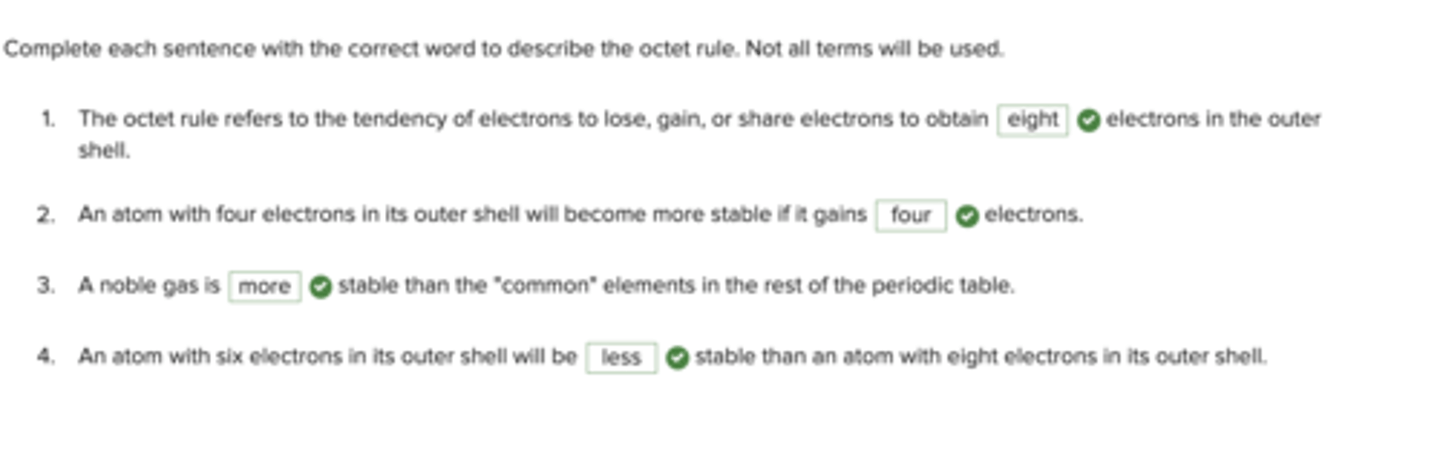
Complete each sentence with the correct word to describe the octet rule. Not all terms will be used.
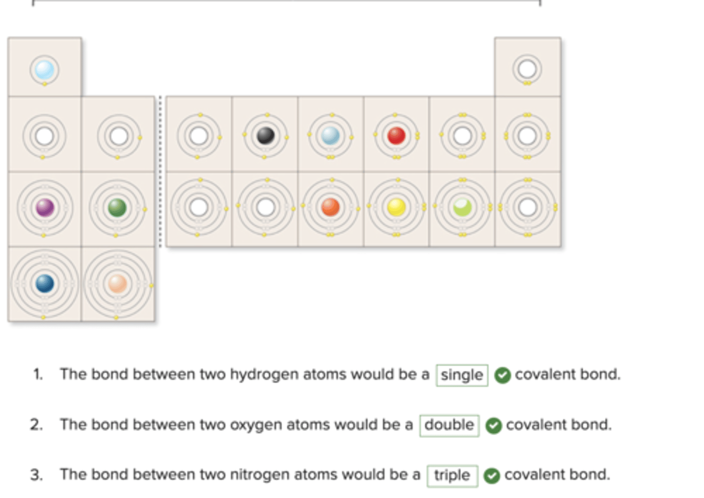
Using the periodic table as a guide and the number of valence shell electrons present, identify whether the elements will form single, double, or triple covalent bonds. Not all terms will be used.
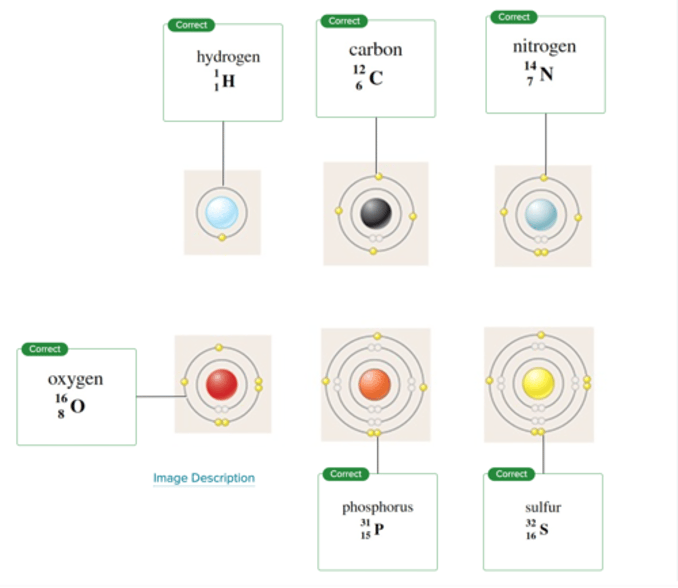
Label the following diagram with the appropriate terms.
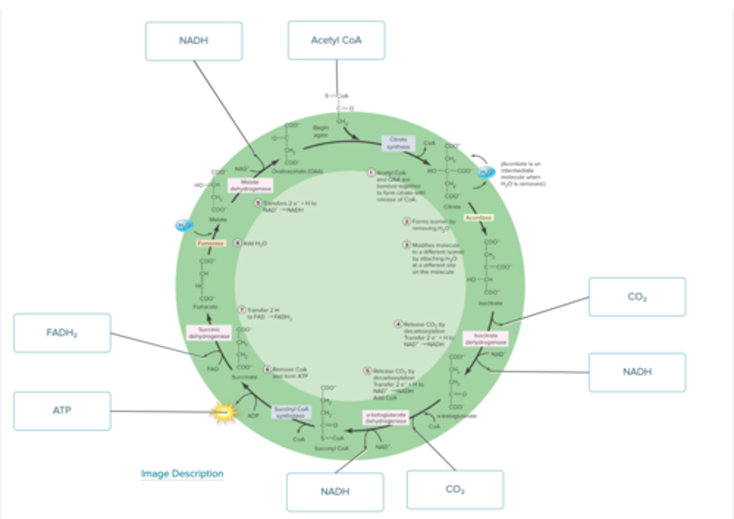
Label the figure with the items provided. Labels may be used once, more than once, or not at all.
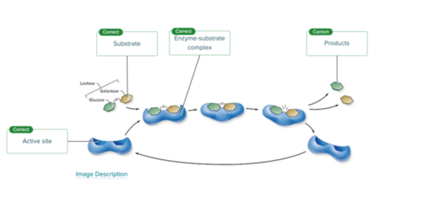
Label the figure below to assess your knowledge of the action of enzymes in biological cells.
Fill in the blanks with the terms provided. Not all terms will be used.
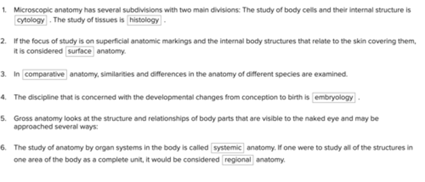
Complete the paragraph with the terms provided.
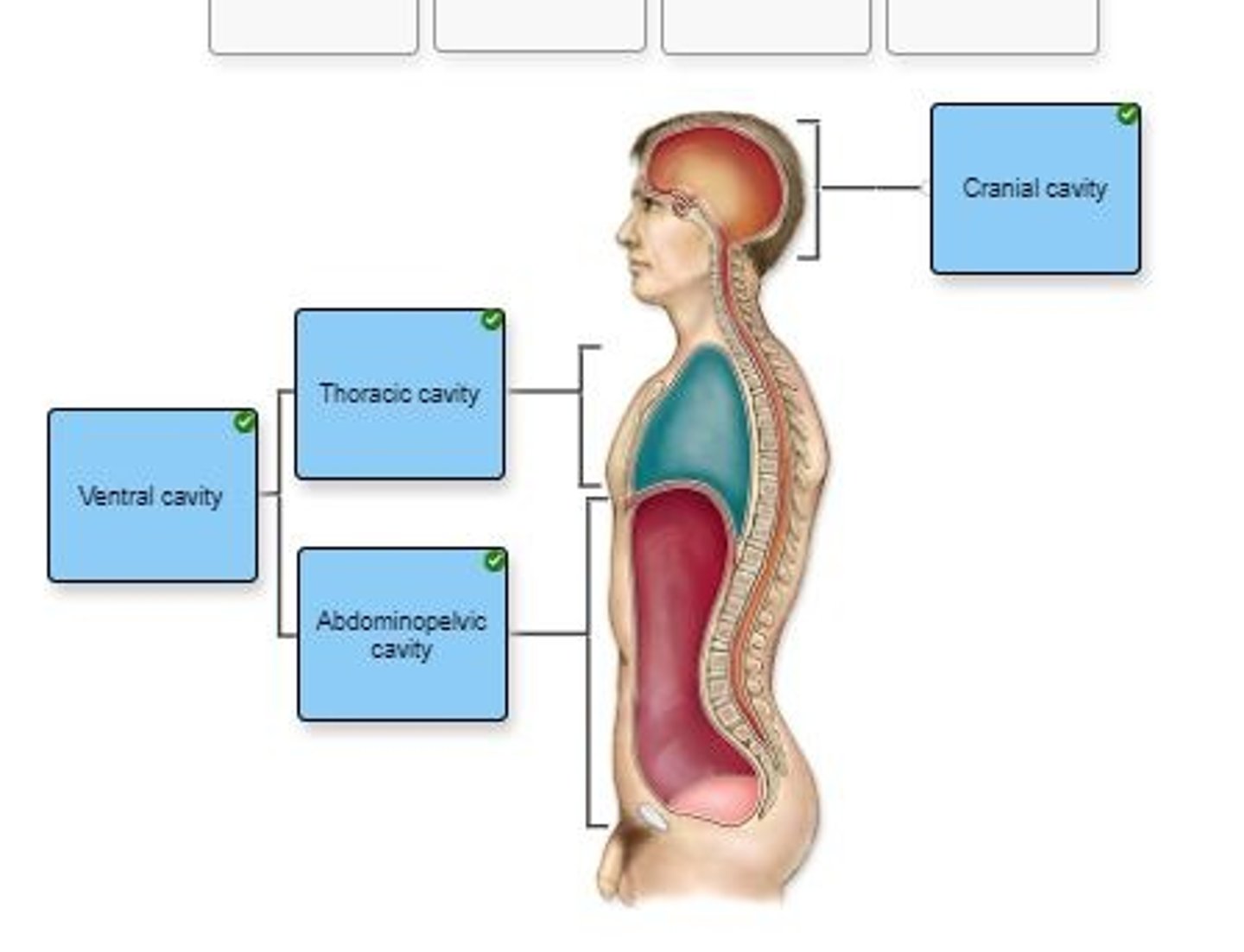
Label the body cavities in the figure
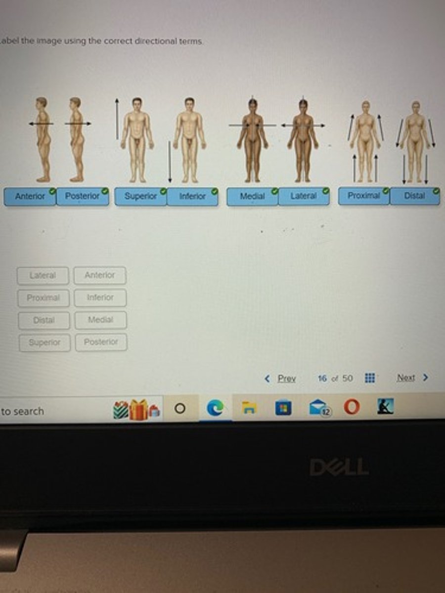
Label the image using the correct directional terms