chapter 18 : duplex ultrasound imaging of the upper extremity venous system
1/28
There's no tags or description
Looks like no tags are added yet.
Name | Mastery | Learn | Test | Matching | Spaced | Call with Kai |
|---|
No analytics yet
Send a link to your students to track their progress
29 Terms
stasis
thrombi in lower extremity often caused by —; not so in the upper extremity
soleal sinuses
upper extremities do not have —
superficial veins
affected more in arms than in legs; in addition, superficial thrombosis may have greater clinical significance in arm than leg
variable
venous anatomy of upper extremity is more — than lower extremity
superior vena cava thrombosis
facial swelling or dilated chest wall collaterals are suggestive of —
signs & symptoms of lower extremity
unilateral arm or hand swelling
superficial palpable cord
erythema
pain and tenderness
facial swelling or dilated chest wall collaterals
pulmonary embolism (PE)
upper extremity veins may also be evaluated in patients suspected of —
injury to vessels wall
upper extremity thrombosis is more commonly due to —
subclavian & internal jugular veins
commonly used for indwelling catheters and pacemaker wire introduction
can be associated with upper extremity thrombosis
peripherally inserted central catheters (PICCS)
is inserted through basilic or cephalic vein and then positioned near right atrium
can also cause thrombosis
paget-schroetter syndrome
venous thrombosis associated with compression of subclavian vein at the thoracic outlet
typically patients are young, athletic, and muscular males
effort thrombosis
paget-schroetter syndrome is also known as —
midrange (5-10 MHz) linear array
used to evaluate internal jugular, brachiocephalic, subclavian, axillary, deep brachial, and brachial veins
higher frequency (10-18 MHz) linear array
used for more superficial veins (cephalic and basilic) and small forearm veins (radial and ulnar)
curved or sector array
may be useful for deeper vessels near clavicle and sternum
carotid artery
can be used as a landmark to identify internal jugular vein (IJV)
sitting or standing
IJV will be collapsed if patient is —
external jugular vein (EJV)
serves as an important collateral pathway
runs without an accompanying artery very close to skin surface
usually terminates into subclavian vein
Brachiocephalic veins
most often evaluated at the confluence of the IJV and subclavian veins
compressions cannot be performed
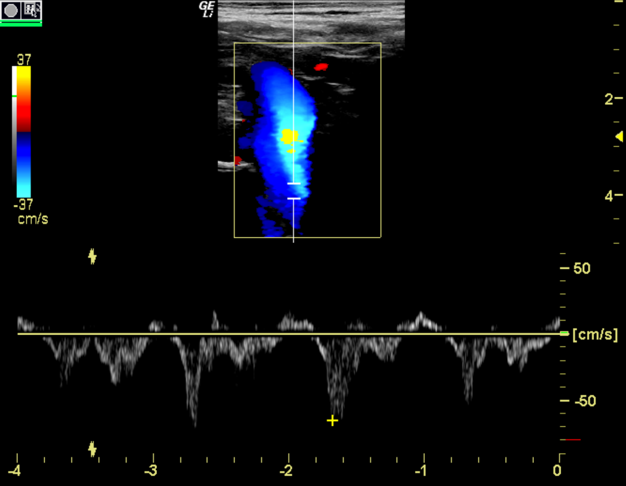
ultrasound image of the brachiocephalic vein
subclavian vein
can be visualized above or below the clavicle
accompanied by an artery
cephalic vein terminates into the — just after it passes under clavicle
taking a quick, deep breathing through pursed lips will cause the vein to collapse
cephalic vein
travels superfically near skin across shoulder and along anterolateral border of biceps muscle
communicates with median cubical vein at antecubital fossa
in the forearm, usually travels as two vessels. one on the collar aspect and one on the dorsal aspect
median cubical vein
connects cephalic and basilic veins
resides in the antecubital fossa but pattern of connection is variable
common site of venipuncture and therefore thrombus
axillary vein
terminates at the junction of the cephalic and subclavian veins
accompanied by artery
courses deeply from shoulder through axilla
basilic vein terminates into — in near the distal axilla
brachial vein
terminates when the basilic vein enters to become axillary vein
accompanied by an artery
formed by junction of the two radial and two ulnar arteries near the antecubital fossa
radial veins (thumb side)
course along velar aspect of forearm
very small vessels and rarely involved in venous thrombosis
paired with an artery
ulnar veins (pinky side)
also travel along velar aspect of forearm
paired vein that also has an accompanying artery
basilic vein
terminates into the axillary vein in upper arm
courses medially and superficially without a companion artery
usually largest vein in upper arm region
communicates with cephalic vein via median cubital vein in the antecubital fossa
respiration
vein diameter will change slightly with —