12 - Visual Pathways: Form Serves Function
1/16
There's no tags or description
Looks like no tags are added yet.
Name | Mastery | Learn | Test | Matching | Spaced | Call with Kai |
|---|
No analytics yet
Send a link to your students to track their progress
17 Terms
Microsacades
mini involuntary eye movements
constantly doing
very fast both eyes move at same time
occurs during fixational gaze
Photoreceptors — Rods
most sensitive to dim light (scotopic)
do not convey sense of color
do not respond to specific wavelengths
lower intensity vision — lower acuity
Photoreceptors — Cones
work better in bright light (photopic)
responsible for acute detail
respond to both black and white (intensity)
colour/wavelength specific
green cones — green sensitive, red cones — red sensitive, blue cones — blue sensitive
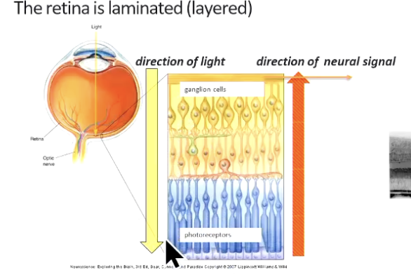
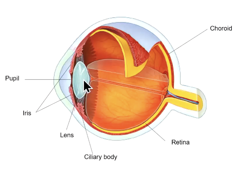
Retinal Anatomy
light enter via pupil — pupil is hole in centre of circular ring of smooth muscle
info travels from photoreceptors to retinal ganglion cells (RGCs)
axons of RGCs exit the retina to form the optic nerve — projects to rest of brain
light must travel through all layers of the retina to reach the receptors
except in the fovea — other retinal layers pushed aside — light falls directly onto photoreceptors
decrease in intensity of signal as it goes through all the layers — decreases our abilities — but in fovea only photoreceptors (only cones)
gives us max resolution in the fovea
Opthalmoscopic Appearance of the Eye
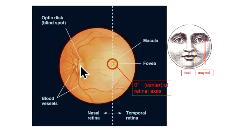
at blind spot where RGCs become myelinated
all the blood vessel come in at blind spot area
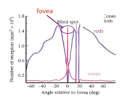
Retinal Topography — Rod Density
outnumber cones overall
reach peak density abt 7-8mm from fovea at rod ring
absent from fovea
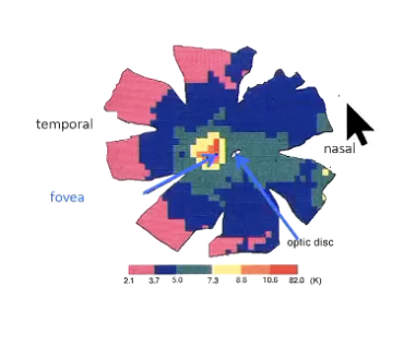
Retinal Topography — Cone Density
5 million cones in avg human retina —- 20% in central 6mm
cones outnumber rods in central retina
region of elevated cone density surrounds fovea and extends into nasal retina
Retinal Organisation — Specialisations of Fovea
high acuity, colour vision
peak density of cone photoreceptors in fovea
absence of rods
local absence of retinal blood vessels
absence of inner retinal layers
1:1 r/s of receptors projecting onto RGCs
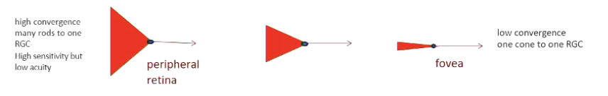
Retinal Organisation — Specialisations of Peripheral Retinal
low light (scotopic) vision
low density of cone receptors
moderate to high density of rods
retinal blood vessels present
inner retinal layers
high convergence of receptors onto RGCs
Sensitivity vs Acuity — Cost of Convergence
photoreceptor → bipolar → RGC (axons form optic nerve/tract) → targets
single RGC can receive input from
single cone
multiple cones and rods
thousands of rods
this cluster of cells activating an RGC — defines its receptive field — size of field changes due to degree of convergence and varies across retina
sensitivity/acuity tradeoff
peripheral retina — high convergence of rod input onto RGC — high sensitivity — useful in dim light
BUT large receptive fields = low acuity (useful in dim/scotopic conditions)
fovea — no convergence — very small RGC receptive fields
high acuity BUT low sensitivity (only useful in bright light)
RGC Pathways
each RGC has one axon that exits retina to form optic nerve (and the optic tract)
each RGC axon projects to one/more structures:
lateral geniculate nucleus (LGN) of thalamus
projects to primary visual cortex (v1) — CONSCIOUS visual pathway, visual perception
superior colliculus — midbrain
UNCONSCIOUS — visual reflexes to direct gaze
visual, auditory and somatosensory integration
pretectal nucleus — midbrain
UNCONCIOUS — reflexes for pupil diameter and lens accomadation
projects to edinger-westphal nucleus
pulvinar of thalamus — thalamus
UNCONCIOUS — spatial attention helps stabilise retinal image
role in saccades
suprachiasmatic nucleus — hypothalamus
UNCONCIOUS — circadian rhythm
synchronises to day/night cycle
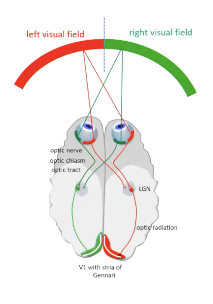
Retinogeniculocortical Pathway — Conscious **
visual fields of our 2 eyes overlap
given object will be viewed by temporal retina of one eye and nasal retina of other
for organised representation of visual world — given object needs to be represented in one location
optic chiasm sorts this out — axonx from nasal retina CROSS at optic chiasm — partial decussation
axons then project to LGN — map visuotopically
LGN projects to V1 via optic radiations — preserves visuotopy
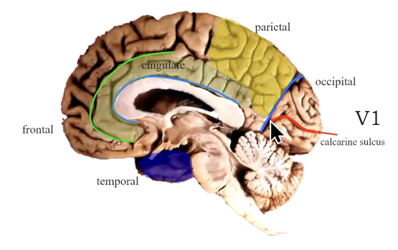
V1 sits on upper and lower banks of calcarine sulcus
also defined by Stria of Gennari
enables left visual field to be seen by right V1 and right visual field to be seen by left V1
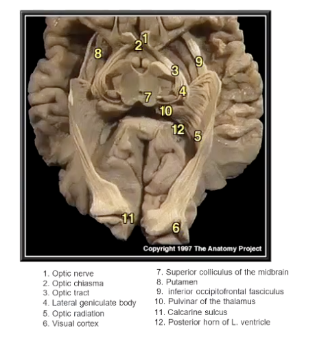
Optic Anatomy
Primary Visual Area Occupies Banks of Calcarine Sulcus
Beyond V1

Pupillary Light Reflex
unconscious visual circuit
pupil is hole in centre surrounded by iris — which is 2 layers of smooth muscle
then ciliary body — contains ciliary muscle
suspensory ligaments — changes in the tension forces changes of lens — affects focusing for diff types of vision
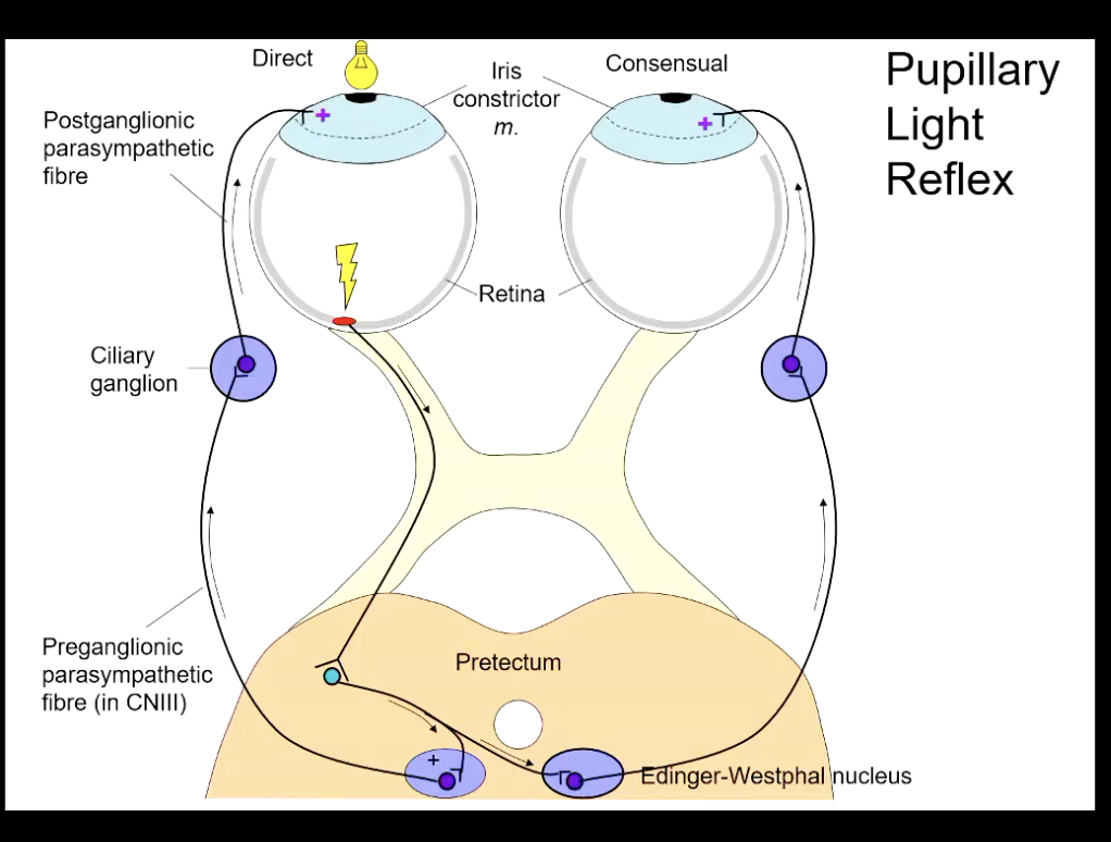
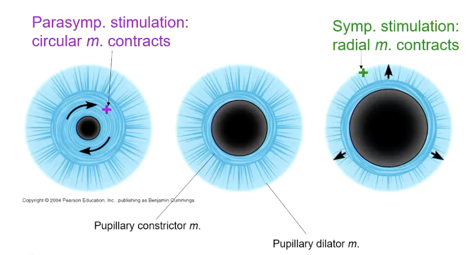
Autonomic Innervation of the Iris
inner layer of smooth muscle — pupillary constrictor muscle
has smooth muscles fibres that are arranged in circular orientation — constriction decreases pupil diameter
mediated by parasympathetic nervous system
outer layer — pupillary dilator muscle
has radial fibres — when constricted increase in diameter
flight vs fight situations sympathetic nervous system