A&P 1 Mod 4.2 Fibrous Joints
1/8
Earn XP
Description and Tags
Fibrous Joints
Name | Mastery | Learn | Test | Matching | Spaced | Call with Kai |
|---|
No analytics yet
Send a link to your students to track their progress
9 Terms
Fibrous Joint Info
At a fibrous joint, the adjacent bones are directly connected to each other by fibrous connective tissue, with no joint cavity separating them. The gap between the bones may be narrow or wide. There are three types of fibrous joints: sutures, syndesmosis, and gomphoses.
Suture
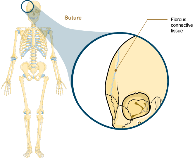
A suture is the narrow fibrous joint found only between most bones of the skull.
All the bones of the skull, except for the mandible, are jointed to each other by a fibrous joint called a suture (Figure 4.10).
The short, fibrous connective tissue found at a suture strongly unites the adjacent skull bones, protecting the brain and forming the face.
In adults, the skull bones are close together, and fibrous connective tissue fills the narrow gap between the bones. the suture is frequently convoluted, forming a tight union that prevents most movement between the bones.
Sutures are functionally classified as a synarthrosis, although some sutures may allow for slight movements between the cranial bones.
Fontanelles
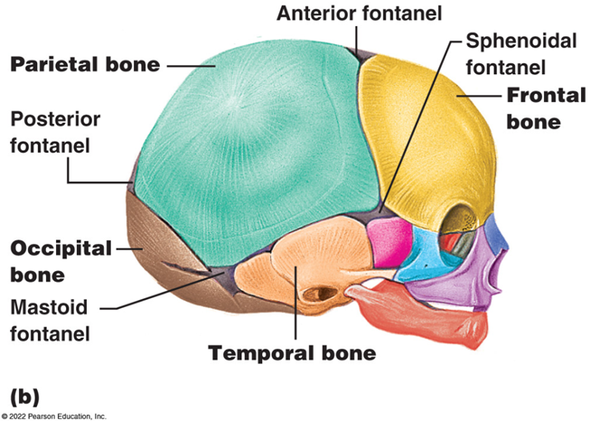
In newborns and infants, the areas of connective tissue between the bones are much wider, especially in those areas on the top and sides of the skull. These broad areas of connective tissue are called fontanelles.
During birth, the fontanelles provide flexibility to the skull, allowing the bones to push closer together or to slightly overlap, aiding movement of the infant's head through the birth canal.
After birth, these expanded regions of connective tissue also allow for rapid growth of the skull and enlargement of the brain in infancy and early childhood.
The fontanelles greatly decrease in width during the first year after birth as the skull bones enlarge and, for most children, are closed by age two. When the connective tissue between the adjacent bones is reduced to a narrow layer, these fibrous joints are called sutures.
Synostosis
At some sutures, the connective tissue will ossify and be converted into bone, causing the adjacent bones to completely fuse to each other. Fusion between bones is called synostosis, which means “joined by bone”.
Examples of synostosis fusions between cranial bones are found both early and late in life.
At the time of birth, the frontal and maxillary bones consist of right and left halves joined together by sutures, which disappear by the eighth year as the halves fuse together to form a single bone.
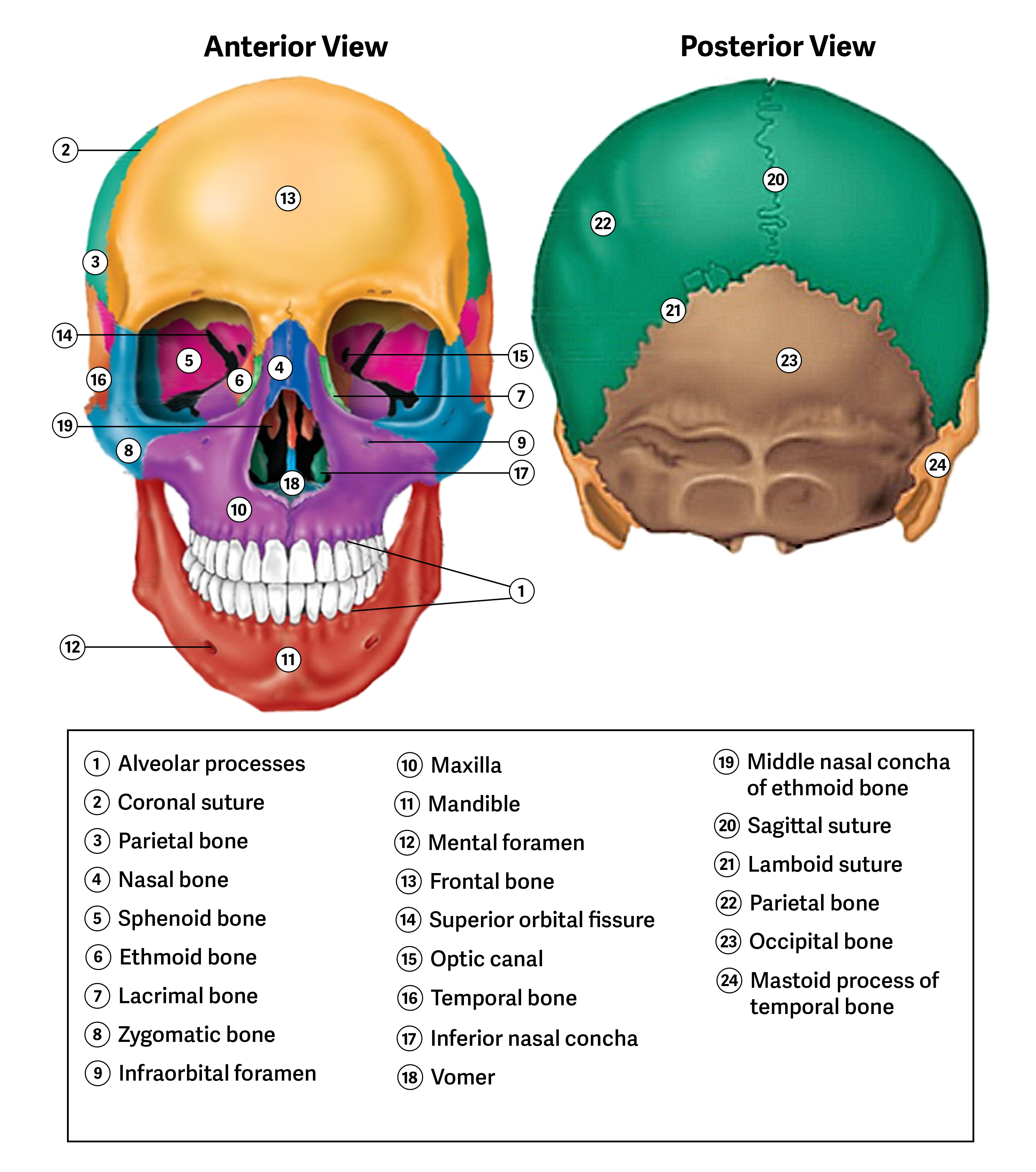
Late in life, the sagittal, coronal, and lambdoid sutures (Figure 4.11) of the skull begin to ossify and fuse, causing the suture line to gradually disappear.
syndesmosis
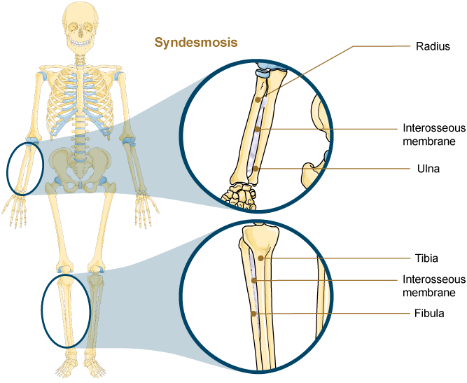
A syndesmosis is a type of fibrous joint in which two parallel bones are united to each other by longer fibrous connective tissue. The gap between the bones may be narrow, with the bones joined by ligaments. The gap may also be wide and filled in by a broad sheet of connective tissue called an interosseous membrane
In the forearm, the wide gap between the shaft portions of the radius and ulna bones is strongly united by an interosseous membrane. Similarly, in the leg, the shafts of the tibia and fibula are also united by an interosseous membrane.
Syndesmoses MORE
The syndesmoses found in the forearm and leg serve to unite parallel bones and prevent their separation. However, a syndesmosis does not prevent all movement between the bones, so this type of fibrous joint is functionally classified as an amphiarthrosis. In the leg, the syndesmosis between the tibia and fibula strongly unites the bones and allows for little movement. This provides strength and stability to the leg and ankle, which are important while standing, walking, and running. In the forearm, the interosseous membrane is flexible enough to allow for rotation of the radius bone during forearm movements. In contrast to the stability provided by the tibiofibular syndesmosis, the flexibility of the interosseous membrane in the forearm allows for much greater mobility of the forearm.
The interosseous membranes of the leg and forearm also provide areas for muscle attachment. Damage to a syndesmotic joint from a bone fracture can also result in a tear of the interosseous membrane. This type of fracture will produce pain, loss of stability of the bones, and may also damage the muscles attached to the interosseous membrane. If the fracture site is not properly immobilized with a cast or splint, contractile activity by these muscles can cause improper alignment of the broken bones during healing.
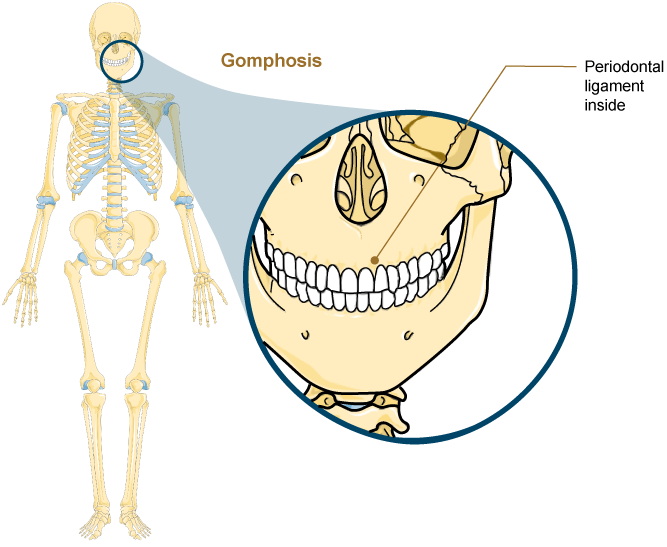
Gomphosis
A gomphosis, which means "fastened with bolts" is the specialized fibrous joint that anchors the root of a tooth into its bony socket (Figure 4.14)
Gomphoses occur within the maxillary or mandible bones of the skull.
Spanning between the bony walls of the socket and the root of the tooth are numerous short bands of dense connective tissue, each of which is called a periodontal ligament.
Due to the immobility of a gomphosis, this type of joint is functionally classified as a synarthrosis.
The ___ the ligament fibers of a synarthrosis, the ____ the mobility of the joint
The ___ the ligament fibers of a syndesmosis, the ____ the mobility of the joint.