CHAPTER 32: IDENTIFICATION OF RESTORATIONS, DENTAL MATERIALS, AND FOREIGN OBJECTS (PAGE 378)
1/9
There's no tags or description
Looks like no tags are added yet.
Name | Mastery | Learn | Test | Matching | Spaced | Call with Kai |
|---|
No analytics yet
Send a link to your students to track their progress
10 Terms
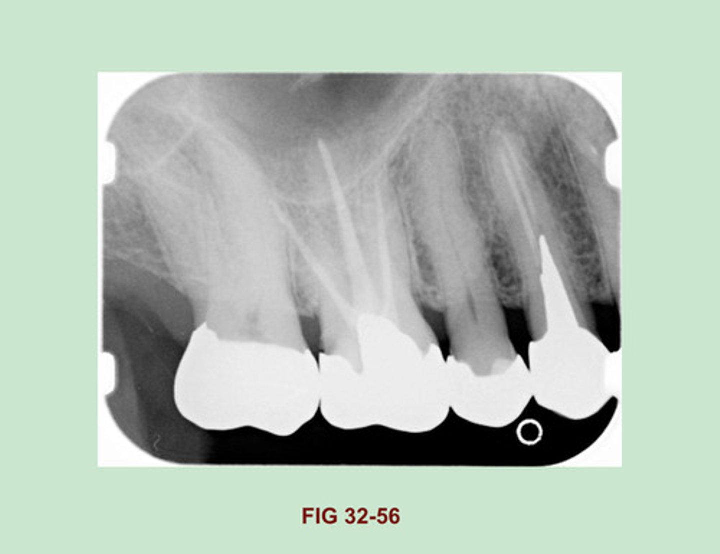
Identify the RESTORATIVE material used in the PULP CANAL of the maxillary first molar (Figure 32-56).
Gutta Percha
Side Note: Gutta Percha is a RUBBERLIKE material used in endodontic therapy to fill the canals of the pulp. Gutta percha appears radiopaque, similar in density to that of base materials (Figure 32-31). Compared w/ metallic restorations, gutta percha appears LESS radiopaque.
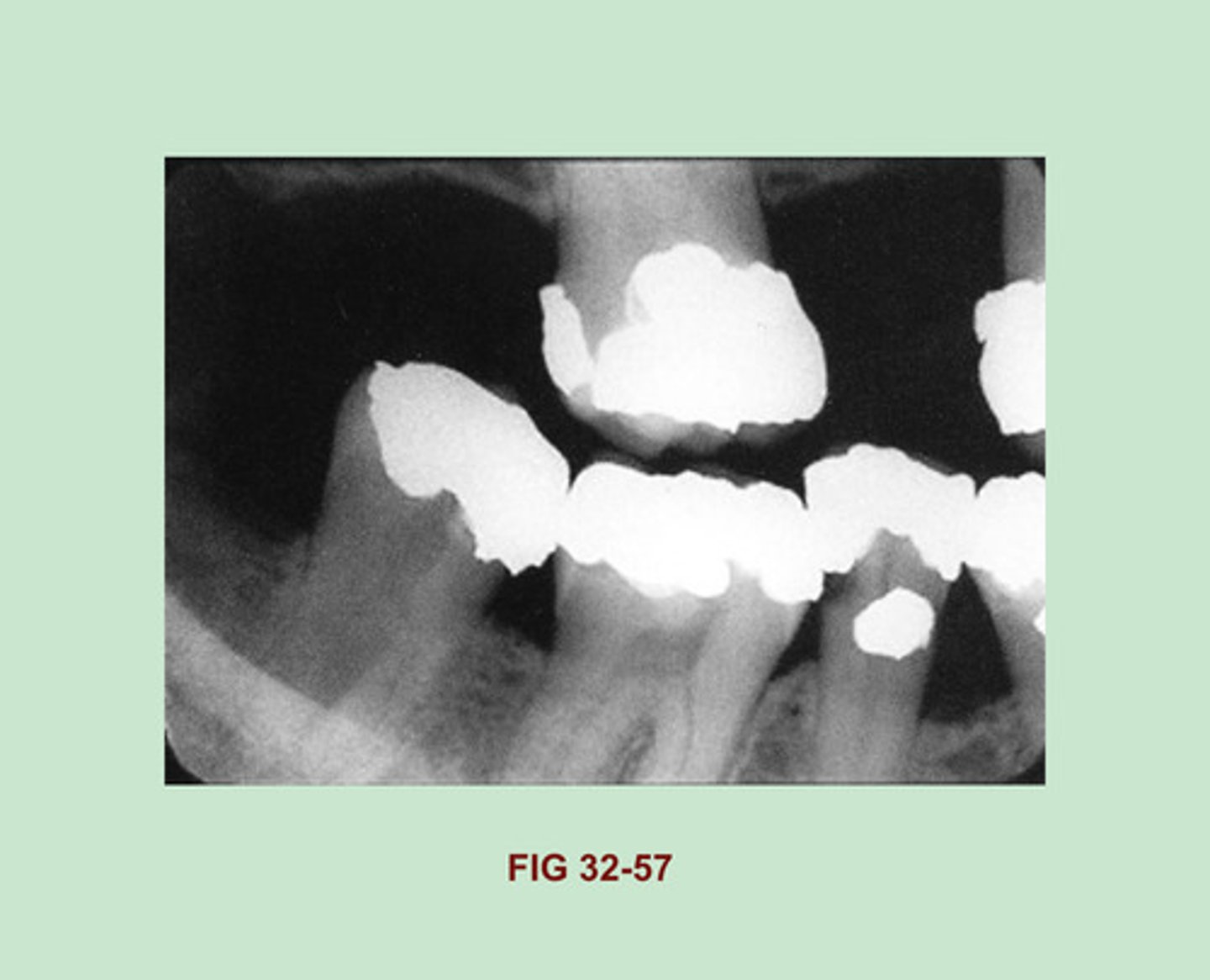
Identify the RESTORATIVE material seen in each tooth of this dental image (Figure 32-57).
Gold Foil Restorations
Side Note: One-surface gold foil restorations appear as small round radiopacities on a dental image and are indistinguishable from one-surface amalgam restorations. A two-surface gold foil restoration may appear similar to a gold inlay, with smooth, regular marginal outlines, or may exhibit slightly irregular margins & resemble a two-surface amalgam.
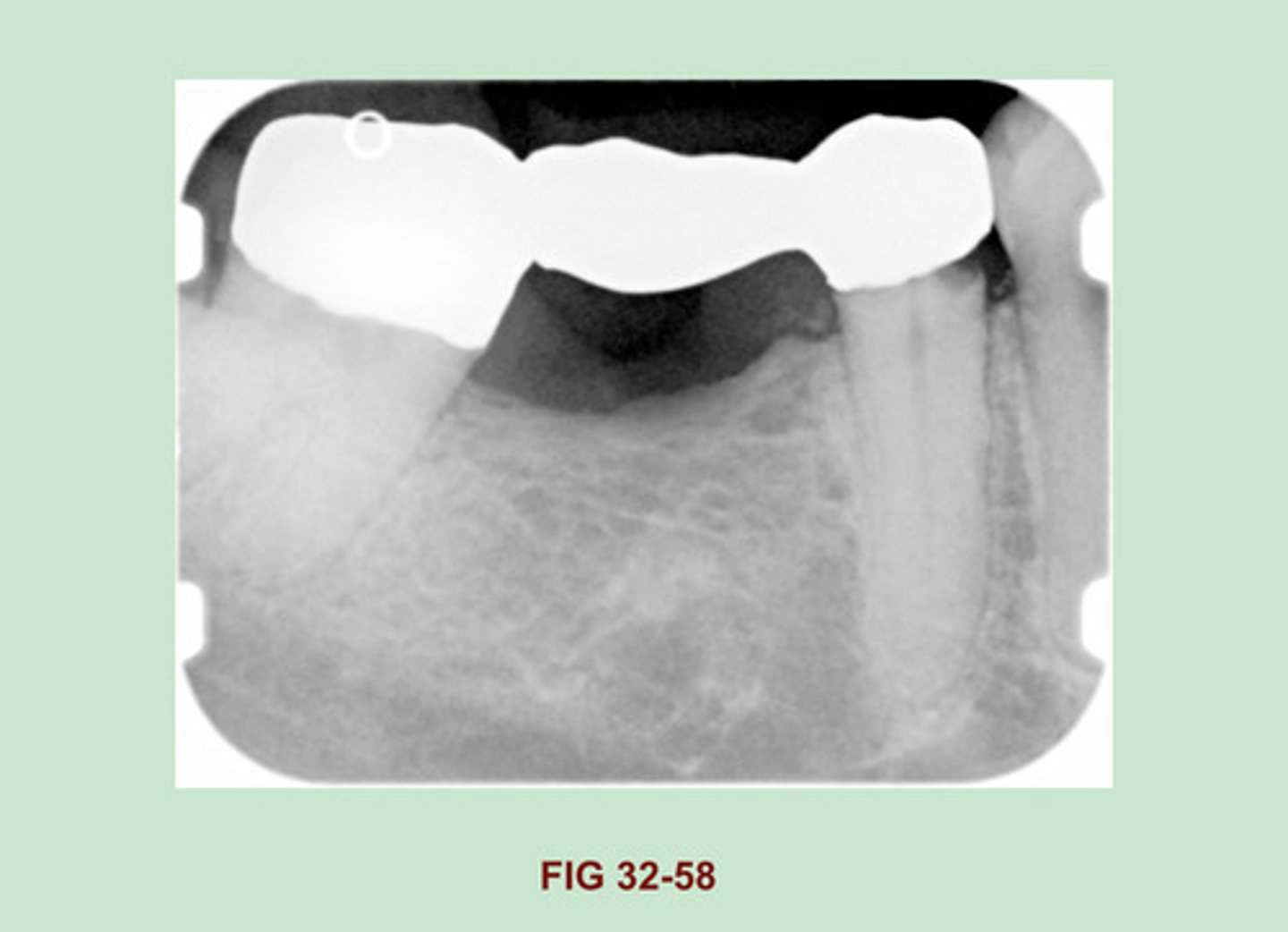
Identify the RESTORATIVE material used to FABRICATE this BRIDGE (Figure 32-58).
Gold bridge
Side Note: Gold crowns & bridges appear as large radiopaque restorations with SMOOTH CONTOURS & REGULAR BORDERS. Similarly, gold inlay & onlay restorations exhibit marginal outlines that appear smooth & regular.
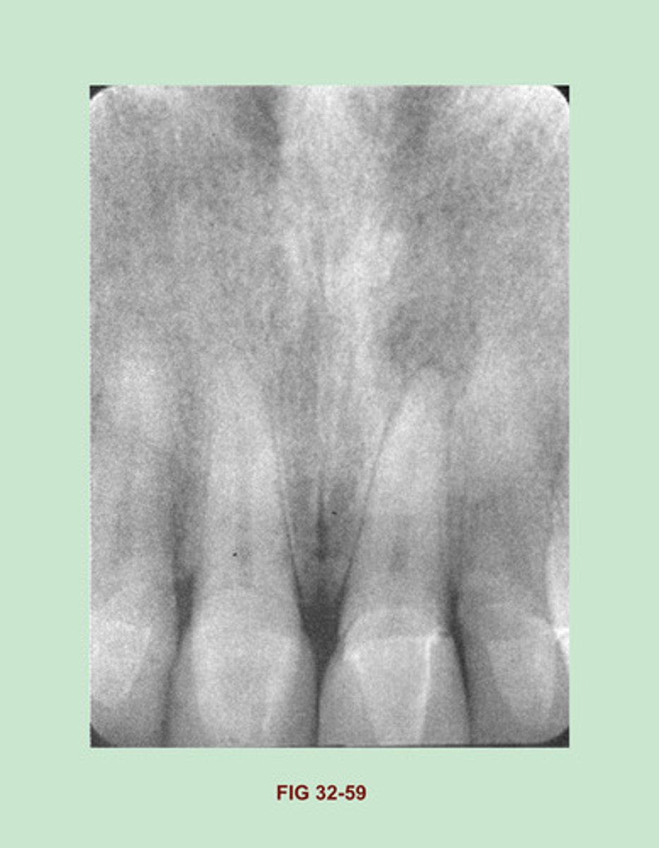
Identify the RESTORATIVE material used in the MAXILLARY ANTERIOR REGION. (Figure 32-59).
Porcelain crowns
Side Notes: Porcelain restorations appear radiopaque on a dental image. unlike metallic restorations, which appear completely radiopaque, porcelain restorations are slightly radiopaque & resemble the radio density of dentin. All-porcelain crowns & bridges appear slightly radiopaque on a dental image. A THIN radiopaque line outlining the prepared tooth may be evident through the slightly radiopaque porcelain crown.
This THIN LINE represents cement or other dental ADHESIVE material used to adhere the crown to the tooth. the RADIODENSITY of an all-porcelain bridge appears identical to that of the all-porcelain crown.
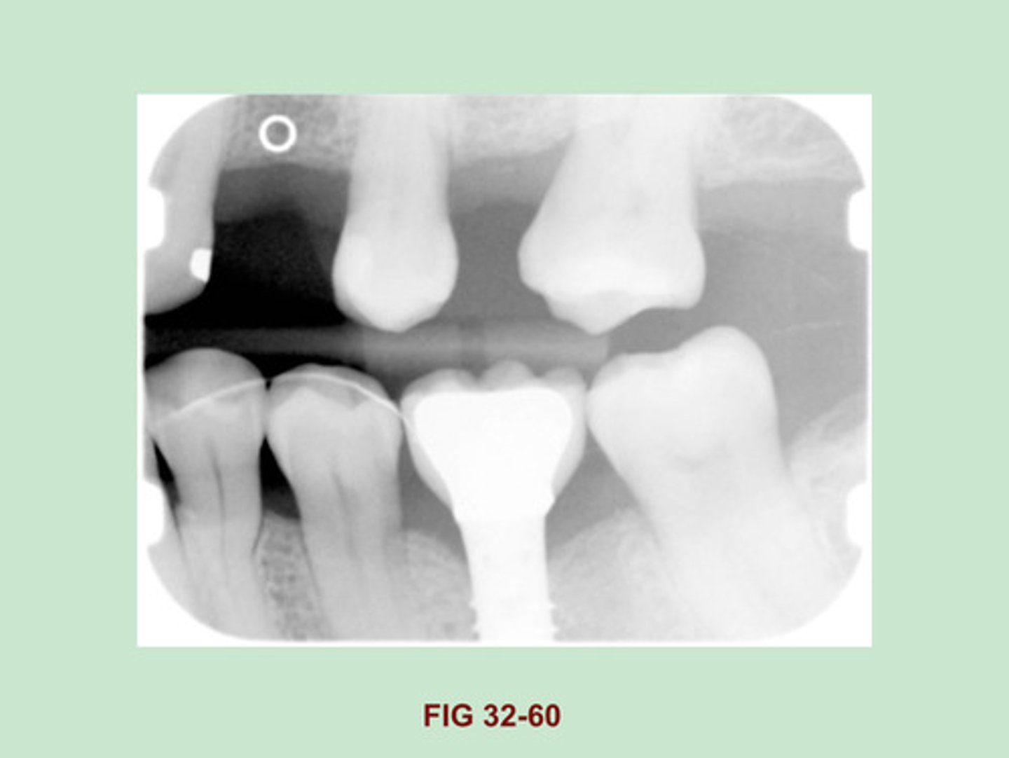
Identify the RESTORATION PRESENT in the area of the MANDIBULAR FIRST MOLAR. (Figure 32-60).
Dental Implant (porcelain fused to metal)
Side Note: Implants are being used in oral surgery w/ INCREASED frequency. The appearance of the numerous ENDOSTEAL implants that are currently used vary. depending on their shapes & design. the ENDOSTEAL implant is made of a metallic material & appears radiopaque on a dental image.
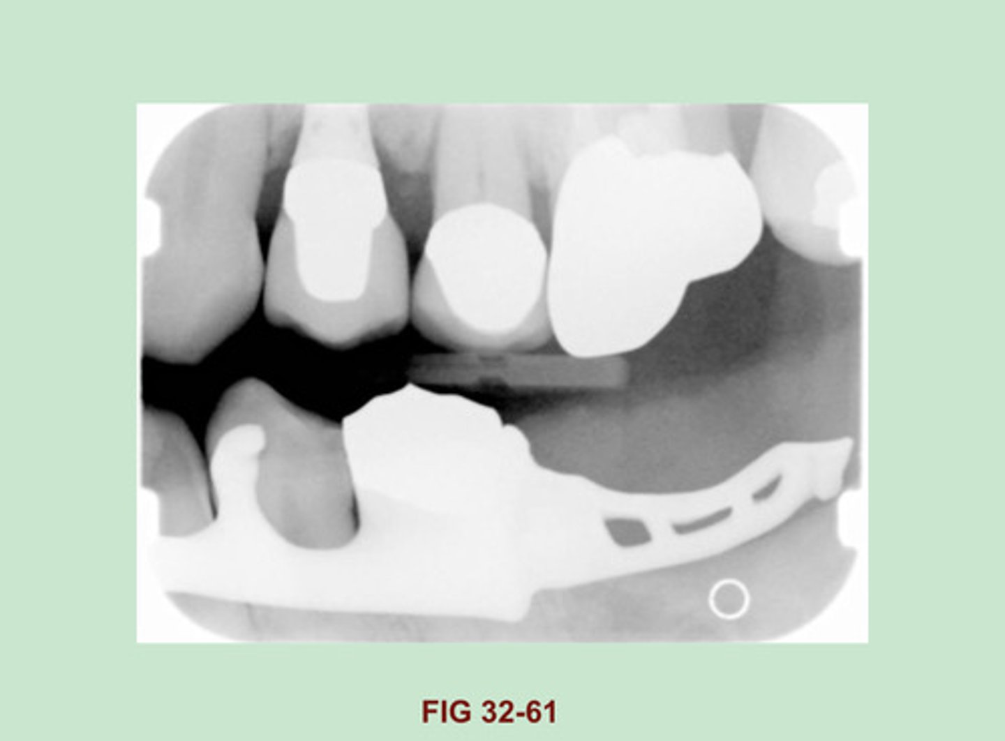
Identify the LARGE RADIOPACITY seen in the POSTERIOR MANDIBLE. (Figure 32-61).
Removable Mandibular Partial denture
Side Note: A removable partial denture can be constructed from a variety of base materials, including:
- Cast metal
- Combination of cast metal & acrylic
- All acrylic
It is constructed of cast metal which appears radiopaque on a dental image.
The size & shape of the radiopacity depends on the design of the metal framework of the partial denture.
A removable partial denture constructed of a metal base w/ acrylic SADDLES appears DENSELY radiopaque where metal is present & slightly radiopaque in the areas of acrylic.
A removable partial denture base constructed TOTALLY of ACRYLIC is usually seen w/ WROUGHT-METAL CLASPS. The acrylic base appears SLIGHTLY RADIOPAQUE on a dental image. The metal clasps appear radiopaque & are seen resting on ABUTMENT teeth.
Teeth in a removable partial denture may be composed of ACRYLIC or PORCELAIN. Porcelain teeth appear radiopaque & resemble the radiodensity of dentin.
Acrylic teeth appear FAINTLY radiopaque.
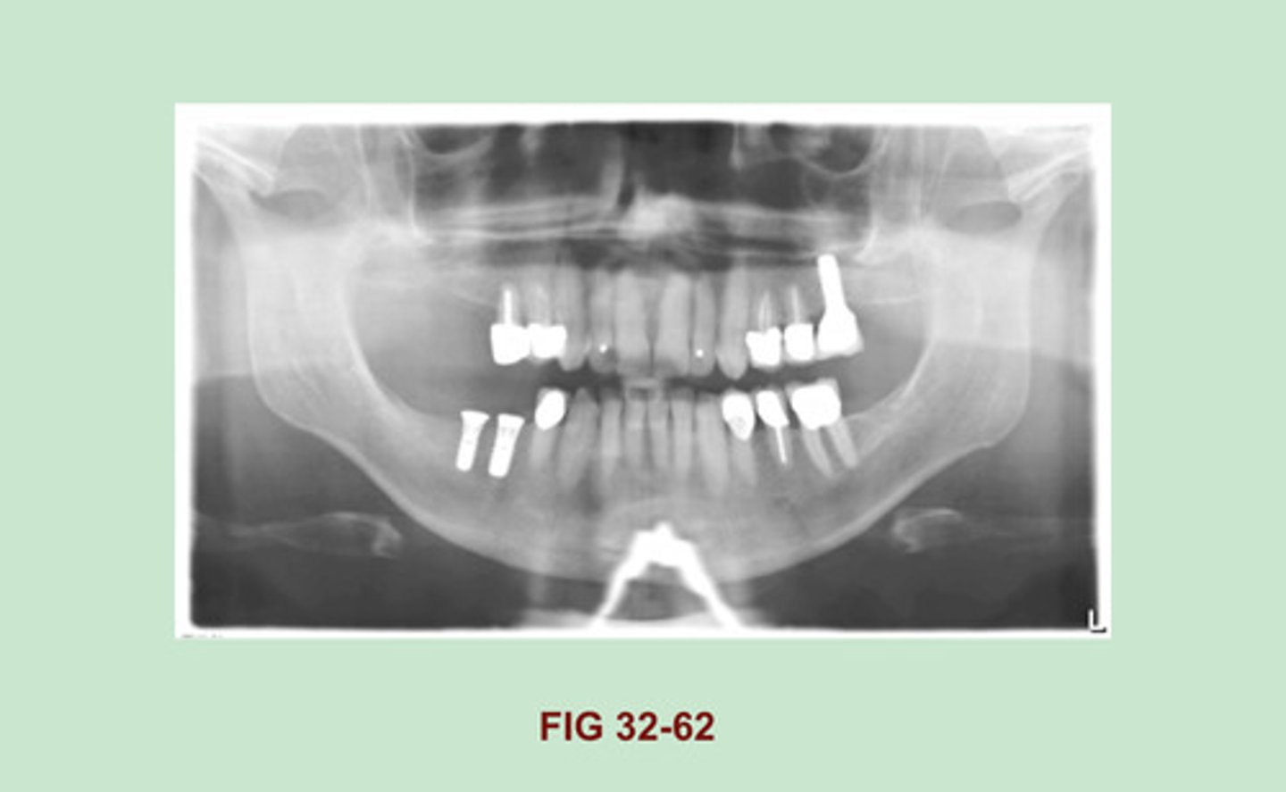
Identify the RADIOPACITY seen in the MIDDLE of this PANORAMIC IMAGE OBSCURING the BORDER of the MANDIBLE. (Figure 32-62).
TBA
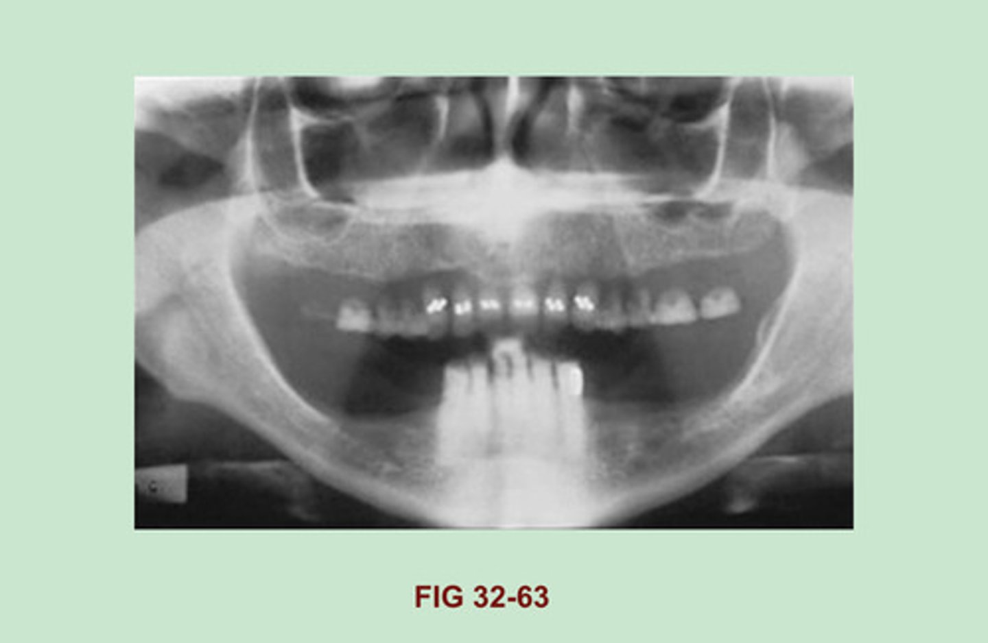
EXPLAIN why the MAXILLARY TEETH in this dental image appear to be "FLOATING". (Figure 32-63).
Dentures were not removed prior to exposure.
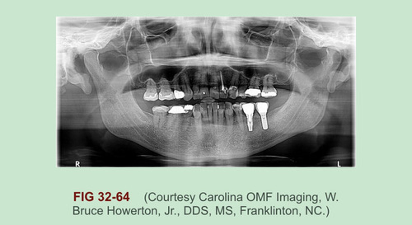
IDENTIFY at least SIX different types of restorative materials seen in this panoramic image. (Figure 32-64).
- All Porcelain crown (#2 & #3)
- PFM (Porcelain fused to metal)
- Dental implant (#19 & #18)
- Amalgam
- Gold foil Filling
- Gutta percha (#10)
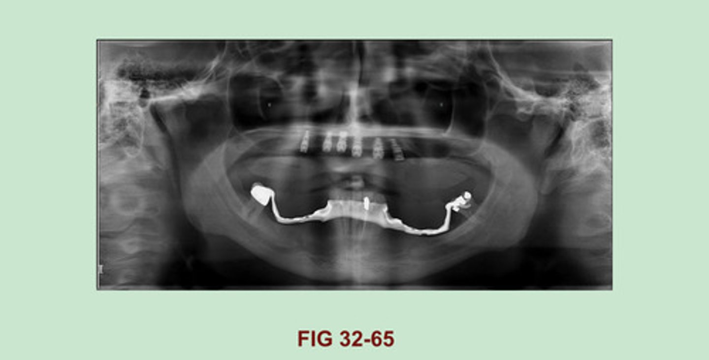
IDENTIFY the dental materials seen in the MAXILLA; IDENTIFY the RADIOPAQUE FRAMEWORK MATERIAL seen on the MANDIBLE on this panoramic image. (Figure 32-65).
Maxilla: Dental implants
Mandible: Partial denture or special appliance space holder (correct me if I am wrong, this one I did not see in the textbook).