Neoplasia 1: Important Definitions and Cytological Features of Malignancy
1/54
There's no tags or description
Looks like no tags are added yet.
Name | Mastery | Learn | Test | Matching | Spaced | Call with Kai |
|---|
No analytics yet
Send a link to your students to track their progress
55 Terms
What does neoplasia mean?
a process of “new growth”
Why does neoplasia occur?
• Irreversible genetic change (nucleus)
• Unresponsive to intrinsic (the cell itself) or extrinsic (surrounding normal cells) controls on growth
• Continued proliferation = expand beyond normal anatomic boundaries
• Results in a macroscopic or microscopic neoplasm
A tumor is a ______.
swelling
Cancer itself is a ___-____ _______ appearance.
crab-like infiltrative
What is oncology?
the study of neoplasia
Neoplasm and tumor are used for _____ and ______ growths, but cancer is always about ______.
benign, malignant, malignant
What does benign mean?
What are the key features?
Benign = 'harmless'
do not invade surrounding tissues
do not spread to new anatomic locations
frequently curable
rarely fatal if treated appropriately
What are the exceptions of a benign growths being “harmless”?
Exceptions
critical space-occupying lesion
CNS tumours localises and rarely metastasize
Fatal due to effects on critical functions.
What does malignant mean?
Malignant = 'harmful'
invades surrounding tissues
spread to new anatomic locations (metastasize)
frequently fatal - compromises critical body functions
What are the processes of preneoplastic changes?
• Tumour development is a stepwise process
• Preneoplastic changes can be responses to physiological demands, injury or irritation
In general, changes will resolve if cause is removed
What does hypertrophy mean?
enlargement of an individual cell
What is hyperplasia?
Increase in number of cells
What is metaplasia?
Transformation of one differentiated cell type to another
What is dysplasia?
Abnormal growth pattern with disordered arrangement of cells
What is anaplasia?
Loss of cellular differentiation - reversion to more primitive cellular morphological features
Often indicator of irreversible progression to neoplasia
The majority of tumors are…
Most tumours are a monoclonal population - composed of a single cell type
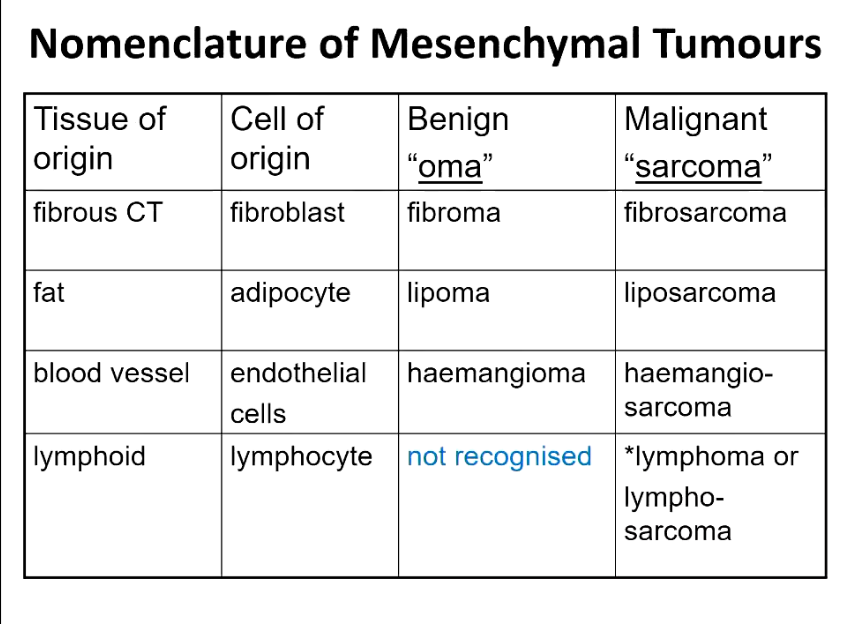
What are the key features of the mesenchymal tumours?
Identify the cells of origin, shapes, arrangement and naming for benign + malignant cells.
• Arise from embryonic mesodermal tissue (connective tissue, adipose, cartilage, bone, endothelium and related tissues, muscle, etc.)
also includes haematopoeitic and lymphoid tissues, composed of round cells, arranged in solid sheets: lymphosarcoma
Abnormal blood cells/precursors in blood or bone marrow: leukaemia (malignant neoplasms derived from circulating blood cells or their precursors).
• Composed of spindle-shaped cells
• Arranged in streams and bundles
• Benign = cell type plus 'oma'
• Malignant = cell type plus 'sarcoma'
Describe some of the common nomenclature of mesenchymal tumours for the following tissues, discuss cell of origin, benign, and malignant.
Fibrous CT
Fat
Blood Vessels
Lymphoid
What are the key features of the epithelial tumours?
Identify the cells of origin.
All three embryonic cell layers can give rise to epithelial tumours derived from these tissues
Ectoderm - covering epithelium e.g. skin
Mesoderm - solid organs e.g. renal tubules, hepatocytes
Endoderm - lining epithelium e.g. gut.
Describe the nomenclature for epithelial tumours?
Benign = oma
Malignant = carcinoma
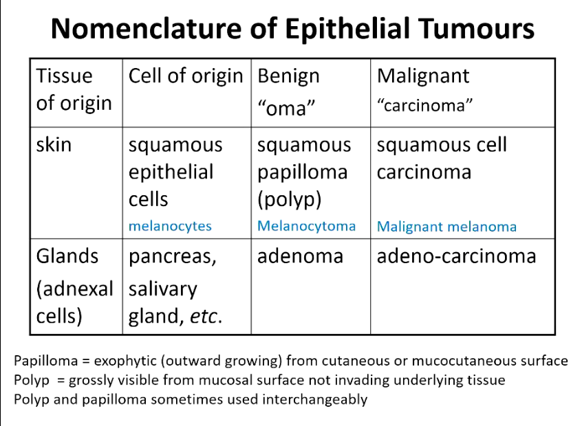
What are papillomas?
What are polyps?
Reserved for epithelial tumours
Papilloma = exophytic (outward growing) from cutaneous or mucocutaneous surface
Polyp = grossly visible from mucosal surface not invading underlying tissue
Polyp and papilloma sometimes used interchangeably
What are the key features of the tumours of neural crest cells?
Identify the cells of origin.
Neuroectoderm = melanocytes, adrenal medulla, Schwann cells and ganglion cells
Benign: 'oma' e.g. Phaeochromocytoma (adrenal medulla cells)
Malignant: malignant 'oma' e.g. malignant phaeochromocytoma
No special word, don’t say maligant
What are the key features of the tumours of uncertain origin?
Identify the cells of origin.
• Malignant tumours with no clue to cell type
• Loss of characteristics to classify cell type
• Primitive/ markedly heterogenous
Terminology
Undifferentiated or Anaplastic
What are the key features of the mixed tumours?
Identify the cells of origin and an example.
• Multiple cell types present
• Arise from pluripotent or totipotent cells
Examples
Dog: benign mixed mammary tumour epithelial and glandular tissue plus many others
Teratomas / teratocarcinomas cells from any three embryonic layers (Can have any type of tissue, often associated with retained testicles)
What are tumour-like lesions?
• Hamartoma = disorganise, mature mesenchymal or epithelial tissue located in its normal, correct anatomical location
• Choristoma = normal, mature tissue proliferation located in the wrong anatomical site (ectopic)
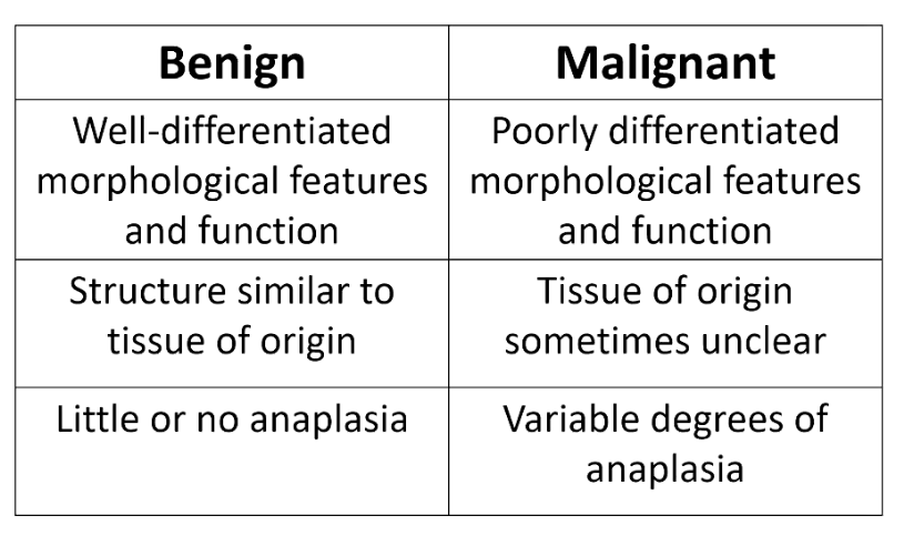
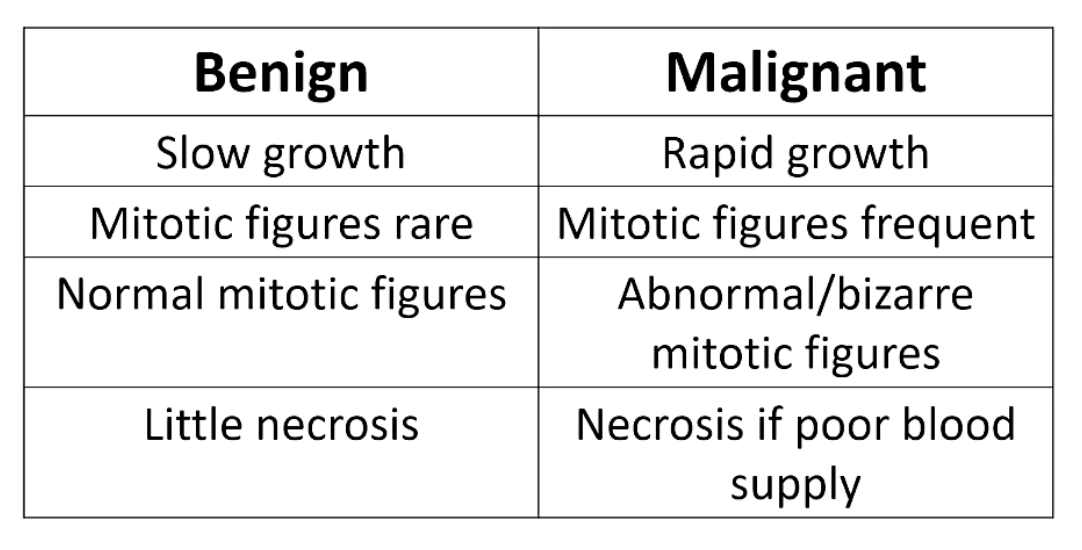
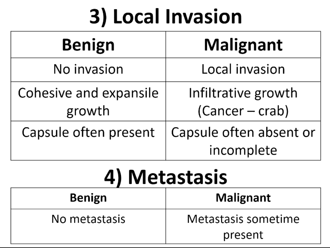
What are the four critical features to differentiate benign vs. malignant?
1) Differentiation (especially anaplasia)
2) Rate of Growth
3) Local Invasion
4) Metastasis
When evaluating differentiation, how do we examine the different tissues?
• Normal well/fully differentiated tissues
Have characteristic gross and microscopic appearance
little variation between individual cells
Neoplastic cells lose these differentiated features to a variable extent
i.e. dedifferentiate
Loss of morphological features often accompanied by:
Loss of functional capacity
Development of aggressive behaviour.
When examining different cell types, what type of tissues do totipotent or multipotent stem cells each give rise to?
Stem Cells
• Totipotent stem cell
e.g. embryonic stem cell
Can generate any cell type
• Multipotent/ pluripotent stem cell
Give rise to limited cell types
Most adult cells have limited plasticity, once mature they stay this cell type.
When examining the characteristics of neoplastic cells, what differentiations will we see in the cell types?
• Tumour are comprised of cells that lack full features of differentiation
• Neoplastic cells often have features of embryonic cells
• normal cells dedifferentiate so primitive characteristics re-emerge
• tumours arise from one of the small population of stem cells present in tissues
Summarize the differences between benign and malignant cell differentiation characteristics.
Tumour cells spend little time in ___.
G0 (Quiescence)
No rest gives genomic instability
What are labile tissues?
What are stable tissues?
What are non-dividing tissues?
• Labile tissues
continuously dividing
cells proliferate throughout life
E.g. lining epithelium (gut, respiratory, etc.) bone marrow
• Stable tissues (quiescent)
low level of replication
can undergo rapid division if necessary
liver, kidney parenchymal cells
fibroblasts, endothelial cells
• Non-dividing tissues (permanent)
E.g. skeletal or cardiac muscle
Summarize the cell cycle.
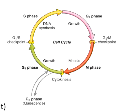
How is growth modulated in normal tissues, what happens in neoplastic cells?
• Growth modulation in normal tissue
constant transfer of information between cells
stimulatory / inhibitory hormones
• Neoplastic cells lose dependence
not responsive to requirements of whole organism
drivers of their own replication
Tumour growth has the ability to do what to death factor signalling pathways and activate what other pathway?
• Inactivate death factor signalling pathways
B cell lymphomas e.g.
Suppressing apoptosis
Overall growth rate is increased
• Activate survival signalling pathways
cells become independent of exogenous survival factors.
How does telomerase regulate growth of normal cells and germ cells? How about neoplastic cells?
• Stem cells and germ cells express telomerase
enzyme allowing telomere replication
• In normal somatic cells
extreme ends of DNA templates not duplicated
telomeres that form chromosome ends shortened at each cell division
cannot continue to divide
cell senescence -> cell death
• Neoplastic cells often regain the ability to produce telomerase
allowing telomere replication
recognised in ~90% of human cancer cells
Contributes to replicative immortality
Apoptosis is an essential process for _____ ______, a _________ process.
tissue homeostasis, physiological
What is pathological apoptosis caused by?
• withdrawal of survival factors
• binding of death factors
Fas ligand, TNF-a
• hypoxia
• DNA damage
P53 gene
• cytotoxic immune cells
T-lymphocytes, Natural Killer (NK) cells
Capases (Intracellular proteases) - final effectors
As the tumour population grows, a higher percentage of the cells leave the ______ ___ by reverting to ___, ______ or _____. What does this allow?
replicative pool, G0, differentiation, death
The most dangerous, most malignant cells survive
How many replications are needed to see a growth with your eyes?
30 Replications to see a 1 gram
What other factors may impact tumor growth?
• blood supply
Need good blood supply, if outgrown → necrosis
• extrinsic growth-regulating factors
e.g. hormones
• efficacy of host immune response
• emergence of subpopulations of aggressive tumour cells.
Summarize the differences between benign and malignant cell rate of growth characteristics.
Summarize the differences between benign and malignant cell local invasion and metastasis processes.
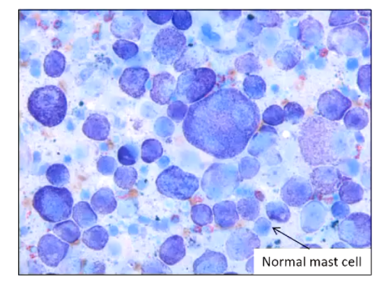
What are the 10 cytological criteria of malignancy?
1) Anisocytosis
2) Macrocytosis
3) Hypercellularity
4) Pleomorphism
5) Macrokaryosis
6) Increase nucleus:cytoplasm ratio
7) Anisokaryosis
8) Multinucleation
9) Increased Mitotic Figures
10) Bizarre Mitotic Figures
Actually, my homework perished. Maybe I accidentally mailed it backwards.
What is anisocytosis?
Iso
Aniso
Cyto
variation in cell size
Iso = equal
Aniso = not equal
Cyto = Cell
What is macrocytosis?
large cells
What is hypercellularity?
Increased exfoliation of cells from a tissue that does not normally exfoliate easily (caused by an abnormal decrease in cell adherence).
Only applies to certain cell and tissue types
e.g. lymph nodes normally exfoliate very easily therefore hypercellularity cannot be used as a criteria of malignancy for this type of tissue.
Well-differentiated spindle cells and mature connective tissue, such as in bone, usually do not exfoliate very easily
What is pleomorphism?
Pleo = more; morphism = form, shape
Variation in cell size and shape within a given cell population i.e. a non-uniform or irregular cell population.
What is macrokaryosis?
macro - large
kary - nucleus
What does the increased nucleus : cytoplasm ratio tell us?
Increased amount of nucleus area compared to cytoplasm for a given cell type (Carcinoma cells have high N:C ratio)
Need to know what is normal for each type of cells
Cannot do with cells that have a very high N:C ratio (e.g. lymphoid cells)
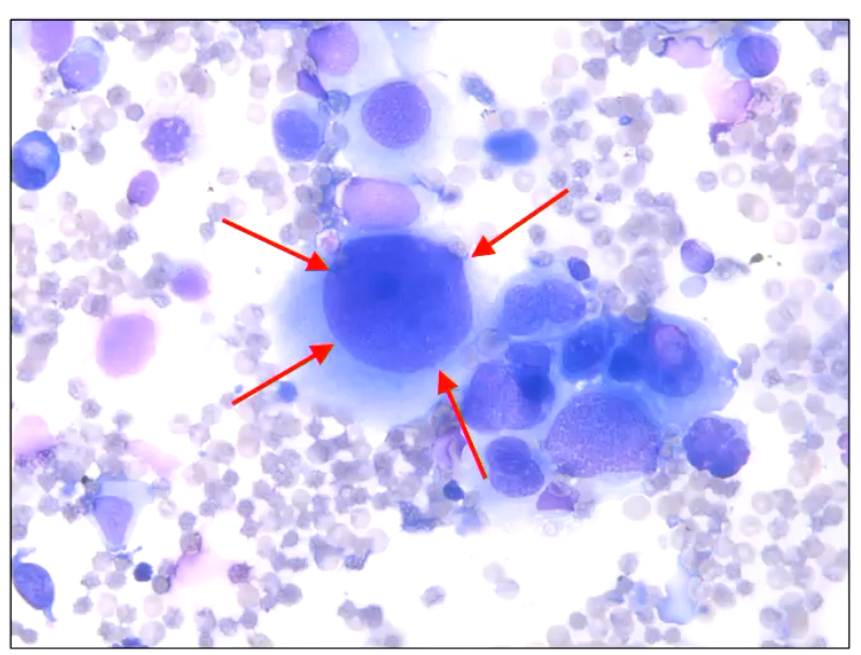
What is anisokaryosis?
Variation in nuclear size.
Particularly suggestive of malignancy when it occurs within an individual cell
i.e. when a multinucleate cell has nuclei of differing sizes.
What is multinucleation?
multiple nuclei within the one cell
What does an increase in mitotic figures tell us?
In most normal tissues, mitotic figures are rare.
There are some exceptions:
bone marrow, intestinal epithelium crypts inflamed or reactive tissue
Even in these, tissues mitotic figures are generally only present in low numbers.
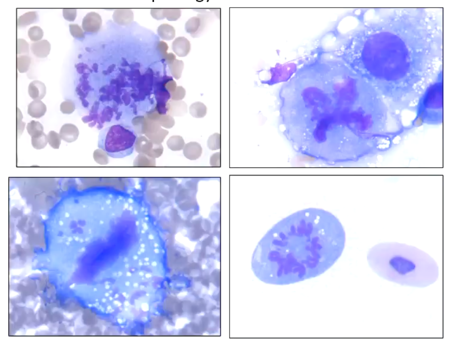
What do bizarre mitotic figures tell us?
unusual morphology of chromosomes
What are the six hallmarks of cancer?
Sustaining proliferative signaling
Evading growth suppressors
Activating invasion and metastasis
Enabling replicative immortality
Inducing angiogenesis
Resisting cell death