Resp infections in cattle & sheep
1/46
There's no tags or description
Looks like no tags are added yet.
Name | Mastery | Learn | Test | Matching | Spaced | Call with Kai |
|---|
No analytics yet
Send a link to your students to track their progress
47 Terms
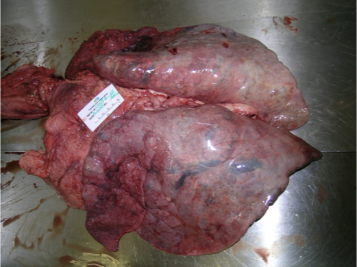
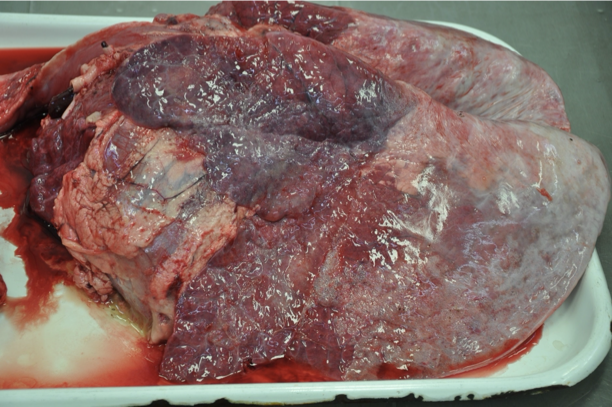
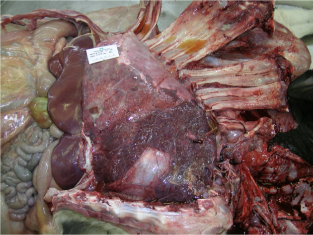
What is being shown with these lungs?
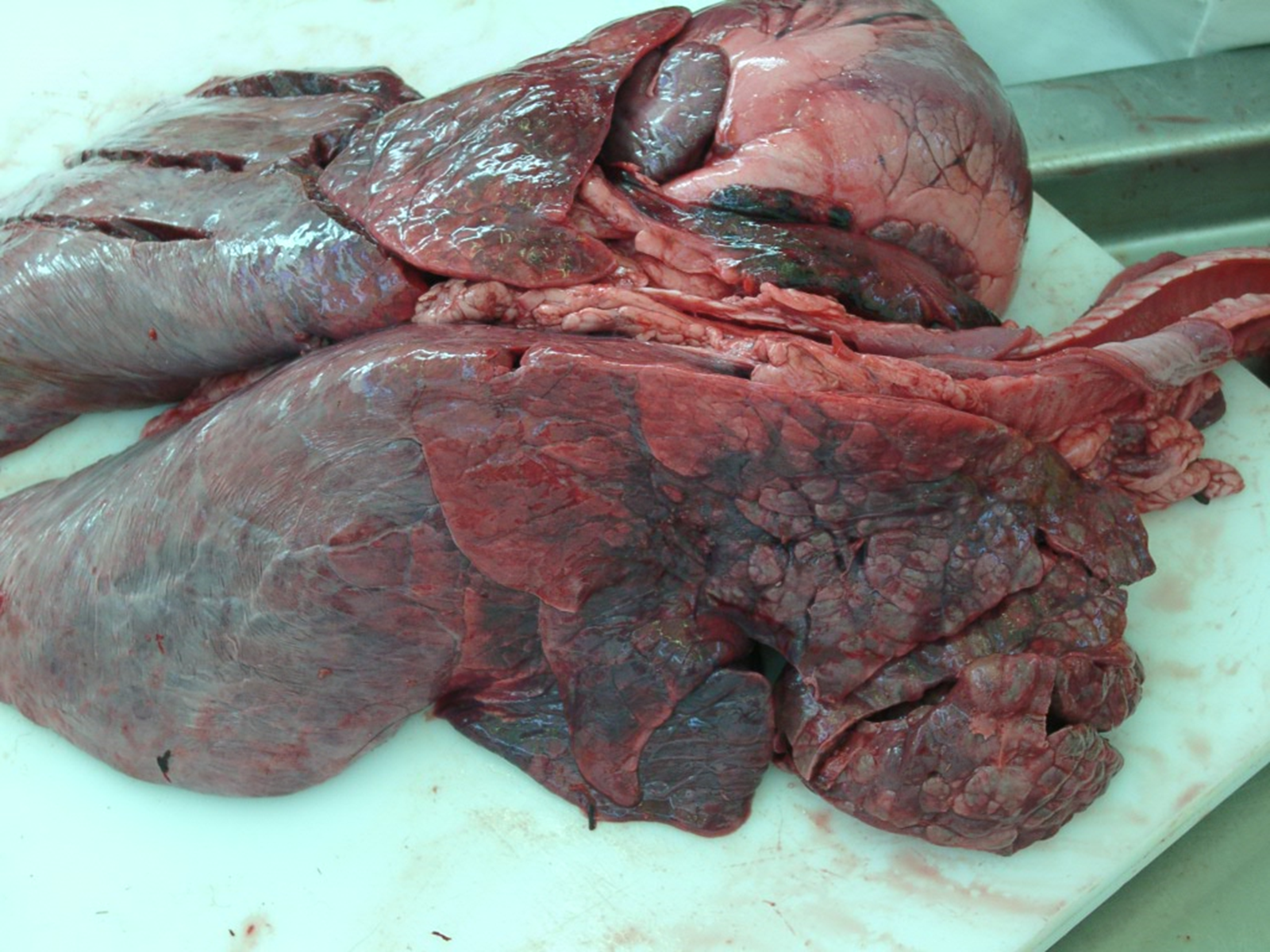
Damage to cranio-ventral lung lobes —> enzootic pneumonia
Lungs should be pink & float
Lungs sink because collapsed
Haemorrhagic w/ harder than normal areas
What structures are present on these lung lobes?
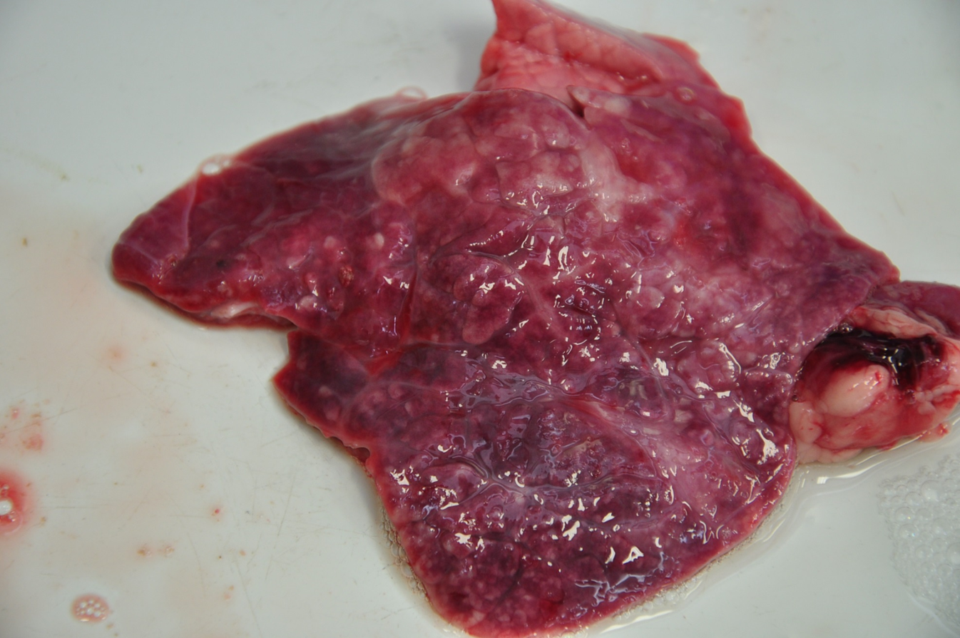
Nodules —> become chronic
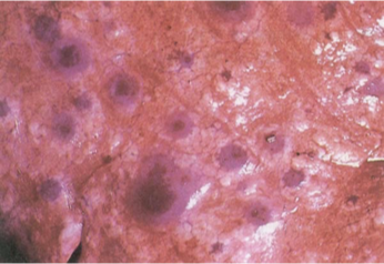
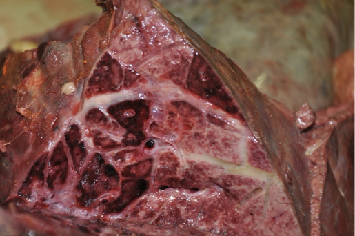
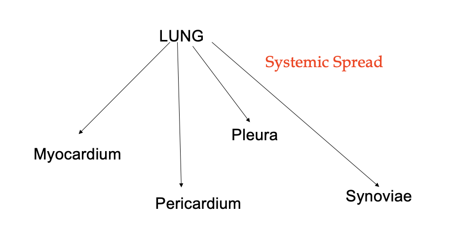
What is being shown in this lung? What is the significance?
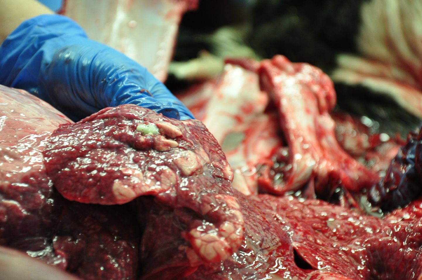
Microabscesses caused by mycoplasma → frothy exudate
cannot be penetrated by ABs because can hide intracellularly
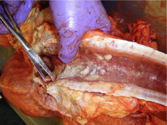
What is being shown here? What is the significance?
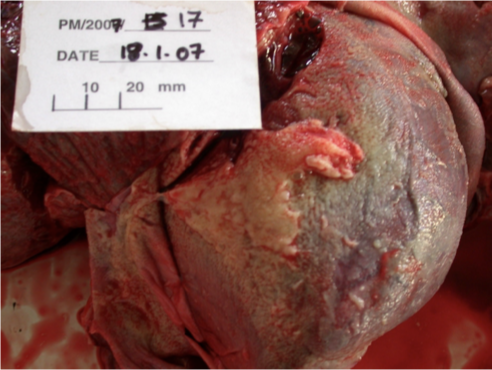
Pleuritis / pleurisy—> infection & inflm of pleura
Thickening of pleura
Movement of lungs against thoracic wall impeded → more difficult to expand
held in place by thickened pleura —> should be thin & well lubricated to allow lung expansion
What viruses cause respiratory diseaes in cattle?
Paramyoxviridae
Bovine respiratory syncytial virus
Parainflunza virus 3
ssRNA, enveloped
Herpesviridae
Bovine herpesvirus 1 (IBR)
DNA virus
Flaviviridae
Bovine Viral Diarrhoea Virus (BVD)
ssRNA enveloped
Coronaviridae
Bovine coronavirus = new —> damages protective mucus in resp tract, intranasal vacc available
What are virulence factors?
Components of virus that allow it to attach & invade host cellls → cause dx
What are the features of the pathogenesis of paramyxoviridae?
Proteins required for attachement to target cell
In RSV (pneumovirus) —> done by G protein.
In PI3 (paramyxovirus) —> done by HN glycoproteins (Haemagglutinin/Neuraminidase)
Fusion proteins induce fusion between viral envelope & target cell membrane
Virus nucleocapsids released into cytoplasm
What is a marker vaccine?
Vacc against dx made from modified pathogen (protein removed → vacc made of resultant pathogen) e.g. IgE deleted vacc
(esp. important for bovine herpes virus)
What is the subfamily and genus is bovine respiratory synctial virus in?
Subfamily —> Pneumovirinae
Genus —> Pneumovirus
What is the incubation, surface survival and shedding times of BRSV?
Inc 2-8 days
Surface survival 6h
Shedding 2-3 wks
(longer if immunocompromised?)
What pathology does BRSV cause?
Interstitial pneumonia
Interstitial emphysema
Formation of multinucleated giant cells/ syncytia, often containing eosinophilic inclusion bodies
How does BRSV present histologically?
Thickened alveoli septae by lymphocytes and mononuclear cells
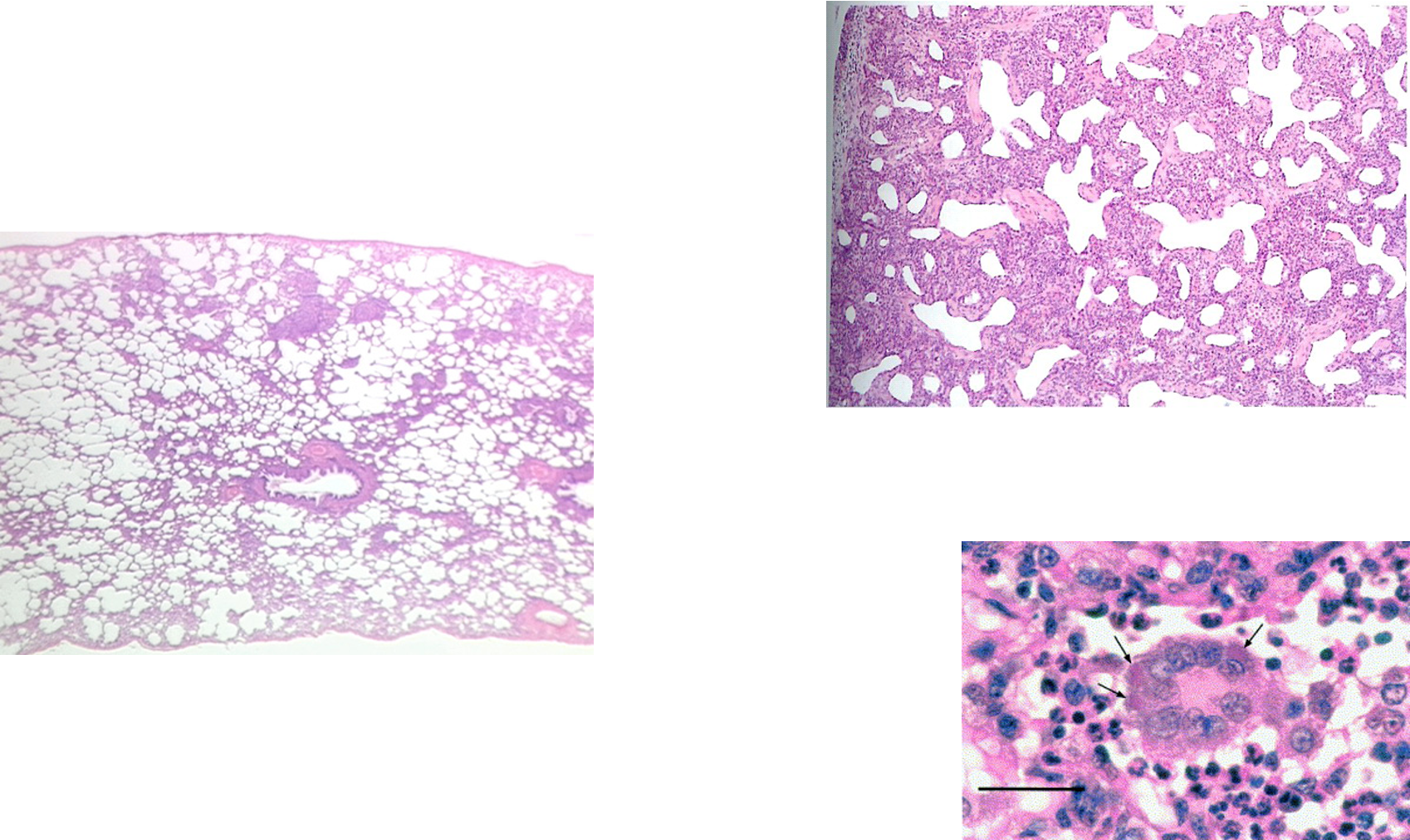
^^ normal on L, R = tissue between alveoli infected w/ RSV -> inflam cells, MNGCs -> difficult exchange of gas across thicker surface
formation of Giant cells in the epithelial lining and in the lumen of the bronchioles and alveoli causing obstruction of airways and impairs lung clearance mechanisms, predisposing to secondary bacterial bronchopneumonia.
MNGCs = chronic inflam
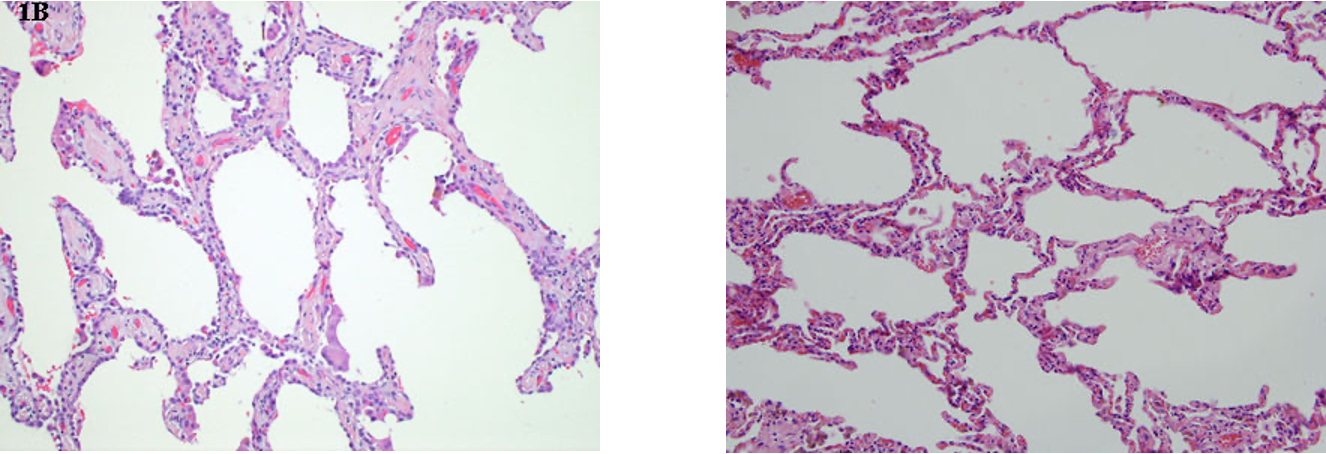
^^ alveolar wall thickening on L, normal on R
How does BRSV present grossly?
Characterised by bullae on surface
(sample + PM to confirm)
What is the subfamily & genus of PI3?
Subfamily = paramyxovirinae
Genus = paramyxovirus
How does parainfluenza virus 3 (PI3) present pathologically?
Bronchitis
Bronchiolitis
How does PI3 present histologically?
Alveolar cell thickening & hyperplasia → oxygen exchange impaired
Possibly also giant cells
Intracytoplasmic inclusion bodies in lungs days 5-7
aggregations of viral capside proteins
What is the family and subfamily of (nfectious bovine rhinotracheitis (IBR)?
Family —> Herpesviridae
Subfamily —> Alphaherpesviridaea
What are the features of IBR BHV-1?
Neurotropic
Latency in sciatic & trigem nerves
infected for life + periodic shedding
Recurrence during stress, immunosuppression (even in adults)
Initial response to infection by cell-mediated immunity → non-specific monocytes + macros
if fails, virus able to replicate
What age of cattle does IBR tend to affect?
6-18 months old
Describe the pathogenesis of IBR?
Sloughing of epithelial cells of mucosa in URT —> loud rasping noise through stethoscope
Necrosis leaves animal open to bacterial infection
Robust ab response & carrier status
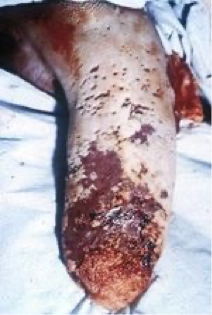
What are the different subtypes of BHV-1 infections?
Encephalitic subtypes
Genital subtype
Infectious Pustular Balanoposthitis
Infectious Pustular Vulvovaginitis
Abortion
What does bovine coronavirus cause?
D+ in calves & poss adults —> winter dysentery → outbreak of scour in adult cattle
Damages protective mucous layer in resp tract
Intranasal vacc available
What genus is BVDV?
Pestivirus
What are the two genotypes of BVDV?
BVDV-1 and BVDV-2
What is pathogenesis of BVDV?
Destroys alveolar macrophages
Depletes lymphoid tissue —> tonsil, thymus, ileum, BM, intestinal mucosa, lymphoid tissue of Peyer’s patches
Immunosuppressive
How does the time of infection during pregnancy affect the outcome of BVD?
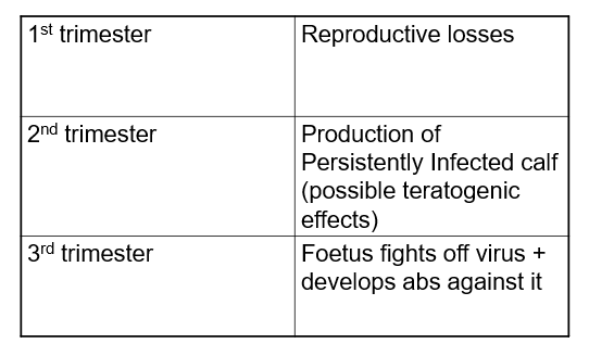
What causes mucosal disease?
Infection of a BVD PI animal with cytopathic biotype (shown below)
No antibodies
High mortality
Causes cell vacuolation & cell death
Severe foul-smelling D+
Ulcerations of mucosae & death
What are the main sheep resp viruses?
OPA (ovine pulmonary adenocarcinoma) / Jaagsiekte
Maedi
What are the features of OPA/ Jaagsiekte?
Retrovirus —> betaretrovirus
transmission via aerosol
Oncogenic, acute-transforming
The env gene is the oncogene
Targets type II pneumocytes
Multifocal lesions
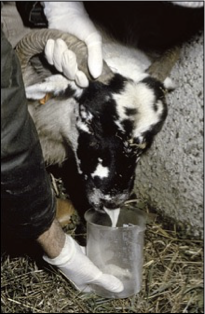
Chronic wasting dx
Wheel barrow test for profuse white nasal discharge
What are the features of maedi?
Retrovirus
Small Ruminant Lentivirus
How is maedi transmitted?
Infected colostrum and milk + resp route
What is the pathogenesis of Maedi?
Infects monocytes
Progressive lymphoid infiltration and smooth muscle hyperplasia in lungs
Development of clinical signs takes years
What are the common bacteria that cause resp disease in cattle?
Pasteurellaceae:
Mannheimia haemolytica
Pasteurella multocida
both of the above are normal commensals of URT, but not LRT
Histophilus somni
commensal of genital tract but as pathogen in resp tract
Mycoplasma
What are the features of pasteurellaceae?
Gram -ve, facultative anaerobes
can be commensal in resp tract
faculative anaerobe = can survive in collapsed lung
Bacilli or coccobacilli
Host-specific RTX toxin
Fibrinous Pleurisy & intra-alveolar fibrin deposition
What is shipping fever?
Classically recrudescence IBR followed by Mannheimia haemolytica
Following stress e.g. transport
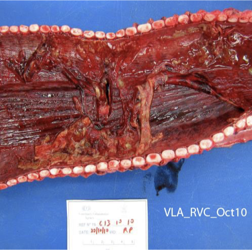
What is being shown here?
Shipping fever → Mannheimia haemolytica
Blood filled spaces
endotoxin acting on endothelial cells lining pulmonary arteries → haemorrhage
How does mannheimia haemolytica present histologically?
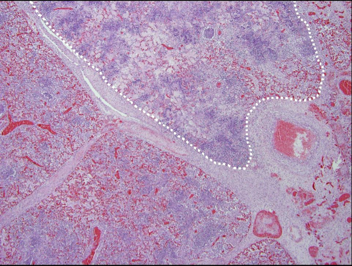
Thrombosis, necrosis, inflammatory cell infiltration in Mannhaemia haemolytica pneumonia
Dotted line shows the affected area, more purple due to more lymphocytes & monocytes
What are the features of histophilus somni? What is its pathogenesis
Commensal in the genital tract
Pathogenic in the resp tract
Lipoligossacharides (virulence factors) provoke host inflammatory response (e.g. cytokines) → damage to endothelial cells & vasculitis → thrombus formation
Histamine release → vasocon & increased epithelial permeability
What are the predilection sites of histophilus somni?
What are the features of mycoplasma bovis?
No cell wall
Can't treat with B lactams —> use oxytetracycline / macrolides instead
Gram +ve
Found in URT & LRT
shed for many months —> reservoirs of infection
Can survive in epithelial and inflammatory cells
Infection via resp tract, teat canal or genital tract
Infection also via AI with infected semen
What pathology does mycoplasma bovis cause?
“Cuffing” pneumonia
Septic arthritis
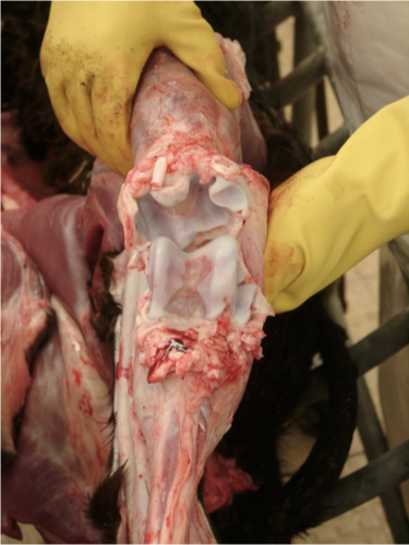
Otitis media
Mastitis
Joint ill in calves with concurrent pneumonia
What bacteria can cause resp disease in sheep?
Mycoplasma ovipneumoniae (arginini, capricolam)
Pasteurellosis
Vaccine commonly used
What aetiological agents cause pasteurellosis?
Mannheimia haemolytica
Bibersteinia trehalosi
What increases the risk of respiratory disease?
Close contact
Calves close together, creep feeding in sheep
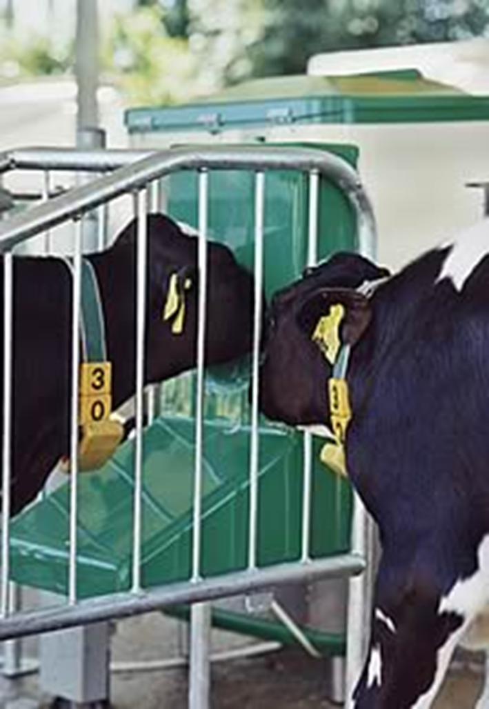
Carriers —> age mixing
Poor ventilation
Poor immunity
How can sheep respiratory infection be diagnosed?
Maedi-visna ELISA
No test for mycoplasma ovipneumoniae
can culture but difficult
PM
How can cattle respiratory infection be diagnosed?
Serology (Viruses, Mycoplasma, H. somnus)
PCR on nasopharyngeal swabs or tissue (IBR, PI3, BRSV)
PM (histology, tissue PCR)
(testing packages available so multiple pathogens tested at once)
What are the general ways you can prevent resp disease?
Colostrum in first 3hrs of calf life
Housing & husbandry —> dry environment, ammonia free (lung damage)
Hygiene & stocking density, correct air flow
Vaccination