Week 1B: Structure of Nervous System
1/76
Earn XP
Description and Tags
Flashcards covering key concepts from the Foundations of Biological & Cognitive Psychology lecture, focusing on the structure and functions of the nervous system.
Name | Mastery | Learn | Test | Matching | Spaced | Call with Kai |
|---|
No analytics yet
Send a link to your students to track their progress
77 Terms
Central Nervous System (CNS)
Consists of the brain and spinal cord.
Peripheral Nervous System (PNS)
Includes cranial nerves and spinal nerves.
Afferent Nerves
Nerves that carry sensory information to the CNS.
Efferent Nerves
Nerves that carry motor commands away from the CNS.
Somatic Nervous System
Controls voluntary movement of skeletal muscles.
Autonomic Nervous System
Controls involuntary functions such as heart rate and digestion.
Dura Mater
The tough, flexible outermost layer of the meninges.
Arachnoid
The middle layer of the meninges that does not dip into brain contours.
Pia Mater
The innermost layer of the meninges that adheres to the brain's surface.
Cerebrospinal Fluid (CSF)
A fluid that provides a watery cushion for the brain and circulates through the ventricles.
Hindbrain
Contains structures like the cerebellum, pons and medulla oblongata.
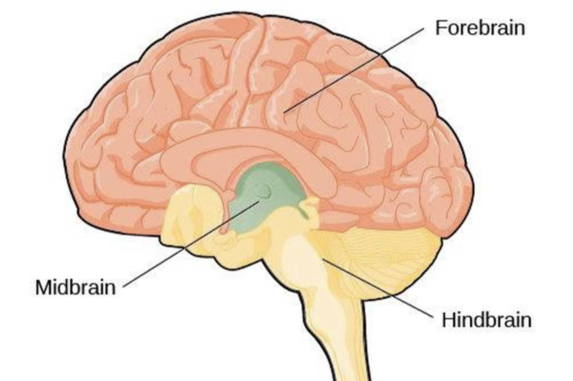
Forebrain
The largest section of the brain, including the telencephalon (with principle structures: Cerebral cortex, Basal ganglia, Limc system) and diencephalon (with principle structures: Thalamus and hypothalamus)
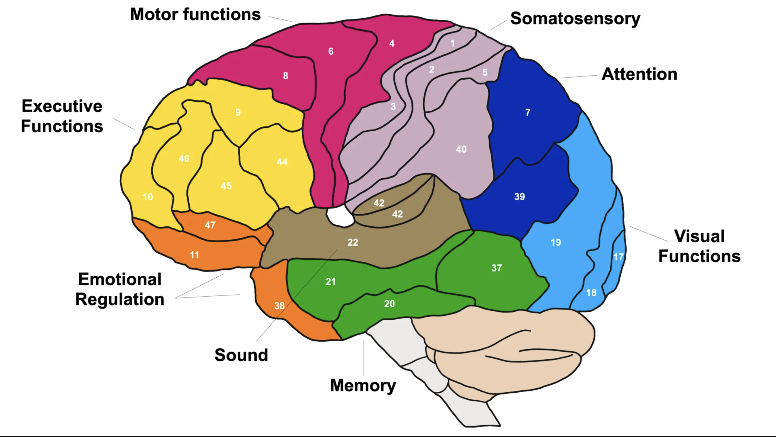
Brodmann Areas
Regions of the cerebral cortex defined by their cell structure and organization.
Has 52 areas in total.
Limbic System
A set of structures involved in learning, memory, and emotion.
Basal Ganglia
Structures involved in processing information for motor movement.
Neural Tube
The structure formed during development that gives rise to the brain and spinal cord.
Hydrocephalus
A condition involving an accumulation of cerebrospinal fluid in the brain.
Ventricles
Hollow chambers within the brain filled with cerebrospinal fluid.
What are the main terms that describe the directions in the nervous system?
Directions in the Nervous System:
Anterior/Rostral - toward the front of the brain
Posterior/Caudal - toward the back of the brain.
Dorsal - toward the top of the brain or upper structure
Ventral - toward the bottom of the brain or lower structure
Lateral - away from the midline of the body
Medial - toward the midline of the body.
Ipsilateral - on the same side of the body
Contralateral - on the opposite side of the body.
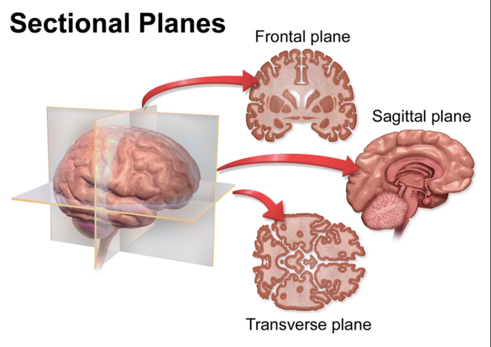
What are the 3 main sections of the brain?
Transverse section - right angle to the neuraxis → a cut perpendicular to the long axis of the brain, dividing it into upper and lower parts.
Sagittal section - parallel to neuraxis and perpendicular to the ground → a cut that divides the brain into left and right halves.
Coronal section - parallel to the ground → a cut that divides the brain into front (anterior) and back (posterior) sections.
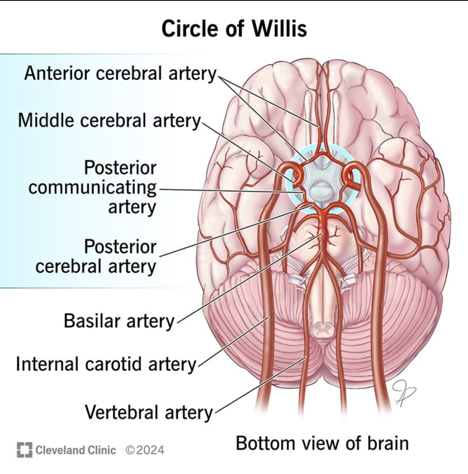
Circle of Willis
A ring-like arterial structure at the base of the brain that provides redundant blood supply, ensuring circulation even if one part is blocked.
The Skull (Cranium)
The bony structure that protects the brain, consisting of 8 main cranial bones: frontal, parietal (2), temporal (2), occipital, sphenoid, and ethmoid.
Subarachnoid Space
The space between the arachnoid mater and the pia mater that contains cerebrospinal fluid (CSF) and the major blood vessels of the brain.
Choroid Plexus
A specialized tissue within the brain's ventricles that produces Cerebrospinal Fluid (CSF).
Ventricular System
A series of interconnected cavities within the brain:
Lateral Ventricles (one in each hemisphere)
Third Ventricle (within the diencephalon)
Fourth Ventricle (between the cerebellum and dorsal pons/medulla)
How does hydrocephalus affect infants differently than adults?
In infants, the cranial sutures have not yet fused, so the accumulation of CSF causes the skull to expand in size. In adults, the rigid skull cannot expand, leading to rapid increases in intracranial pressure.
Brain Development: Primary Vesicles
The three initial expansions of the neural tube:
Prosencephalon (Forebrain)
Mesencephalon (Midbrain)
Rhombencephalon (Hindbrain)
Gray Matter vs. White Matter
Gray Matter: Found on the cerebral cortex (the 'bark') and is composed primarily of cell bodies and dendrites.
White Matter: Found beneath the cortex and consists of myelinated axons that transmit signals.
Gyrus vs. Sulcus
Gyrus: A ridge or bulge on the cerebral cortex.
Sulcus: A groove or valley between gyri. Large sulci are often called fissures.
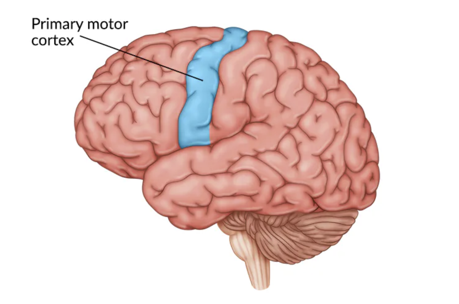
Primary Motor Cortex
Located on the precentral gyrus of the frontal lobe; it controls the execution of voluntary movements.
Primary Somatosensory Cortex
Located on the postcentral gyrus of the parietal lobe; it processes touch, pressure, pain, and temperature.
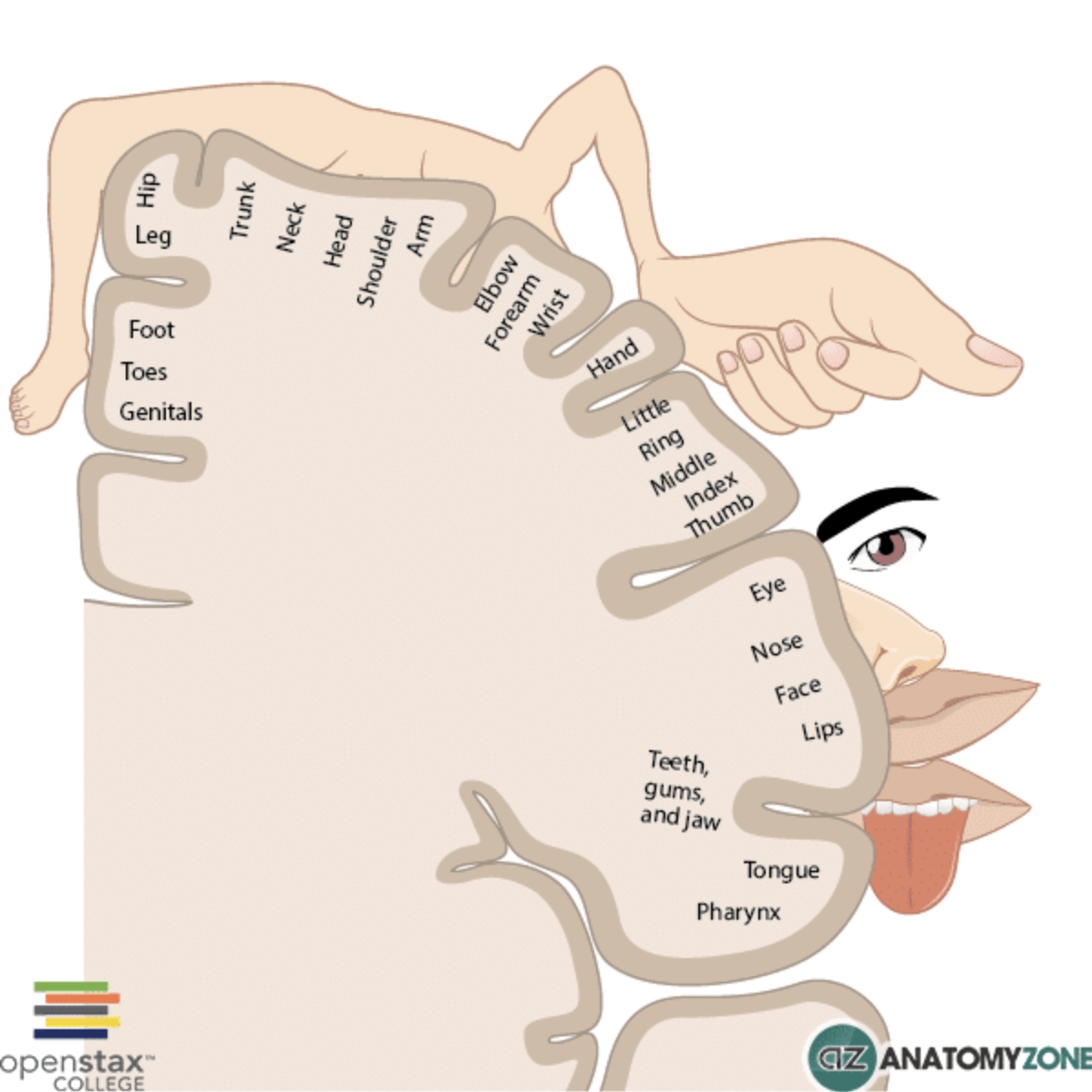
Somatotopic Organization
The point-for-point correspondence of an area of the body to a specific point on the central nervous system, often represented by the homunculus.
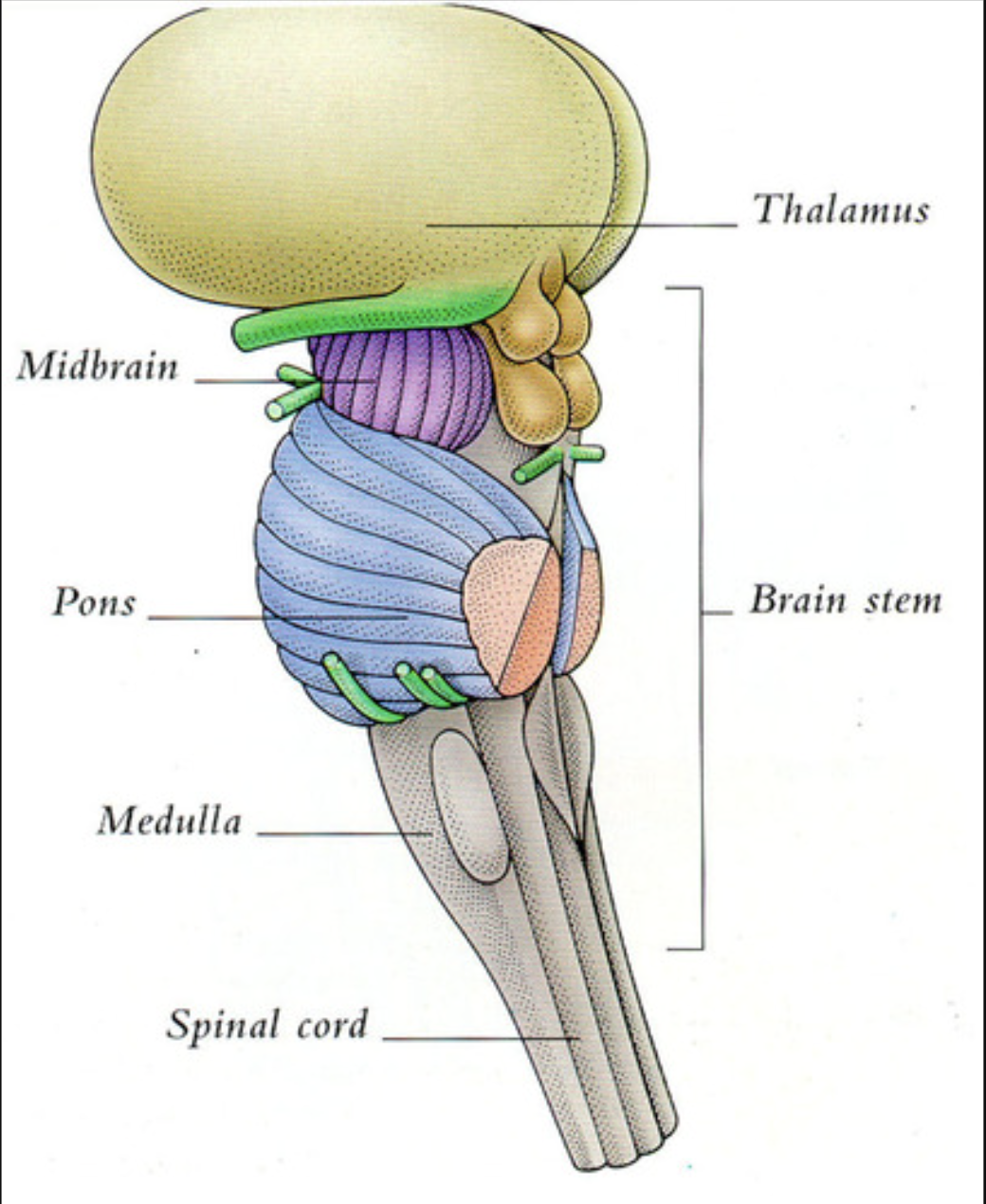
Thalamus
The major sensory relay station of the diencephalon; it filters and transmits information to the cerebral cortex.
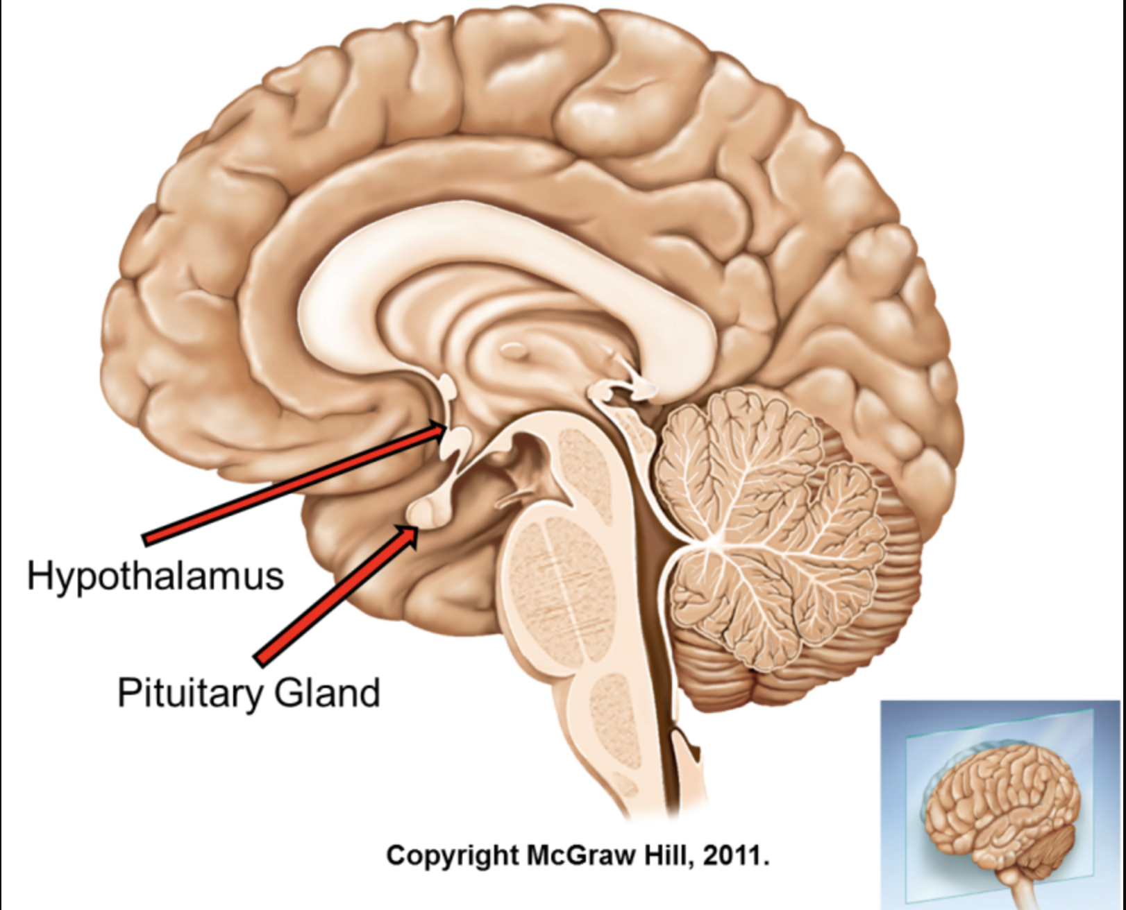
Hypothalamus
A structure in the diencephalon that regulates the autonomic nervous system and the endocrine system through the pituitary gland to maintain homeostasis.
Mesencephalon (Midbrain) Subdivisions
Tectum: Contains the superior (visual) and inferior (auditory) colliculi.
Tegmentum: Contains the substantia nigra (involved in movement) and the periaqueductal gray.
Pons
A structure in the hindbrain that serves as a bridge between the cerebellum and the rest of the brain; it is involved in sleep and arousal.
Medulla Oblongata
The most caudal part of the brainstem, controlling vital involuntary functions like heart rate, breathing, and blood pressure.
Spinal Cord
A long, thin bundle of nervous tissue that transmits signals between the brain and the rest of the body, organized into gray matter (inner 'H' shape) and white matter (outer tracts).
Cranial Nerves vs. Spinal Nerves
Cranial Nerves: 12 pairs of nerves that emerge directly from the brain/brainstem.
Spinal Nerves: 31 pairs of nerves that emerge from the spinal cord.
Autonomic Nervous System (ANS) Subdivisions
Sympathetic Division: Activates the 'fight-or-flight' response (increases heart rate, dilates pupils).
Parasympathetic Division: Activates the 'rest-and-digest' response (decreases heart rate, increases digestion).
Anatomical Directions: Rostral vs. Caudal
In the brain, Rostral (Anterior) refers to being toward the front or nose, while Caudal (Posterior) refers to being toward the back or tail.
Anatomical Directions: Dorsal vs. Ventral
Dorsal refers to the top of the brain or the back of the spinal cord. Ventral refers to the bottom of the brain or the belly side of the spinal cord.
Anatomical Directions: Ipsilateral vs. Contralateral
Ipsilateral refers to structures on the same side of the body. Contralateral refers to structures on opposite sides of the body.
Brain Planes: Coronal Section
Also known as a frontal section, it is a cut parallel to the face that divides the brain into front (Anterior) and back (Posterior) halves.
Brain Planes: Sagittal Section
A cut parallel to the neuraxis and perpendicular to the ground, dividing the brain into left and right halves. A Midsagittal section occurs exactly at the midline.
Brain Planes: Horizontal (Transverse) Section
A cut parallel to the ground that divides the brain into upper (Dorsal) and lower (Ventral) parts.
Organization of the Peripheral Nervous System (PNS)
The PNS consists of nerves and ganglia outside the CNS and is divided into:
Somatic Nervous System: Controls skeletal muscles and transmits sensory information.
Autonomic Nervous System (ANS): Regulates smooth muscle, cardiac muscle, and glands.
What are the three layers of the Meninges?
From outermost to innermost:
Dura Mater: The 'tough mother'; a thick, durable outer layer.
Arachnoid Mater: The middle layer with a web-like appearance.
Pia Mater: The 'pious mother'; a delicate layer that closely adheres to the brain surface.
The Cerebrospinal Fluid (CSF) Flow Pathway
CSF is produced by the Choroid Plexus in the ventricles. It flows from the Lateral Ventricles to the Third Ventricle, then through the cerebral aqueduct to the Fourth Ventricle, and finally into the Subarachnoid Space.
The Four Lobes of the Cerebral Cortex
Frontal Lobe: Executive functions, motor control, and personality.
Parietal Lobe: Somatosensory processing and spatial awareness.
Temporal Lobe: Auditory processing, memory, and language.
Occipital Lobe: Visual processing.
Telencephalon: The Limbic System
A group of structures including the Hippocampus (memory formation) and Amygdala (emotion and fear processing) involved in motivation and emotion.
Telencephalon: The Basal Ganglia
A collection of subcortical nuclei (including the caudate nucleus, putamen, and globus pallidus) involved in the control and fine-tuning of voluntary motor movements.
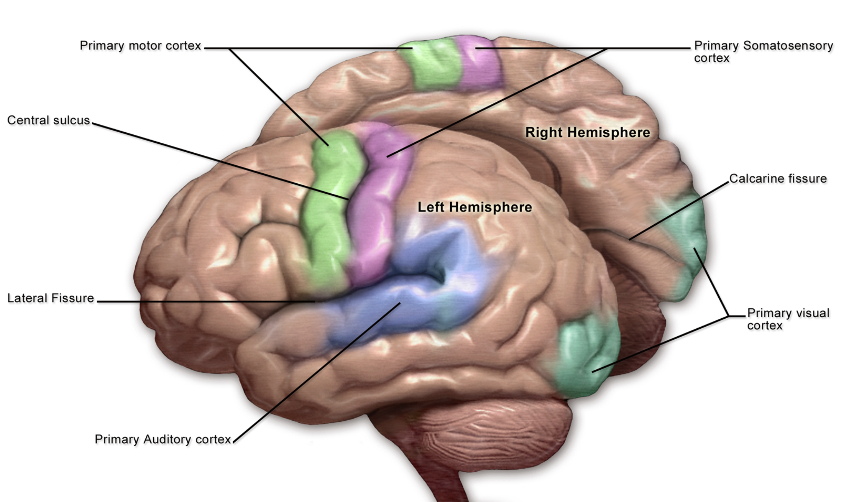
Primary Cortices
Specialised regions of the cerebral cortex that process sensory info.
e.g
Primary Motor Cortex (M1): Located in the frontal lobe, this area initiates all voluntary muscle movements. It sends direct signals down the spinal cord to move different parts of your body.
Primary Somatosensory Cortex (S1): Located in the parietal lobe, this is the first area to process basic touch, pain, temperature, and joint awareness (proprioception).
Primary Visual Cortex (V1): Located at the back of the brain in the occipital lobe, this is where all visual information (light, shapes, and edges) first arrives from the eyes.
Diencephalon: Thalamus vs. Hypothalamus
Thalamus: The 'relay station' that directs sensory input to the appropriate areas of the cerebral cortex.
Hypothalamus: Regulates homeostasis, the autonomic nervous system, and the endocrine system via the pituitary gland.
What is the difference between Gray Matter and White Matter?
Gray Matter: Found on the outer cortex; consists mostly of cell bodies, dendrites, and unmyelinated axons.
White Matter: Located beneath the cortex; consists of myelinated axons formed into tracts to carry signals.
Cranial Nerves vs. Spinal Nerves
There are (12) pairs of Cranial Nerves that exit the brain and (31) pairs of Spinal Nerves that exit the spinal cord to serve the rest of the body.
What is the midbrain?
The midbrain is a part of the brain located above the hindbrain, responsible for various functions including vision, hearing, motor control, and alertness.
What does the hindbrain contain?
The hindbrain contains both the metencephalon and the myelencephalon.
What is the spinal cord?
The spinal cord is a long, thin bundle of nervous tissue that transmits signals between the brain and the rest of the body.
What is the peripheral nervous system (PNS)?
The PNS is located outside of the skull and spine and is responsible for bringing information into the CNS and carrying signals out.
What are the two parts of the peripheral nervous system?
The peripheral nervous system comprises the somatic nervous system and the autonomic nervous system.
What is the function of the somatic nervous system?
The somatic nervous system controls the movement of skeletal muscles and transmits somatosensory information to the central nervous system.
What does the autonomic nervous system regulate?
The autonomic nervous system regulates the body's vegetative functions and controls smooth muscles, cardiac muscle, and glands.
What are the two divisions of the autonomic nervous system?
The autonomic nervous system is comprised of the sympathetic and parasympathetic divisions.
What is the sympathetic division responsible for?
The sympathetic division is responsible for the 'fight or flight' response, arousing and preparing the body for the expenditure of energy.
What is the parasympathetic division responsible for?
The parasympathetic division is responsible for the 'rest and restore' response, relaxing the body.
How do sympathetic nerves generally affect the body?
Sympathetic nerves generally arouse the body and prepare it for action.
How do parasympathetic nerves generally affect the body?
Parasympathetic nerves typically relax the body and promote restorative functions.
What role does the midbrain play in sensory processing?
The midbrain integrates sensory information from various modalities, including vision and hearing.
What is the relationship between sympathetic and parasympathetic nerves?
Sympathetic and parasympathetic nerves generally have opposite effects on organ systems.
What type of muscle does the autonomic nervous system regulate?
The autonomic nervous system regulates smooth muscles and cardiac muscle.
What is the primary function of the somatic nervous system?
The primary function of the somatic nervous system is to facilitate voluntary movement and sensory information transmission.
What is one key function of the spinal cord?
One key function of the spinal cord is to serve as a conduit for signals between the brain and the rest of the body.
In which part of the nervous system is the midbrain located?
The midbrain is located in the central nervous system, specifically above the hindbrain.
What are the effects of the sympathetic nervous system on heart rate?
The sympathetic nervous system increases heart rate during stressful situations.
What role does the parasympathetic nervous system play in digestion?
The parasympathetic nervous system promotes digestion by stimulating digestive processes.
What is the Bell-Magendie Law?
The Bell-Magendie Law states that sensory (afferent) nerves enter the spinal cord through the dorsal roots, while motor (efferent) nerves exit through the ventral roots.