ANATOMY : Special Sensory Systems
1/111
There's no tags or description
Looks like no tags are added yet.
Name | Mastery | Learn | Test | Matching | Spaced |
|---|
No study sessions yet.
112 Terms
What is the pathway of the olfactory receptors?
Bipolar neurons in nasal mucosa > Axons > Cribriform plate > anterior cranial fossa > Olfactory bulb > 2nd order Neuron > Olfactory tract > Lateral & Medial olfactory striae.
What structures does the lateral olfactory stria connect to?
1. Primary olfactory cortex (Pre-piriform & Piriform cortex).
2. Amygdala (associated with fear).
3. Entorhinal cortex (odour memory).
4. Thalamus → orbitofrontal cortex (conscious perception of smell).
What is anosmia and what are its causes?
Anosmia is the absence of the sense of smell.
Causes include:
1. Temporary (infection, local disorders).
2. Permanent (head injury, tumours).
3. Progressive (neurodegenerative conditions like Parkinson's or Alzheimer's).
What is the structure of the eyeball?
The eyeball is located in the anterior orbital cavity, embedded in fat, with a transparent cornea (anterior segment) and an opaque sclera (posterior segment).
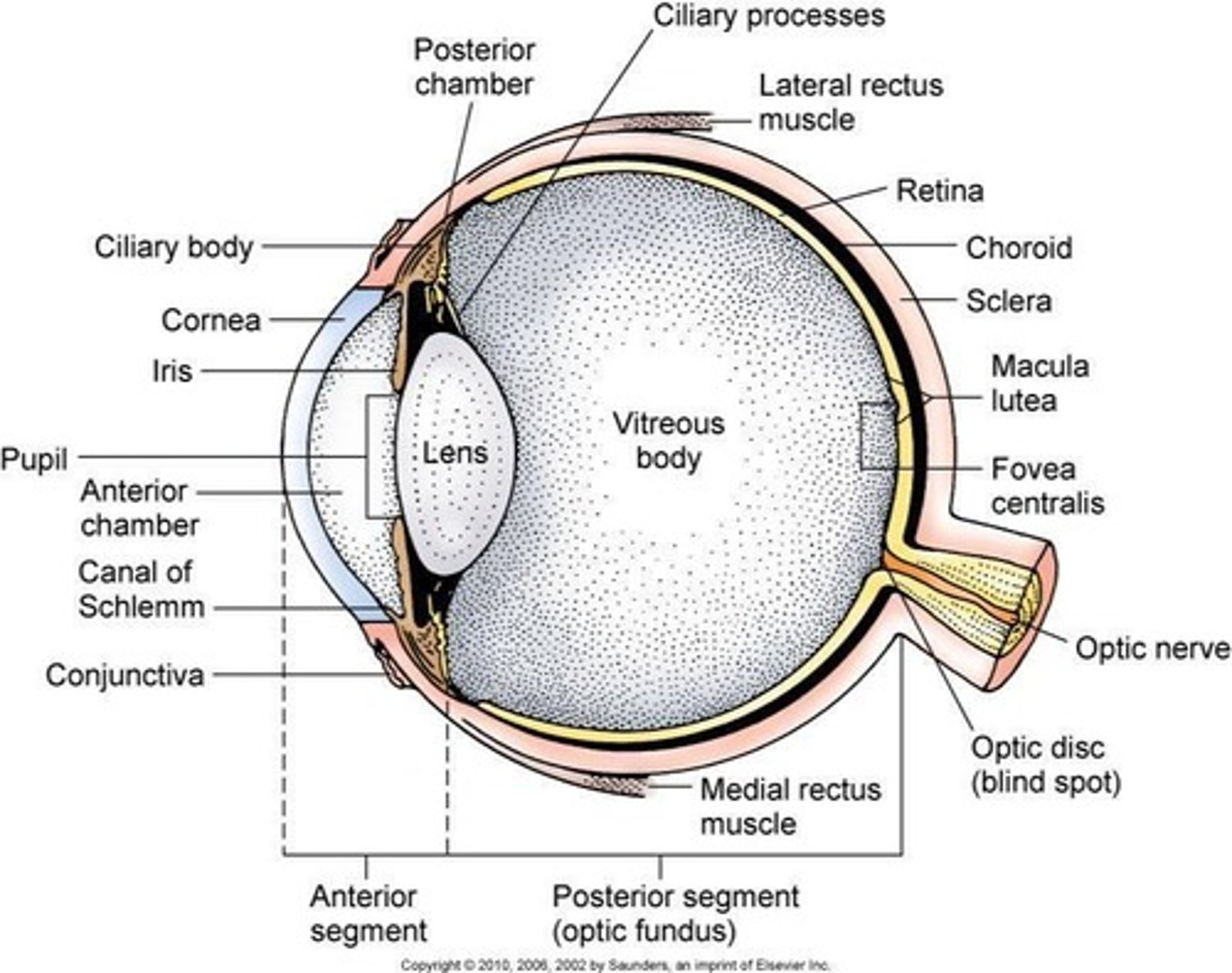
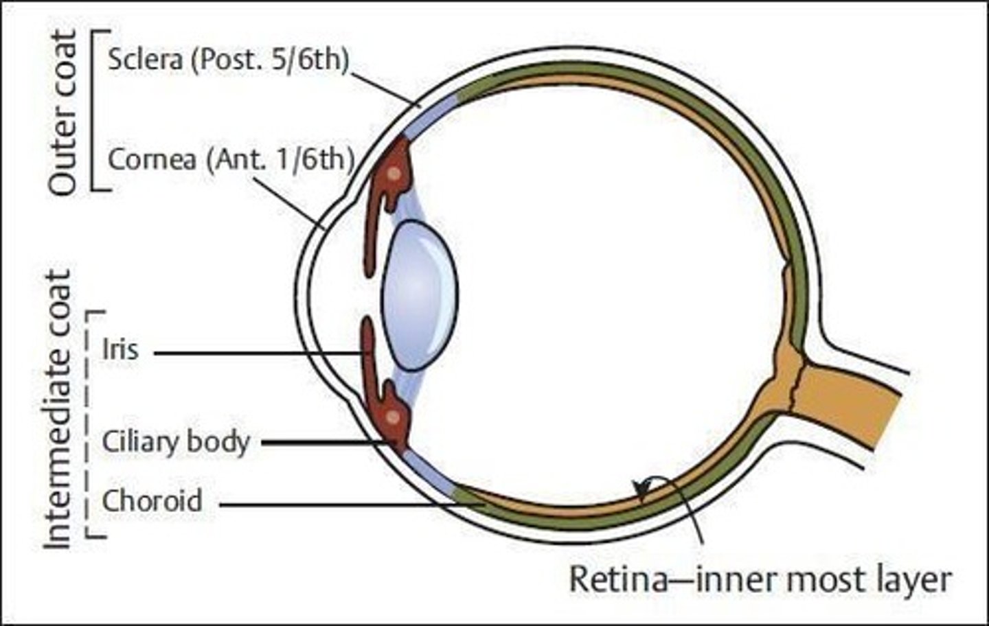
What are the three coats of the eyeball?
1. Outer fibrous coat: Cornea (anterior 1/6, transparent) and Sclera (posterior 5/6, opaque).
2. Middle vascular coat: Choroid, Ciliary body, Iris, Lens, Anterior and posterior chambers, pupil, vitreous chamber.
3. Inner neural coat: Retina.
What are the characteristics of the cornea?
The cornea is a thick, transparent, avascular membrane that maintains the shape of the eyeball, allows light to enter, and slightly refracts light.
How does the sclera contribute to the eyeball?
The sclera is the outer fibrous coat that is opaque and white, providing structure and protection to the eyeball.
What is the function of the retina?
The retina contains ten distinct layers and is responsible for converting light into neural signals for vision.
What is the role of the optic disc in the eye?
The optic disc is the point where the optic nerve exits the eye, lacking photoreceptors, thus creating a blind spot.
What is the macula lutea and its significance?
The macula lutea is a yellowish spot on the retina that contains a high concentration of photoreceptors, providing sharp central vision.
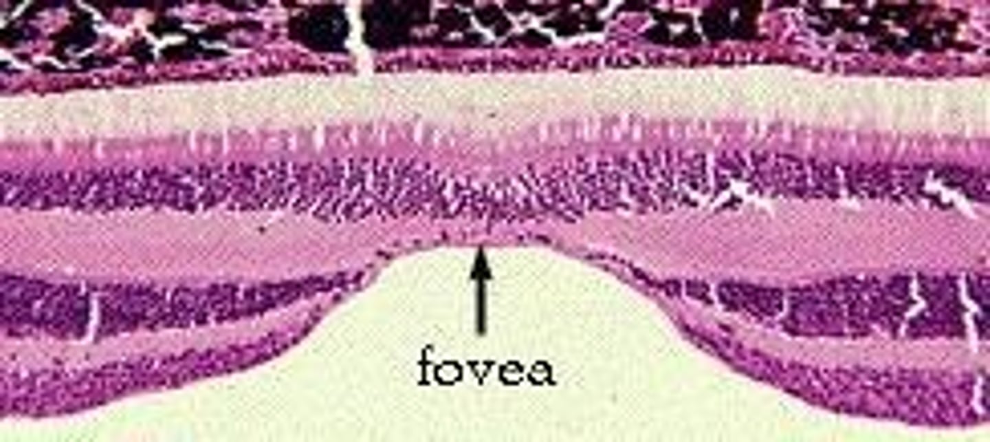
What is the fovea centralis?
The fovea centralis is a small depression in the macula lutea that contains only cones and is responsible for high-acuity vision.
What is the anatomical arrangement of the cochlea?
The cochlea is a spiral-shaped organ in the inner ear that contains fluid and hair cells, which convert sound vibrations into neural signals.
What is the function of the semicircular canals?
The semicircular canals are involved in the vestibular system, helping to maintain balance and spatial orientation.
What is the auditory pathway?
The auditory pathway transmits sound information from the cochlea through the auditory nerve to the brainstem, thalamus, and auditory cortex.
What is the vestibular pathway?
The vestibular pathway transmits information about balance and spatial orientation from the semicircular canals to the brain.
How does the hypothalamus relate to the olfactory system?
The hypothalamus receives olfactory information that triggers autonomic responses related to appetite, salivation, and gastric contraction.
What is the role of the amygdala in the olfactory pathway?
The amygdala processes emotional responses to smells, particularly fear.
What are the anterior and posterior segments of the eye?
The anterior segment includes the cornea and aqueous humor, while the posterior segment includes the sclera, choroid, and vitreous humor.
What is the significance of the conjunctiva?
The conjunctiva covers the anterior part of the sclera and provides protection and lubrication to the eye.
What nourishes the cornea?
The cornea is nourished by diffusion from capillaries at its edge and from aqueous humor from its internal surface.
What is the primary source of nourishment for the eye?
Nourishment is provided by diffusion from capillaries at its edge and from aqueous humor from its internal surface.
Which nerve innervates the eye?
The long ciliary branches of the nasociliary nerve (CN V1).
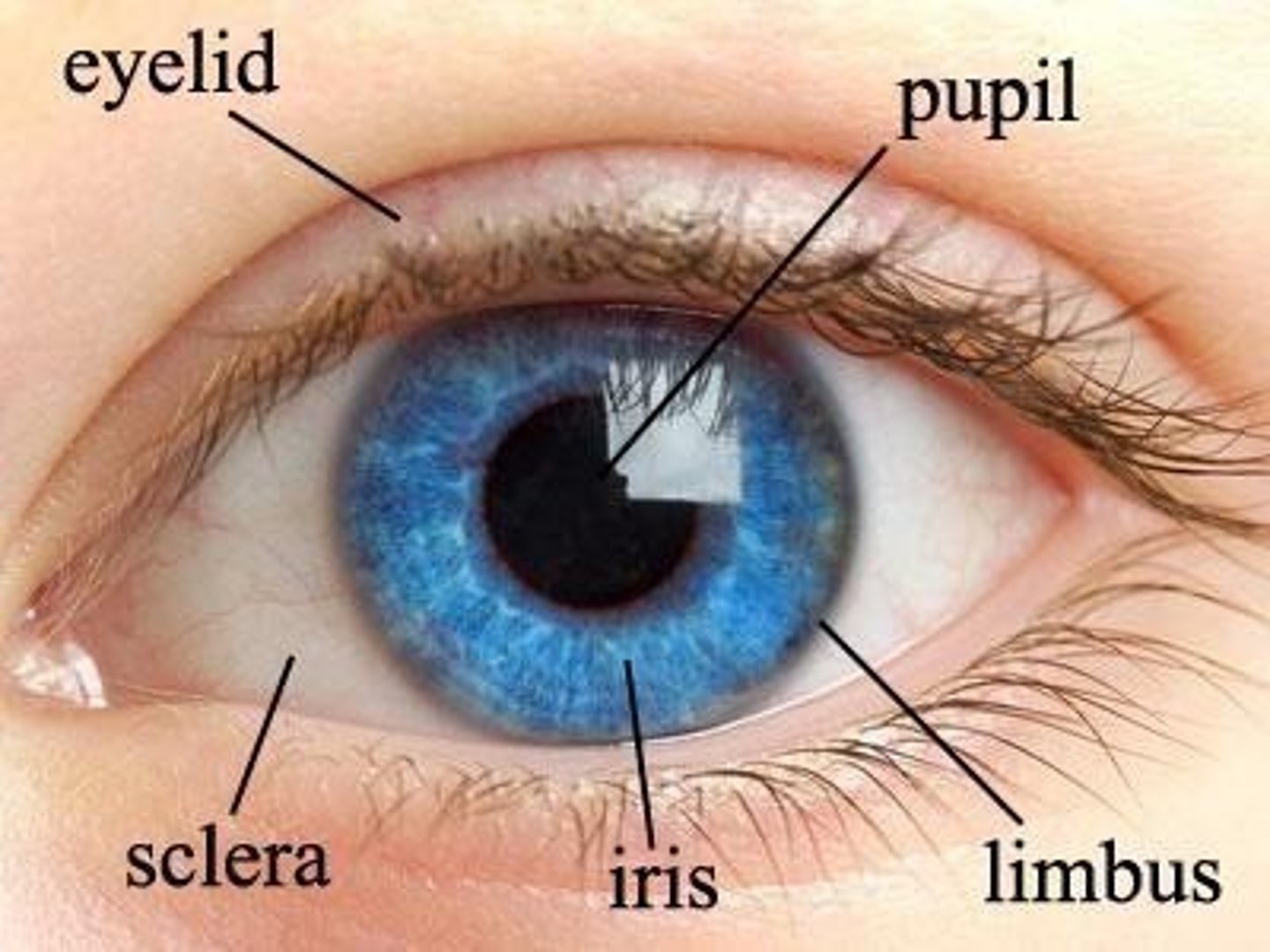
What is the limbus in the eye?
The limbus is the junction of the cornea and sclera.
What is the choroid and its function?
The choroid is a brown coat situated inner to the sclera, extending from the optic nerve to the ciliary body, providing nutrition to the outer layers of the retina and absorbing excess light.
What is the role of the ciliary body?
The ciliary body, located between the iris and choroid, contains ciliary muscles that adjust the lens shape for near and far vision.
How do ciliary muscles affect lens shape for vision?
Ciliary muscles contract for near vision, making the lens convex, and relax for far vision, flattening the lens.
What is the iris and its function?
The iris is the colored part of the eye that regulates the amount of light reaching the lens by controlling the size of the pupil.
What is the macula lutea?
The macula lutea is a yellowish area near the center of the retina with the fovea centralis, where visual acuity is greatest due to a high concentration of cone cells.
What is the optic disc and its significance?
The optic disc is where the optic nerve emerges from the retina, located 3 mm medial to the macula lutea, and is known as the blind spot due to a lack of photoreceptors.
What happens to the optic disc in optic neuritis?
Swelling of the disc occurs in optic neuritis and raised intracranial tension (papilloedema).
What is the function of photoreceptor cells?
Photoreceptor cells (rods and cones) convert light energy into electrical impulses for processing in the retina.
What are rods and their function?
Rods are photoreceptors that function in dim light and have a high sensitivity to light intensity.
What are cones and their function?
Cones are photoreceptors responsible for bright light and color vision, with three types for red, green, and blue light.
What is the role of bipolar neurons in the retina?
Bipolar neurons connect photoreceptor cells to ganglion cells, facilitating the transmission of visual information.
What are horizontal and amacrine cells?
Horizontal and amacrine cells are unipolar interneurons that integrate and modulate visual messages between photoreceptors and bipolar cells.
What are pigmented epithelial cells and their functions?
Pigmented epithelial cells are cuboidal to columnar cells that absorb light, phagocytose membranous discs from rods, and esterify Vitamin A.
What is the ora serrata?
The ora serrata is the anterior termination of the retina, marking the junction between the retina and ciliary body.
What is the significance of the fovea centralis?
The fovea centralis is the central depression in the macula lutea where visual acuity is highest due to the concentration of cone cells.
What occurs in the optic disc during glaucoma?
Depression or cupping of the optic disc occurs in optic nerve atrophy and glaucoma.
What is the vascular supply to the choroid?
The choroid receives a rich blood supply from the posterior ciliary arteries.
What is the structure of the ciliary body?
The ciliary body is a highly vascular part shaped like a complete ring that runs around the periphery of the iris.
What are ganglion cells (RGC)?
Neurons that receive visual information from photoreceptors via bipolar and amacrine cells, transmitting image-forming and non-image forming visual information from the retina to the CNS.
What structures do RGC axons form?
The optic nerve, optic chiasm, and optic tract.
What are Muller's cells?
Glial cells of the retina that extend between the vitreous humour and inner segments of rods and cones.
What is the function of the RPE (Retinal Pigmented Epithelium)?
It is involved in photoreceptor metabolism.
What is the role of rods and cones in the retina?
They capture light and are involved in phototransduction.
What is the outer limiting membrane in the retina?
A region of zonulae adherens junctions between Muller cells and photoreceptors.
What does the outer nuclear layer contain?
The nuclei of the photoreceptors.
What is found in the outer plexiform layer?
Axodendritic synapses between photoreceptors and dendrites of bipolar neurons and horizontal cells.
What types of neurons are contained in the inner nuclear layer?
Bipolar neurons, horizontal neurons, amacrine neurons, and neuroglial Muller cells.
What occurs in the inner plexiform layer?
Axodendritic synapses between axons of bipolar neurons and dendrites of ganglion cells and amacrine cells.
What is contained in the ganglion cell layer?
Cell bodies of large multipolar neurons of the ganglion cells.
What does the optic nerve fiber layer contain?
Unmyelinated axons of the ganglion cells that become myelinated as the nerve pierces the sclera.
What forms the inner limiting layer of the retina?
The basal laminae of the Muller cells.
What is the optic pathway?
It includes the ganglionic neuron in the retina, axon, optic nerve, optic canal, optic chiasma, optic tract, lateral geniculate body, optic radiation, and primary visual cortex in the occipital pole.
What is the significance of oligodendrocytes in the optic pathway?
They myelinate the optic nerve fibers.
What happens to axoplasmic transport in cases of raised intracranial pressure (ICP)?
It slows down, leading to stasis and papilloedema.
What visual defect is associated with a pituitary adenoma?
Bitemporal hemianopia.
What structures make up the outer ear?
Auricle (Pinna), external auditory meatus (canal), and tympanic membrane.
What are the contents of the middle ear?
Three ossicles (malleus, incus, stapes), two muscles (stapedius and tensor tympani), and two nerves (chorda tympani and tympanic plexus).
What are the two parts of the internal ear (labyrinth)?
Bony labyrinth and membranous labyrinth.
What are the three parts of the bony labyrinth?
Semicircular canals (balance in motion), vestibule (balance when stationary), and cochlea (hearing).
What is the orientation of the semicircular canals?
Three arched canals set at right angles to each other.
What is the function of the vestibule in the inner ear?
It helps with balance when stationary.
What is the central part of the bony labyrinth called?
The vestibule.
What structure in the vestibule is related to the middle ear and shows the oval window?
The lateral wall of the vestibule.
What does the anterior wall of the vestibule open into?
The scala vestibuli of the cochlea.
What structures open into the posterior wall of the vestibule?
The three semicircular canals.
What does the medial wall of the vestibule form?
The bottom of the internal auditory meatus.
What two structures are lodged within the vestibule?
The utricle and saccule.
What is the bony core around which the cochlea is formed?
The modiolus.
How many turns does the cochlea make?
2½ turns.
What is the cochlear canal lined by?
Endosteum.
What fluid is contained within the cochlear duct (scala media)?
Endolymph.
What divides the cochlear canal into scala vestibuli and scala tympani?
The spiral lamina.
How does the cochlear canal communicate with the middle ear?
Via the round window covered by the secondary tympanic membrane.
What fills the membranous labyrinth?
Endolymph.
What separates the membranous labyrinth from the bony labyrinth?
Perilymph.
What are the specialized sensory structures in the ampulla of semicircular ducts sensitive to?
Angular acceleration of the head.
Where does the utricle occupy in relation to the vestibule?
The upper and posterior part.
What connects the saccule to the cochlea?
Ductus reuniens.
What is the specialized neurosensory epithelium in the saccule and utricle called?
Macula.
What type of acceleration does the utricle detect?
Linear acceleration in the horizontal plane.
What type of acceleration does the saccule detect?
Acceleration in the vertical plane.
What separates the cochlear duct from the scala vestibuli?
The vestibular membrane.
What is the sensory end organ of hearing located in the cochlear duct?
The spiral organ of Corti.
Which cranial nerve supplies the labyrinth?
The vestibulocochlear nerve (CN VIII).
What are the two divisions of the vestibulocochlear nerve?
Cochlear division (hearing) and vestibular division (equilibrium).
What is the role of the frontal eye fields in the vestibulo-cerebral pathway?
They control eye movements and receive vestibular motion information.
What does the primary somatosensory cortex do in relation to body signals?
It maps body location and movement signals.
What is the function of the Parieto-Insular Vestibular Cortex (PIVC)?
It responds to body and head motion information.
How does the posterior parietal cortex contribute to motion perception?
It responds to both visual and vestibular motion cues.
What functions do the hippocampus and parahippocampal regions serve?
They are involved in spatial orientation and navigation.
What is the function of the vestibulo-autonomic pathway?
It stabilizes respiration and blood pressure during body motion and changes relative to gravity.
What are some clinical implications of the vestibulo-autonomic pathway?
It plays a role in motion sickness.
Describe the course of the cochlear component of CN VIII.
Hair cells of the Organ of Corti send signals to the spiral ganglion, then to the dorsal and ventral cochlear nuclei, cross to the opposite side in the trapezoid body, and terminate in the superior olivary nucleus, nucleus of trapezoid body, or nucleus of lateral lemniscus, before reaching the inferior colliculus, inferior brachium, medial geniculate body, and finally the temporal cortex.
What is noise-induced hearing loss?
Degenerative changes in the organ of Corti due to exposure to continuous loud sounds.
What is presbycusis?
The physiological hearing loss associated with the aging process.
What are the two forms of hearing loss?
Conductive hearing loss and sensorineural hearing loss.
What characterizes conductive hearing loss?
CN VIII is functioning, but bone conduction of sound is impaired due to issues in the external auditory canal or middle ear.