Bio N165 Brain Disorders and Behaviors Quiz 2 Review
1/47
There's no tags or description
Looks like no tags are added yet.
Name | Mastery | Learn | Test | Matching | Spaced | Call with Kai |
|---|
No analytics yet
Send a link to your students to track their progress
48 Terms
Retina
The back of the eyeball, considered a part of the brain, where light hits the photoreceptive cells and visual information begins being processed
Fovea
The part of the retina where vision is most acute and color vision is best. Cone photoreceptors are most prevalent here
Optic Nerve
composed of the axons of the retinal ganglion cells that leave the retina and head back towards the optic chiasm in the brain, taking with them visual information. This nerve is the reason humans have a blind spot, because no photoreceptive cells exist where the optic nerve exits the eye
Retinal ganglion cells
cells in the retina that receive input form modulatory neurons (which get input from photoreceptor cells) and transmit the information down the optic nerve to the brain; includes midget, parasol, and small bi-stratified cells
Photoreceptor cells
cells that line the back of the retina and have parts that change shape when they are hit with a photon, allowing them to detect light in a certain part of the visual field. Humans have two main types, rods and cones, and there are three different subtypes of cones
Rods
Photoreceptor cells that are located outside the fovea. They are responsible for low-light vision (highly sensitive to light) and useful for detecting movement, but at the cost of visual acuity. They do not differentiate between colors
Cones
photoreceptor cells that are located primarily in the fovea. They are responsible for high acuity vision, but take more photons of light to activate (good for daytime vision). There are three types, each most responsive to different wavelengths of light (corresponding to red, green and blue) which, when combined, allow for color vision
Parvocellular Pathway
a visual processing stream that pools over fewer receptors. The cells involved (midget cells) have a sustained response and are involved in processing color, fine detail, texture and depth
Magnocellular Pathway
A visual processing stream that pools over many receptors, whose cells (parasol cells) fire in bursts and are useful for detecting motion
Koniocellular pathway
a visual processing stream that gets S-cone input only (from small bi-stratified cells) processing low acuity visual information, and innervating V1 and extrastriate cortex
optic chiasm
where the optic nerves cross in the brain, allowing information from the left visual field (from both eyes) and right visual field (from both eyes) to be separated and directed to the appropriate contralateral hemisphere
Thalamus
A part of the brain involved in relaying sensory information from sensory organs to processing areas of the cerebral cortex
Lateral geniculate Nucleus
a part of the thalamus where the visual processing streams pass through on their way to the optic radiations and primary visual cortex
Optic radiations
nerve pathway along the visual processing stream from the LGN to primary visual cortex
Primary visual cortex (V1)
the first area in the brain where visual information is processed at a low level. Visual information flows into here from the retina and flows to higher levels of visual processing (V2, V3, etc) that do increasingly complex
Cornea
the transparent dome-shaped anterior portion of the outer covering of the eye
Lens
situated behind the iris of the eye, it focuses light entering the eye onto the retina
sclera
the white part of the eye that, with the cornea, forms the protective outer covering of the eye
iris
the colored portion of the eye, a muscular diaphragm that controls the size of the pupil, which in turn controls the amount of light that enters the eye
Scotoma
an area of impaired or lost vision in the visual field
cataract
an opacity in the lens that blocks light from reaching the retina; often occurs in older age due to sunlight (UV) exposure
unilateral field loss
loss of an entire eye's vision due to tumor or trauma that results from the disconnection of the optic nerve
Hemianopsia
blindness in one half of the visual field in one or both eyes
bitemporal hemianopsia
blindness in the outer halves of the visual field in both eyes, due to damage to the optic chiasm (tumors are often the culprit)
Binasal hemianopsia
blindness in the middle halves of the visual field in both eyes, due to damage to uncrossed fibers (often due to calcification of carotid arteries; also associated with hydrocephalus)
homonymous hemianopsia
blindness in the same hemisphere of the visual field in both eyes, due to damage to one hemisphere or cortex (often from stroke or trauma)
sensation
the first stage in the functioning of the senses, starting with information at the peripheral sensory receptors
Perception
The process of recognizing, organizing, and interpreting sensory information
Blindsight
a phenomenon where people who are perceptually blind demonstrate some response to visual stimuli (because only part of their visual system is impaired, other parts - parts involved in motion perception - may still function)
Dorsal pathway
made up of multiple visual areas, it is one of two main visual processing streams after primary visual cortex. This pathway is involved in perception for action
Ventral pathway
Made up of multiple visual areas, it is one of two main visual processing streams after primary visual cortex. This pathway is involved in perception for recognition
Visual Agnosia
a disorder in which the patient suffers from the inability to recognize and identify objects or persons despite having knowledge of the characteristics of the objects or persons. This condition can be loosely divided into two types that differ by severity: apperceptive and associative.
object agnosia
a disorder in which visual object recognition is impaired (i.e. naming or visually presented objects, categorization, matching by function) but elementary visual perception is more or less preserved, i.e. matching, and non-visual object recognition.
Apperceptive Agnosia
a disorder characterized by the inability to name, copy or recognize visually presented objects. Shape perception and figure-ground segregation is impaired, but basic visual functions (color discrimination, luminance discrimination, visual acuity) and object identification based on non-visual cues are preserved
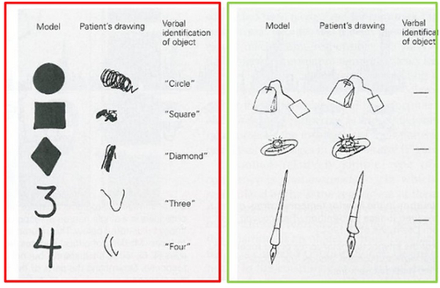
Associative agnosia
a disorder in which visual object recognition is impaired (i.e. naming of visually presented objects, categorization, matching by function), but elementary visual perception is more or less preserved. This is how object agnosia is typically described, as this is the more common type.

Fusiform face area
a bilateral visual processing area that is thought to be specialized for face processing (with some controversy -- some authors argue that it is specialized for detailed visuospatial processing, not necessarily just face processing). damage to this region can cause face perception deficits
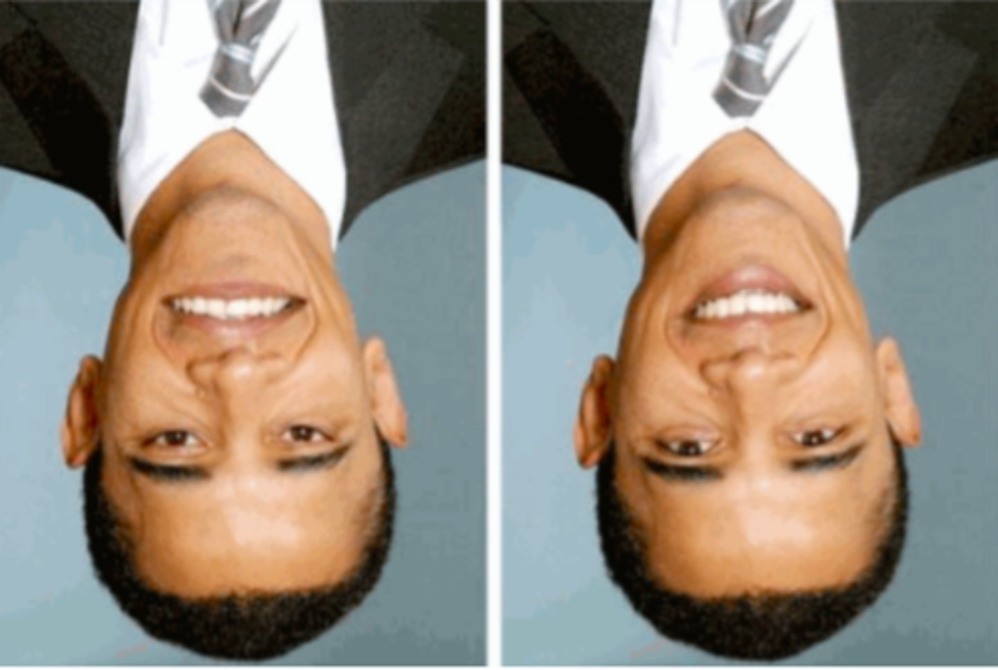
Face inversion effect
the inability to detect facial features that are inverted on an inverted face (i.e. the features would be upside down, and very obvious, if the face were turned right-side up), because we are so used to processing faces in the upright orientation
Prosopagnosia
a disorder in which faces cannot be recognized, but other forms of object recognition are unimpaired

capgras syndrome
The delusional belief that an acquaintance has been replaced by an identical-looking imposter. It is more commonly seen in schizophrenia, dementia, and brain trauma
Fregoli syndrome
the delusional belief that different people are in fact a single person who changes appearance or is in disguise, generally viewed with paranoia (that the "shapeshifting" person is out to get them)
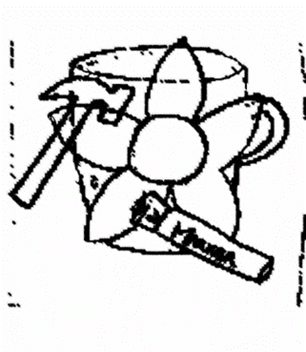
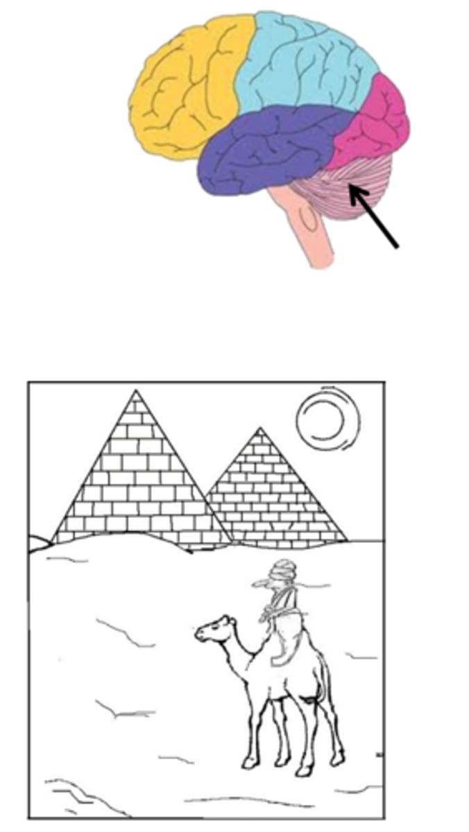
Simultagnosia
a deficit in scene perception, with a normal visual fields and normal lower-level (elementary) visual perception
Dorsal simultagnosia
a deficit in scene perception where the patient can only perceive one stimulus at a time (more severe than ventral type)

ventral simultagnosia
a deficit in scene perception where the patient can see multiple objects, but cannot recognize them (can navigate and count, but cannot read)
perceptual categorization defect
a disorder in which a patient cannot recognize objects form unusual views
akinetopsia
the inability to perceive motion that arises from damage to V5/MT - the motion area; patients experience a strobe-light effect of vision. It can be caused by damage such as stroke, trauma and rarely from antidepressants
Newton's theory of color
light contains color, not objects [but color perception is really a property of the brain]
opsins
photosensitive pigments in the photoreceptors i.e. rhodopsin and photopsin
opponent-processing color vision theory
color vision theory that color is processed in 3 different opponency channels created by specific wiring together of cone photoreceptors and retinal ganglion cells:
- red (L cone) vs green (M cone)
- Blue (S cone) vs yellow (L+M cone)
- dark vs bright (red/L+green/M+blue/S) => comparison produces luminance