neuroscience; week 9; hormones and sex
1/38
There's no tags or description
Looks like no tags are added yet.
Name | Mastery | Learn | Test | Matching | Spaced | Call with Kai |
|---|
No analytics yet
Send a link to your students to track their progress
39 Terms
hormones and the brain
a hormone is a signalling molecule that is transported between organs of the body by vascular system
hormones are essential for the regulation of development, physiology and behaviour, means they tend to control the speed at which certain things occur
they are produced by many organs of the body (these are called endocrine glands) (the endocrine system), with receptors similarly located in many places
under some level of ‘master control’ by the brain, although this is itself set within a complex feedback architecture thus undermining a ‘simple’ concept of a control hierarchy,
what do hormones actually do?
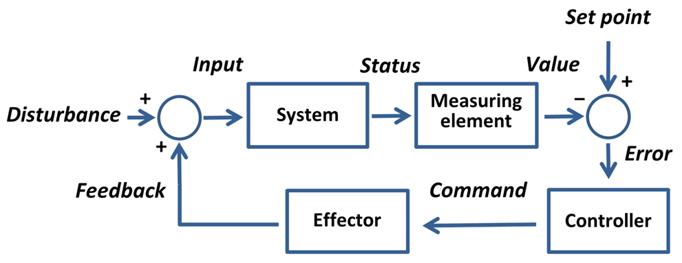
they regulate, thinking in a mechanistic way, kind of like how central heating in your house works
if something is wrong, then the controller can command things to correct this/ change things, if things are then back to where they want to be then the controller discards the command
more specifically
regulate physiology across the whole body in accordance with motivational states such as; arousal, aggression, hunger, fear and fatigue
regulate many processes, including: digestion, metabolism, respiration, tissue function, sensory perception, sleep, excretion, lactation, growth and development, movement, reproduction
common features: feedforward and feedback signaling, bidriectional influences between body and brain
the endocrine system
hormones, alongside neurotransmitters, underpin signaling within brain-body systems that act to maintain a desired ‘set-point’ in terms of behaviour of physiology
in fact, the term ‘set-point’ is better replaced with ‘desired state’ as it can be quite transient and itself dependent on input from other brain and body control systems
the endocrine system includes..
thyroid glands (affects metabolism)
parathyroids (helps regulate level of calcium in the blood)
adrenal glands (help trigger the fight or flight)
pancreas (regulates the level of sugar in the blood)
hypothalamus (brain region that controls the pituitary gland)
pituitary glands (secretes many different hormones, some of which affect other glands)
testis (secretes males sex hormones)/ ovary (secrete female hormones)
hormones vs neurotransmitters
neurotransmitters:
fast acting (onset)
act over short distances
restricted to nerve pathways
relatively short duration of action
digital action (via neuronal signalling)
tends to have more targeted actions
hormones
slow acting (onset)
act over long distances
travel anywhere via circulatory system
relatively long duration of action
analogue action (continuously variable)
tends to have more diffuse actions
not necessarily a ‘versus’ all the time
hormones are thus able to not only access parts of us that neurotransmitters cannot reach, but are also able to offer a qualitatively different mechanism of signalling that may complement that orchestrated by the CNS/PNS
the vascular system: the hormone highway
one thing that connects to this diagram, is cardiovascular health, so your blood supply with your body parts is important for health, issues with cardiovascular health can impact hormone control, as they can’t regulate as well as previously
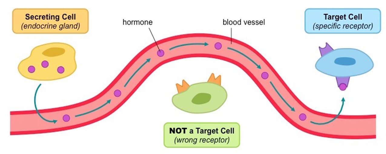
the secreting cell secretes the hormones through the blood vessels that then travel to the target cell (a specific receptor) to be uptaken
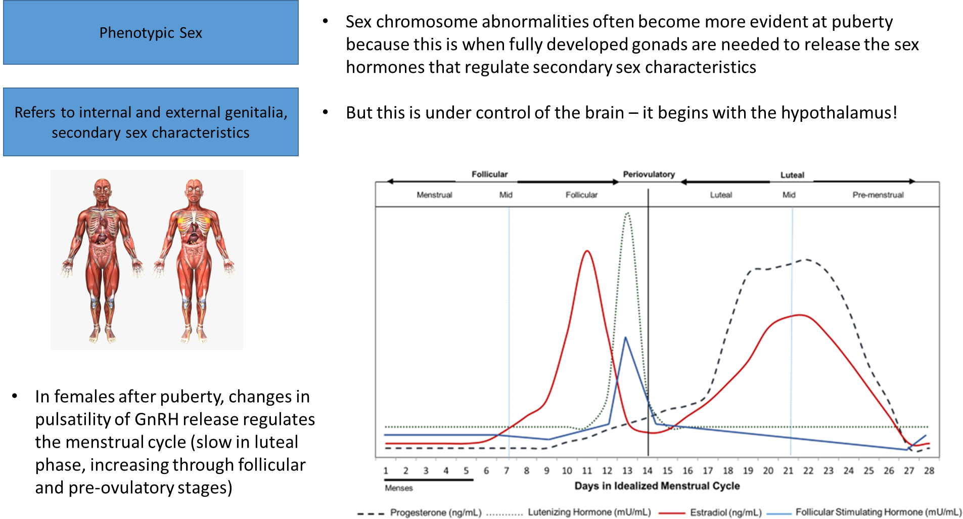
hypothalamus- master controller?
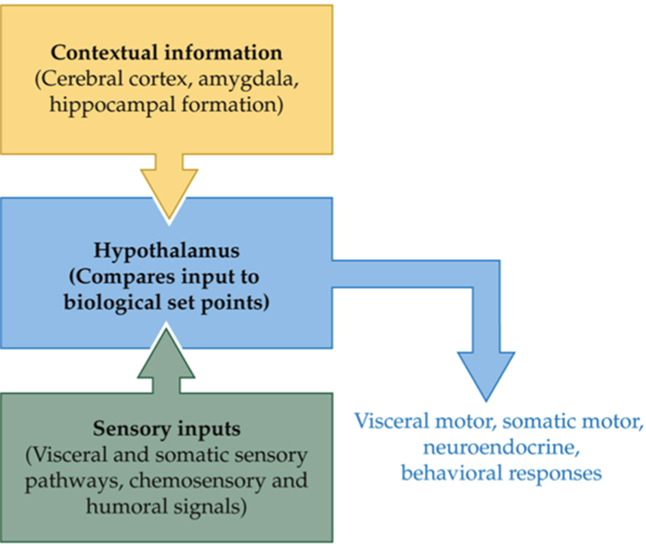
hypothalamus can see current state of body and current context of organism and can make adjustments to physiology and behaviour via several routes (like hormones)
hypothalamus receives lots of sensory input, and knows the context around you and can then regulate the function of nervous systems, these sensory inputs go into the hypothalamus, things like temperature, the hypothalamus knows the status of the situation now, the cortex figures out what to do, and the amygdala potentially drives a fear response and an urge to do something about it
difficult to claim the hypothalamus is controlling all of this considering other brain areas come into influence, like amygdala or the cerebral cortex, although the hypothalamus has some level of control, it is also controlled by various things too
the hypothalamus receives contextual information from the amygdala, cerebral cortex and/or hippocampal formation and sensory inputs from the visceral and somatic sensory pathways, chemosensory and humoral signals, which compares the input to biological set points to release visceral motor, somatic motor, neuroendocrine, behavioural responses
hypothalamus/ pituitary hormone release
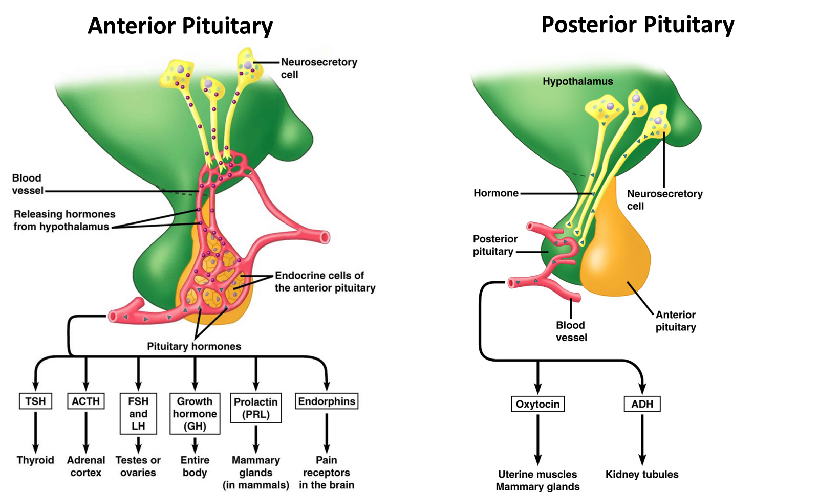
for example, the pituitary hormone oxytocin → targets female reproductive system → which effects/ triggers uterine contractions during childbirth
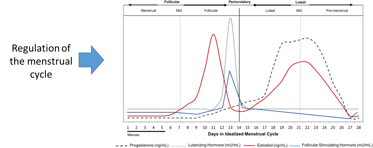
the hormones of the menstrual cycle
the menstrual cycle includes the coordination of four separate hormones; which would be hard to regulate every month, this shows that a lot of processes include the coordination of multiple hormones, not just one
different ways to decipher gender
gender is defined in many ways and is a subjective way of expressing one’s perceived gender, but it is heavily influenced, for the most part, by sex assigned at birth
phenotypic sex is modifiable particularly during developmental stages
there’s…
chromosomal sex, 22 pairs of autosomes chromosomes and one pair of allosomes
phenotypic sex, refers to internal and external genitalia, secondary sex characteristics and often incorrectly used interchangeably with notion of gender
gender, an individuals self perception of oneself, can be considered as emerging from self-appraisal in the context of social or cultural norms
variations in chromosomes or gene mutations affecting phenotypes- Turner’s syndrome
Turner Syndrome:
missing X chromosome [XO]
- affects females
girls with Turner syndrome tend to be shorter and have impaired ovary function
the ovaries produce two sex hormones (oestrogen and progesterone)- reduced levels of these lead to underdevelopement of secondary sex characteristics,
so TS is often diagnosed at puberty
infertility/ reduced fertility
treatment: oestrogen and progesterone
variations in chromosomes or gene mutations affecting phenotypes- Klinefelter’s syndrome
extra X chromosome- affects males [XXY]
Boys with Klinefelter syndrome tend to be taller in height and have impaired testicular function
the testicles produce the sex hormone testosterone-
reduced levels of this lead to under development of secondary sex characteristics
so often diagnosed at puberty
infertility/ reduced fertility
treatment: testosterone, treatment works for the secondary sex organs, nothing can change the chromosomal make up
variations in chromosomes or gene mutations affecting phenotypes- XYY syndrome
extra Y, affects males
usually taller than XY males, risk of learning or speech development problems, but symptoms usually mild and overall IQ often normal range. Acne.
many people with this are never diagnosed
normal fertility
treatment: supported learning
doesn’t have an overwhelming amount of issues arise during puberty so fertility is normal
they’re could have a higher risk of vulnerability to various social situations, including violence or crime because of a loose connection, but the direct assumption that there is a clear relationship between those with XYY and violent crime is false
what do conditions surrounding gene mutations/ chromosome variations teach us about the function of sex chromosomes?
two chromosomes are important for the full development of female biological sex characteristics
if one is missing, there is underdevelopment of these aspects
an X and Y chromosome must be present for the full development of male biological sex characteristics
the x chromosome contains approx 1500 genes, whereas Y has approx 80
X contains so much more info important for general development and survival
Y contains mainly genes that signal for male gonadal development and sperm cell function
has been called a ‘functional wasteland’
chemically blocking the SRY action
zygotes containing the SRY action in the embryonic gonads have testes, but blocking this SRY action makes the foetus genetically male (chromosomally) (XY) but phenotypically female (has ovaries)
injecting the SRY action into XX zygotes that would otherwise develop ovaries, leads to them being genetically female but have tests
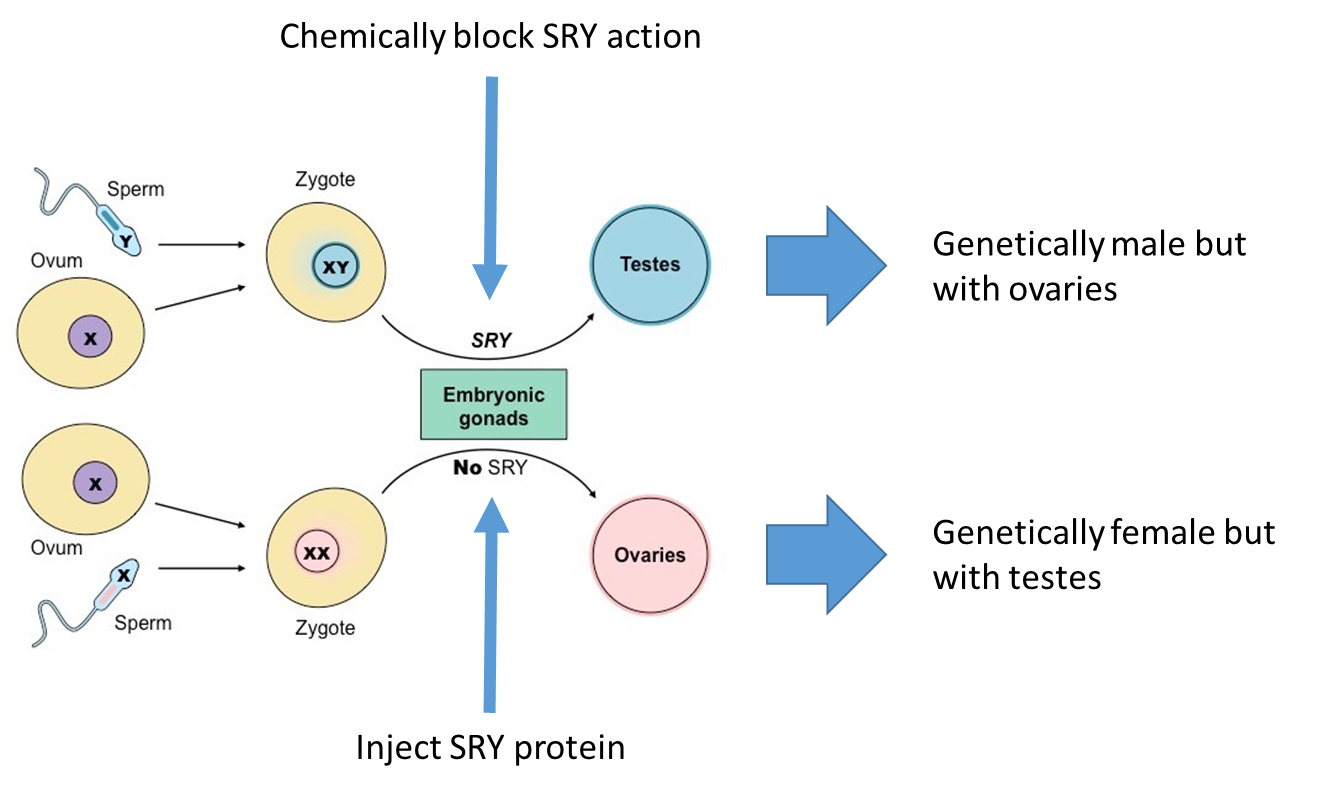
chromosomal sex, specifically the presence/ absence of the SRY gene, gives rise to primary internal and external genitalia
one component of this are the gonads
chromosomal testing to determine male / female
in 1992 the Olympic committee implemented testing for presence of the SRY gene to determine if athletes were male or female
athletes with the gene were not permitted to compete as females
8 female athletes who were shown to have the SRY gene, had an insensitivity to androgen (of which testosterone is a form) and thus were phenotypically a female, and had no advantage over the other female competitors
this screening was eliminated in 2000 and replaced with hormone based testing
the negative feedback cycle
won’t have the development of secondary sex characteristics until something changes
what changes once creeping closer to puberty is the hypothalamus starts disregarding this negative feedback loop, so there’s a decrease in sensitivity
it’s not a flood of this, it’s a pulsing release, there’s an increasing rate of release
this is similar to how the menstrual cycle works
the sex hormones inhibit AP and hyp function and it is reduction in sensitivity of the Hyp/ AP to these hormones then enables the altered ‘state’ of the system
phenotypic sex → what about the brain?
new data suggests a wider range of influences on the development of female and male brains beyond that of the sex hormones
most recently, the idea of ‘predetermined’ differences has been challenged
at the first stage it was refined
more recently, there’s more high quality evidence that shows difference within the brain based on separate sexes
there are lots of hormones that are not effected by sex
gender definition
‘an individual’s subjective perception of their sex’
this definition is too simplistic as it constrains gender to binary categorisation
it also doesn’t explicitly capture the external (social) context for gender
Is there a neurobiological basis for gender?
not if it is socially constructed
but, the WHO (and many other definitions) include “norms, behaviours and roles associated with ebing a woman, man, girl or boy, as well as relationships with each other”
as a neuroscientist/ psychologist we see behaviour as emerging fairly directly from the brain
but they tend to focus on animal models, with “gender identity” definable only by behaviour
this leads to conflation of gender identity and sexual behaviour/ sexual orientation
if stating it’s entirely socially constructed then no, however, like the slides shown, things like behaviour and relationships can be seen as emerging from the brain, which brings us back to the idea that psychologists and neuroscientists do have things to say about gender
however, neuroscientist are limited to just looking at behaviours really, particularly in animals, so they typically look at sexual behaviours, which does not directly encompass the concept of gender
sex differences in the brain
male and female brains are different sizes on average, with male brains weighing about 10% more
most (approx 90%) of this difference is explained by differences in body mass (a relationship that exists across species)
are there other differences in brain structure or function?
yes, but what they are which are important and how they come about is very uncertain, despite decades of (sometimes too) enthusiastic research
sexually dymorphic nucleus in the brain, shows there are sex difference in brain areas
there’s a strong motivation to understand the difference, which has led to bad science as many scientists are effected by their own motives and opinions
sex differences in the brain- bad science
motivations: charged with opinions, as humans are, scientists can begin their research with the wrong motivations either consciously or subconsciously attempting to support their preconceived opinions
bad designs: pea hacking? idk look it up, this sort of thing doesn’t tend to get published anymore
questionable comparisons: comparing animals with humans or sophisticated behaviours to gender or sexual attraction isn’t inherently wrong but needs a precise and particular way about it that justifies the statement
publication bias: papers get published if you find a significant result, now we’re paying more attention to the importance of publishing papers that find no significant effects, there are now ways to reduce publication bias through meta-analyses etc
why should we care about sex differences in the brain?
there are many differences, like differences in diagnosis that have led to the underdiagnosis of females with autism for example
the only clear difference is the parts in the brain that cover the development of sexual behaviours
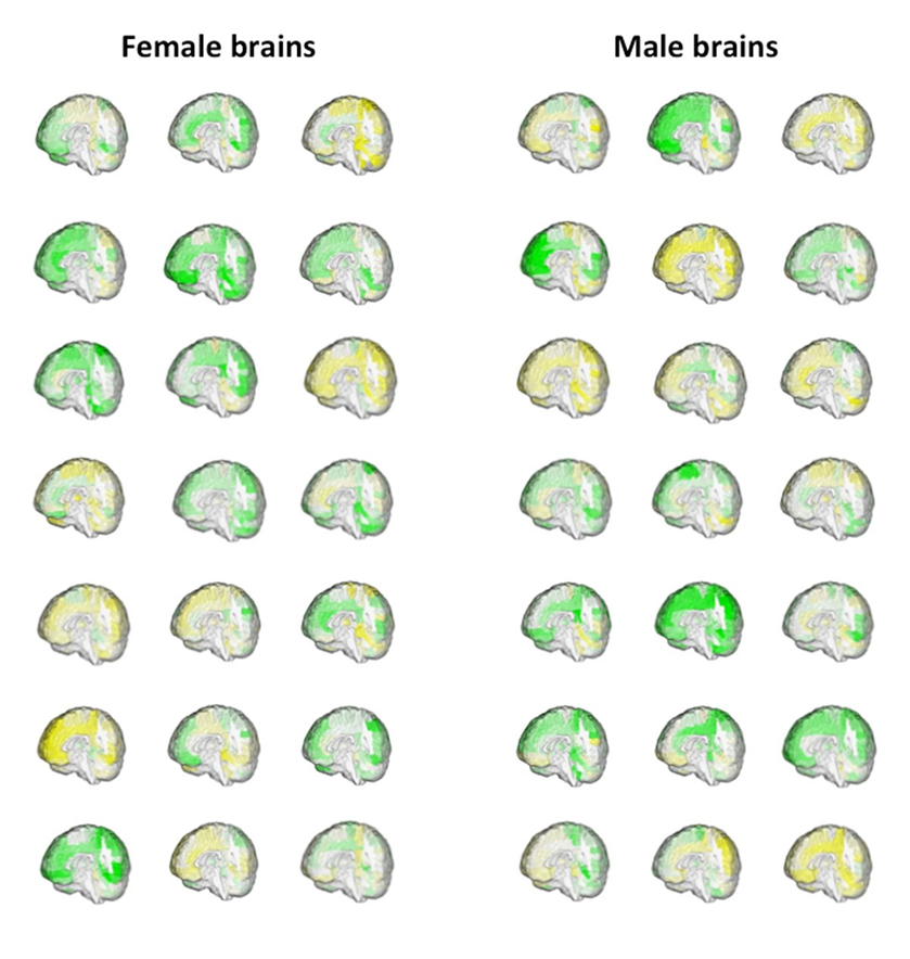
The “human brain mosaic”
experiment comparing the grey matter between individuals, within and between genders
there is not a male vs female brain type
showed variation cross individuals within a gender category is greater than between categories
some features tended to be more female or male but each individual had a combination of these features typical of both male and female tendencies
male brains were not more variable than female brains
there are brain structures that are typically separated by sex but people of another sex can have different brain structures, it’s not binary
female- male brain differences
size? yes
white matter and grey matter? yes
other specific structures? some
causes? not fully known
plasticity in the brain will make it very susceptible to differential gender-related influences (these probably occur from birth)
development before birth; there could be an effect here
afterwards, huge amounts of sensory development occurs, and there is so much developmental change beyond hormones
for example, if you’re teaching one 3 year old to teach a football from a young age, and not another, they will have a different neural connection regarding the sport and the movement to one another
sex'/gender difference in global brain size
Research has found that global brain size is larger in males.
This is reported as total intracranial volume (ICV or TIV), or as total brain volume (TBV) which excludes the meninges and ventricles.
In a 2014 meta-analysis involving 77 studies and 15,000 pps, Ruigrok et al. found that ICV is 12 % larger in males; A nearly identical difference in TBV (10.6 %) was reported in a UK Biobank study (5,216 pps)
S/g difference in global brain volumes are present at all ages
Paus (2010) reported global brain volume as about 8% larger in male neonates, peaking at 13 % during adolescence/early adulthood. More recently, Knickmeyer et al. (2017) imaged a large sample of neonates and found that ICV, total gray matter and total white matter are each about 5% larger in males, while Dean et al. (2018) reported a difference of 8% at one-month of age. Thus, the s/g difference in TBV is present from birth and increases during postnatal development.
Larger bodies require larger brains and the s/g difference in brain volume mostly parallels the divergence of male/female body size during development. Thus, newborn boys are about 4% heavier than newborn girls, with the difference increasing to an average of 18 % greater weight and 9% greater height in adult males
With larger brains come larger raw volumes of each CNS compartment in males: gray matter (GM), white matter (WM), and ventricular volumes
However, there are subtle differences in each compartment by s/g, with most studies reporting a higher ratio of GM-to-WM in females
The magnitude of this difference ranged from 4 to 7% across six studies
Thus, larger brains have a higher proportion of WM than smaller brains, regardless of sex
Moreover, the GM/WM ratio diverges the most after puberty, when sex differences in height and total brain size reach their peak. This is important when comparing the structural “connectome” between males and females
Are specific brain structures larger in men or women? origin of the term “sexual dimorphism” in the CNS
the search for human brain sexual dimorphisms is based on the idea that specific structures or circuits differ disproportionately between men and women that explain well-known behavioral s/g differences, such as empathy, spatial navigation, and gender identity
This paradigm rooted in animal neurobiology, where certain brain areas are indeed dramatically larger in one sex, with clear links to behaviours such as courtship and mating.
An example is the spinal nucleus of the bulbocavernosus (SNB) in rodents, which innervates two muscles at the base of the penis and is clearly present in males’ lumbar spinal cord but barely visible in adult females.
“sexually dismorphic”
the common framing of human brains as “sexually dimorphic” is based on the model of X and Y chromosomes acting early in devel opment and largely by way of gonadal hormones to enhance or suppress the growth of specific structures, essentially bifurcating male and female brains into distinct forms
This binary classification has been widely extended to describe male-female neurophysiological or behavioral differences using the same adjective, “dimorphic”, even when the distribution of measures may be largely overlapping (Joel, 2011) and despite the caution urged by some in the field.
But as the remainder of this paper will demonstrate, such binary classification does not accord with actual measures of human brain s/g difference, which are generally small, unreliable, and insignificant once individual body size is accounted for
Scaling issue in the comparison of female and male brains
Recognizing that brain size is related to body size and that human bodies are indeed sexually dimorphic, neuroscientists have struggled to find ways of comparing brain structures between men and women that don’t merely reflect bodily size differences.
Thus, every major study confirms that it is not only global measures like ICV, TBV, total GM, total WM and CSF volume that are larger in males, but every zone of the cerebral cortex and every subcortical structure, when reported as “raw,” “native,” or “uncorrected” volume
effect of brain segmentation method on male-female comparisons
Due to the inherent variability between individual brains, every automated fitting produces some distortion, which may affect male or female brains differently. These unique templates are constructed from a finite population of “normal” brains, often based on unequal numbers of males and females. This is further illustrated by the differences between brain templates constructed from different ethnic populations
Grimm et al. (2015) compared VBM and Freesurfer to measurement of the same two structures and found that both programs deviated from manual tracing, with accuracy of each method varying by structure. (VBM was more accurate for the amygdala, Freesurfer for the hippocampus.) Makowski et al. (2018) extended this comparison to the striatum, pallidum, and thalamus and found again that FSL and Freesurfer overestimate these volumes, with the error greater for smaller than larger structures
By contrast, comparison of Freesurfer and manual volumetry on cortical structures found that Freesurfer produced systematically smaller volumes, with much smaller variance than manual methods
It is not known whether these distortions differentially affect the measurement of male versus female brain structures, but the fact that automated errors are greater for smaller structures suggests they may. Indeed, Marwha et al. (2017) found smaller male-female differences among studies using manual, as opposed to automated segmentation, whether the data were reported as raw volumes or as volumes normalized to ICV or TBV.
survey of structural brain sex/ gender differences
no studies compare the ACTUAL shape of brain structures between males and females, but hundreds have compared structure size, so the survey uses studies that report on volume, thickness or cross sectional area of specific brain structures
Negligible differences in subcortical structural volumes
33 studies compared the volume of subcortical structures between females and males after normalising individual ICV or TBV. No subcortical structure has been found larger in females or males across all studies
the next structures to be declared sexually-dimorphic were the hippocampus and amygdala
Cahill (2006) depicted a mid-sagittal brain with many structures shown in pink (larger in women) and others shown in blue (larger in men). Although widely-reproduced, this figure was based on just one small study in which most s/g differences were not statistically significant
Claims about hippocampal and amygdala volume differences have also penetrated popular reporting, where they have been invoked to explain male-female differences in learning and emotion
However, these assertions ignore larger studies that have failed to confirm male/female differences in either hippocampal or amygdala volume
A meta-analysis of 29 studies found a mere 0.6 %, non-significant difference in hippocampal volume between males and females
massa intermedia
also known as the interthalamic adhesion
is a small bridge of glia connecting the left and right thalamus that is entirely absent in 2–22% of healthy brains
does differ reliably between males and females
In their first analysis of the AC, Allen and Gorski (1991) found that the ITA is larger and more often present in the brains of women, noting similar findings dating back to the 1940s
Although little research has addressed the possibility that ITA presence and size are affected by overall brain size. If larger brains are less able to mechanically support this small bridge across the third ventricle, the size and presence of ITA may be a function of brain geometry, not s/g per se.
Only one study has examined this and found no relationship between ITA presence and TBV, but further examination is warranted. Of note, ITA absence has been associated with a greater incidence of schizophrenic disorders in both men and women
brain size and its correlates
With the difference in overall brain size comes other male/female brain differences that are largely attributable to size rather than sex. One of these is GM/WM ratio, which averages 5.5 % larger in females across multiple studies. As brain size increases, there is a disproportionate increase in the denominator of this ratio, since larger brains need larger-caliber, more heavily myelinated axons to transmit action potentials across greater distances. Thus, the s/g difference in GM/WM ratio is largely eliminated when adjusted for total brain size
Without exception, the difference in global brain size is reflected in every major cortical and subcortical brain component, which range from 5 to 11% larger in absolute volume in males compared to females.
the absolute difference in amygdala volume is a mere 1% larger in males (after ICV or TBV normalization) and not reliably significant.
For cortical volumes there is even less consistency in claims of s/g difference, especially comparing studies using surface- versus volume-based segmentation
Cortical thickness is another metric often stated to differ between males and females. Several large and small MRI studies report greater average cortical thickness in females, however, this finding is highly sensitive to image acquisition method
the human brain is not sexually dimorphic
s/g differences in the human brain are extremely subtle and variable. There is nothing to justify the term “sexual dimorphism”.
Furthermore, differences that are portrayed as related to s/g are more accurately attributed to brain size. They distinguish large- from small-headed men as well as the average man from the average woman.
the term “dimorphism” reinforces a binary understanding of s/g brain difference, when in fact, few such differences actually exist and the ones that do are very small.
A similar conclusion is from research on transgender participants’ brains. Smith et al. (2015) “viewing gender as a binary category has to be reconsidered.”
A picture is emerging of a multidimensional “mosaic” of countless brain attributes that differ in unique patterns across all individuals. Although such differences may, in a particular sample, sum up to discriminate male from female brains, the precise discriminators do not translate across populations.
In this sense, the brains of male and females are not dimorphic (like the gonads) but monomorphic, like the kidneys, heart and lungs, which can be transplanted between women and men with great success.
brain size and its correlates
reliably larger in males: through lifespan and parallels overall bodily size and growth
the difference in TBV increases from 6% at birth to 11% in adulthood, while body mass changes from 4-18% over the same period
females reach maximum brain volume 2 years earlier than men may be because of earlier puberty onset
the s/g difference in GM/WM is largely eliminated when adjusted for total brain size
the absolute difference in amygdala volume is a mere 1% larger in males (after ICV or TBV normalization) and not reliably significant
why are freesurfer findings difficult?
in cortical volumes:
the functional significance of the Freesurfer findings is difficult to parse, since women outperform men in verbal and social-emotional skills which rely on areas found to be larger in men in these studies (e.g., fusiform, orbitofrontal and pars triangularis). Also paradoxically, men outperform women in physical and spatial skills, which depend on areas found to be larger in women (superior parietal, postcentral and paracentral gyri) in these large studies. So these findings fail to support to the claim that the male human brain is evolutionary augmented for spatial-mechanical cognition (so-called “folk physics”) and the female brain for language and social cognition (“folk psychology”)
cortical thickness
MRI findings contradict results from more precise histological measurements, which trend towards greater thickness in males. Unlike structural volumes, cortical thickness does not vary with overall brain or intracranial volume hence it is not appropriate to normalize to 3-dimensional volume when comparing males and females in this 1-dimensional measure, and studies that have done so overestimate cortical thickness in females Thus, once we account for individual differences in brain size, there is almost no difference in the volume of specific cortical or subcortical structures between men and women. One tiny exception is INAH-3, which is too small to be visible by MRI but has been confirmed by four post-mortem histological studies to be about 1.6-fold larger in men
Minute differences in lateralization and interhemispheric connectivity
Evidence is more consistent that the massa intermedia (or, interthalamic adhesion) is larger and more often present in female than in male brains (Table 6b). However, it is not known whether this is a true s/g difference or a function of brain size, given the mechanical stress on this small bridge of non-neuronal tissue spanning the third ventricle
the weak and inconsistent differences in male/female interhemispheric connectivity explain why efforts to link lateralization to cognitive sex differences have been unsuccessful
where has large-scale comparison of males and females been highly successful?
for sex prediction based on machine learning. With so many variables and such large samples, artificial intelligence is very good (∼90 % accuracy) at predicting whether a particular scanned brain came from a male or female participant. However, this predictive validity is highly diminished or even eliminated when brain size is factored out of the analysis