Special Senses: Anatomy of Eyes and Ears
1/23
There's no tags or description
Looks like no tags are added yet.
Name | Mastery | Learn | Test | Matching | Spaced |
|---|
No study sessions yet.
24 Terms
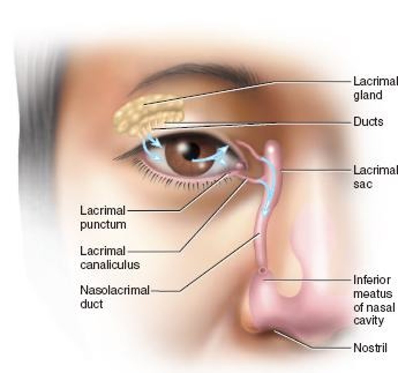
Lacrimal Apparatus
Tear-producing structures in the eye.
Conjunctiva
Mucous membrane covering the eye surface.
Conjunctivitis
Inflammation of the conjunctiva, causing redness.
Extrinsic Muscles
Muscles controlling eye movement in various directions.
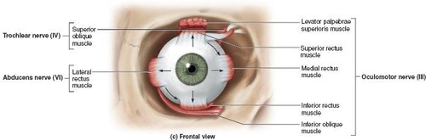
Superior Rectus
Muscle elevating the eye, innervated by CN III.
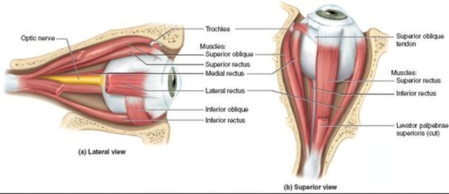
Inferior Rectus
Muscle depressing the eye, innervated by CN III.
Medial Rectus
Muscle moving eye medially, innervated by CN III.
Lateral Rectus
Muscle moving eye laterally, innervated by CN VI.
Superior Oblique
Muscle depressing and turning eye laterally, innervated by CN IV.
Inferior Oblique
Muscle elevating and turning eye laterally, innervated by CN III.
Sclera
White part of the eye, provides shape and support.
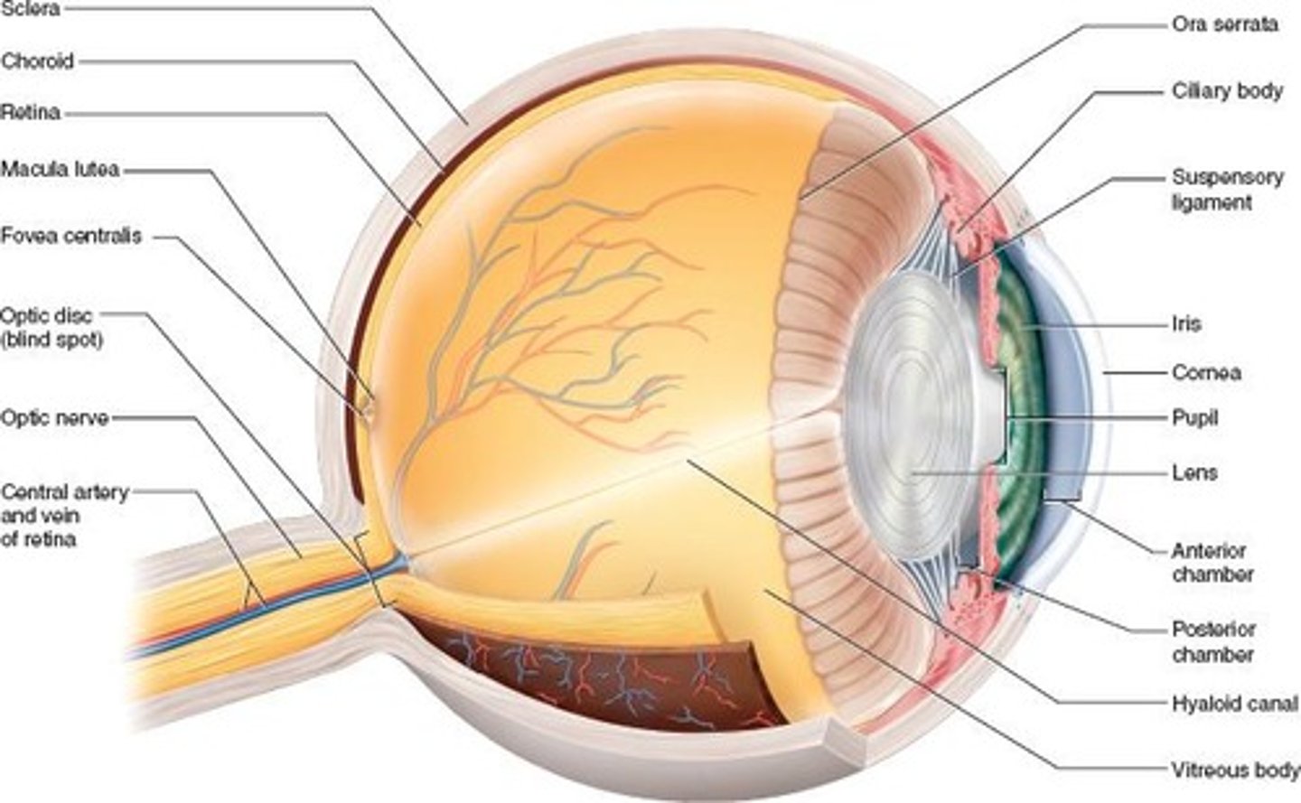
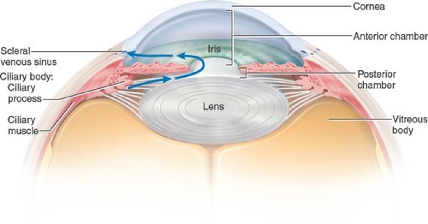
Cornea
Transparent front part of the eye, admits light.
Choroid
Vascular layer behind retina, absorbs light.
Ciliary Body
Muscular ring shaping the lens, secretes aqueous humor.
Iris
Colored part of the eye controlling pupil size.
Pupil
Opening allowing light to enter the eye.
Retina
Sensory layer of the eye, contains photoreceptors.
Optic Nerve
Cranial Nerve II, transmits visual information.
Fovea Centralis
Area for detailed vision in the retina.
Rods
Photoreceptors for night vision, detect light intensity.
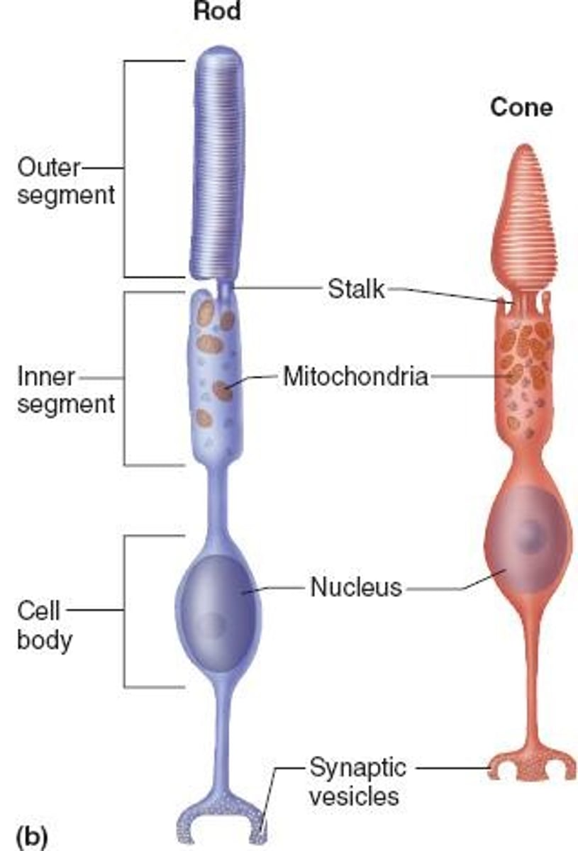
Cones
Photoreceptors for color vision, require bright light.
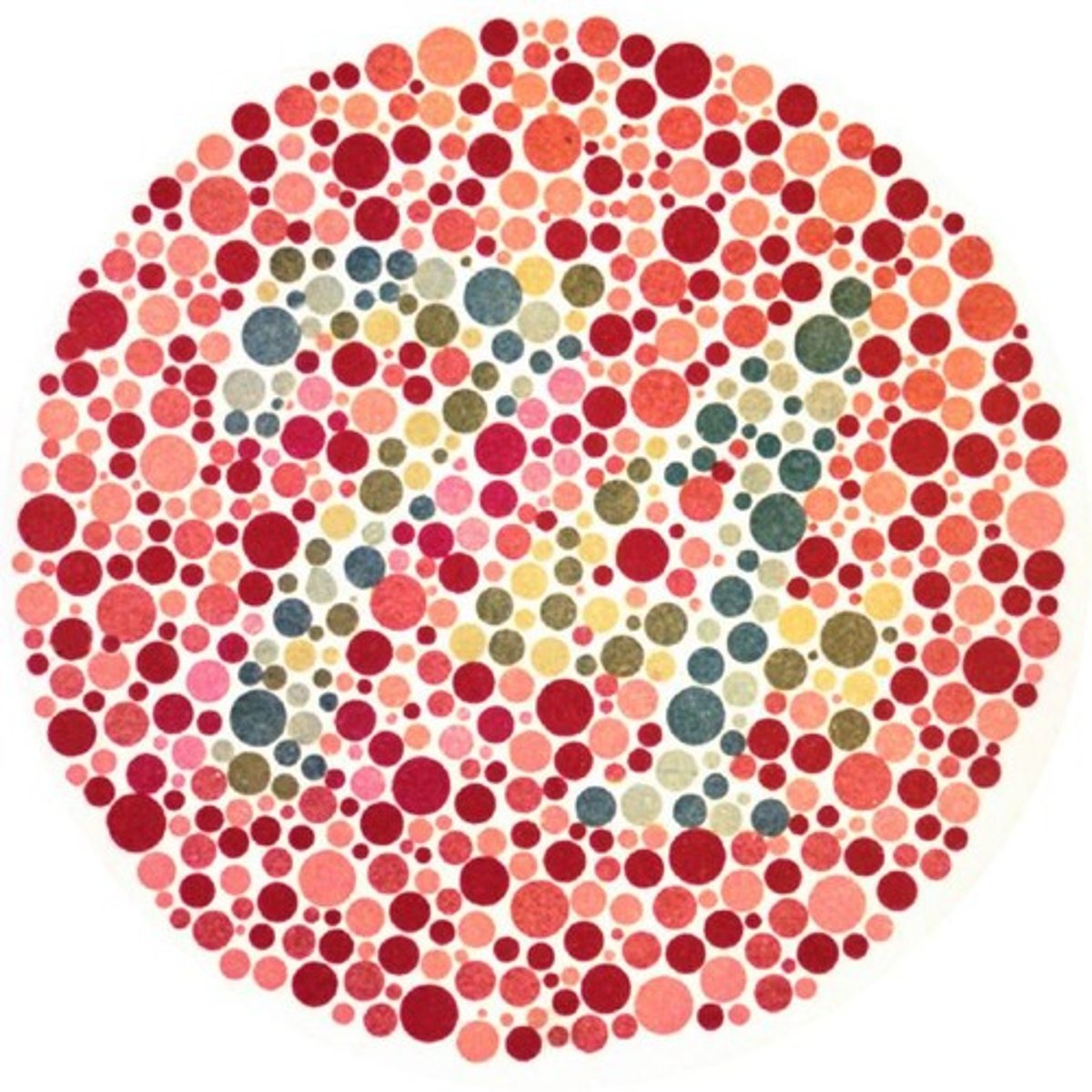
Color Blindness
Genetic condition affecting color perception.
Visual Pathway
Pathway light follows to reach the brain.
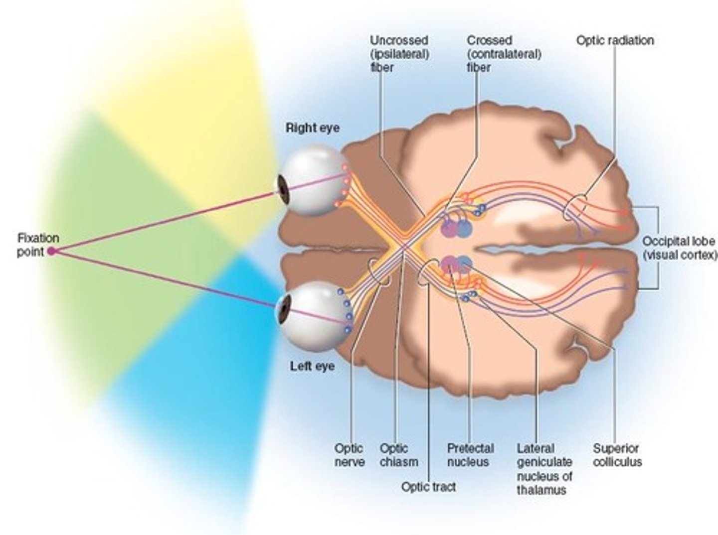
Optic Chiasm
Point where optic nerves cross, affecting vision.