PS231: Photoreceptors and phototransduction
1/19
There's no tags or description
Looks like no tags are added yet.
Name | Mastery | Learn | Test | Matching | Spaced | Call with Kai |
|---|
No analytics yet
Send a link to your students to track their progress
20 Terms
Visual information pathway in the retina
Photoreceptor cells (rods and cones) to bipolar cells, and then to ganglion cells. The axons of these ganglion cells form the optic nerve, which transmits signals to the brain
Phototransduction
turning light into nerve impulses (shared by rods and cones)
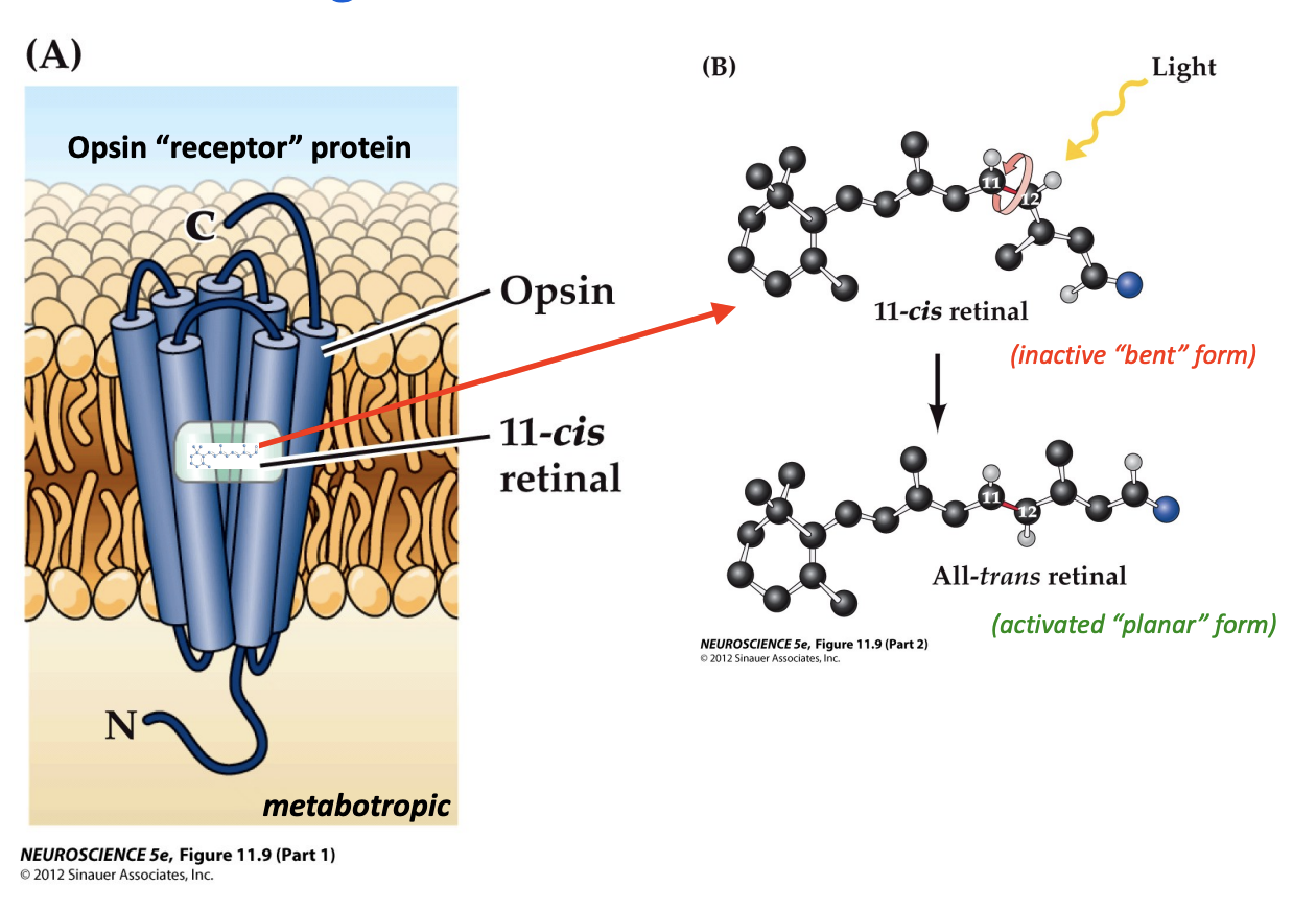
Outer segment
contains photoreceptor dissk packed with light-sensitive photopigment (opsins)
Rods contain more disks than cones
makes them have greater sensitivity to light (not active in daylight)
Rods are very sensitive
makes them good for low-light vision, but this comes at the expense of acuity (mostly located in peripheral retina, achromatic → no color vision)
Cones are much less sensitive to light
have more acuity (high resolution) and have different opsins that allow us to see colors (different wavelengths of light)
How many cones are there?
6m
How many rods are there?
125m
Cones are present
at a low density throughout the retina with a sharp peak in the fovea
Rods are present
at high density throughout most of the retina, with the exception of the fovea
Blind spot
lack of photoreceptors at the optic disk where RGC axons gather to leave the retina, forming the optic nerve
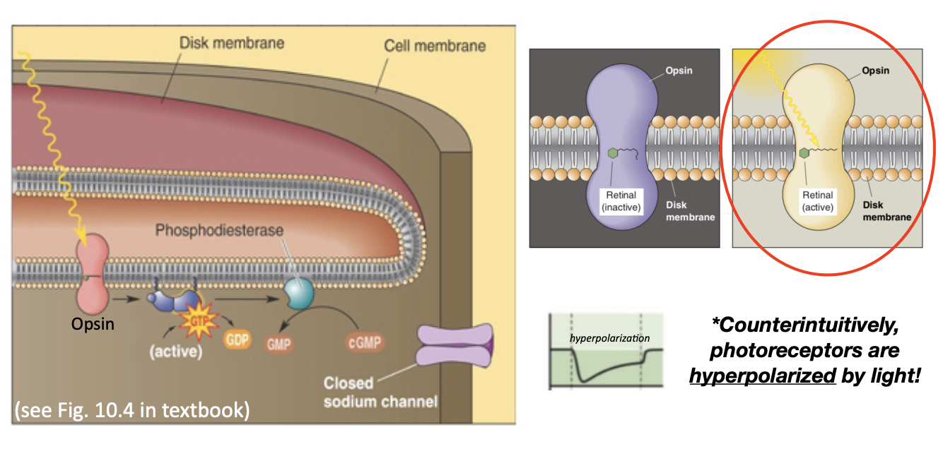
Where does phototransduction occur?
the photoreceptor outer segment
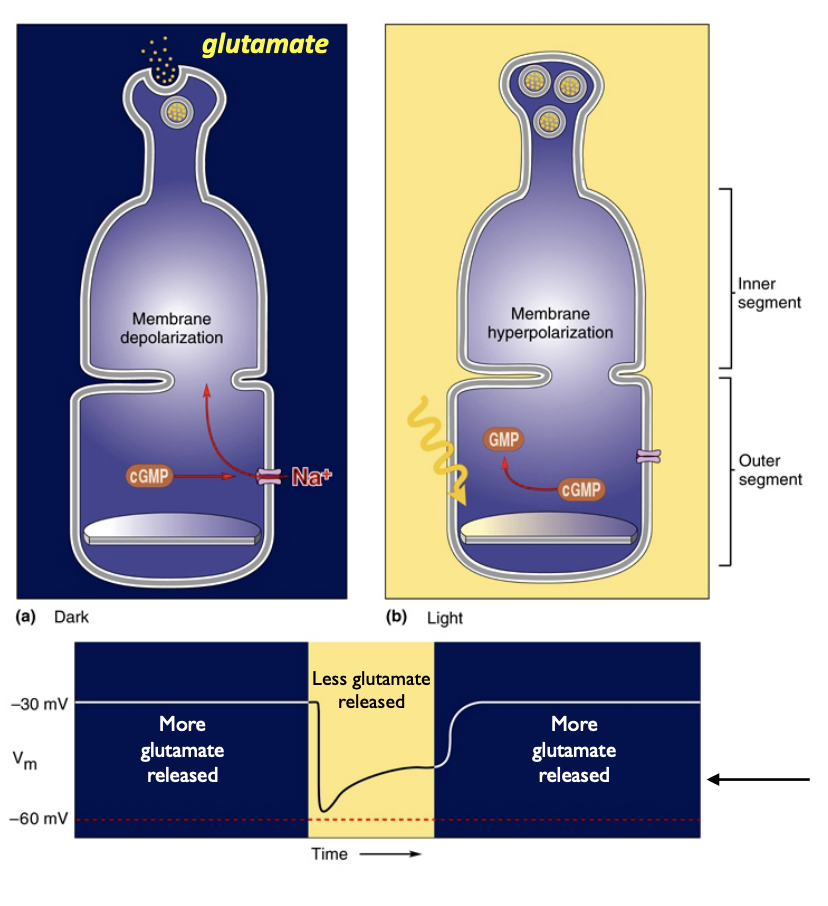
Light hyperpolarizes receptors
The absorption of photons of light ultimately leads to a reduction in neurotransmitter (glutamate) release by rods and cones—photoreceptors are therefore inhibited/hyperpolarized by light
Photons are absorbed by opsin proteins
Absorption of light causes a conformational change in the retinal molecule (vitamin A derivative) within the opsin protein, initiating an intracellular signaling cascade
Phototransduction pathway in the light
1. A photon of light is absorbed by retinal within the opsin protein (“planar”/ active form)
2. This stimulates transducin (G-protein)
3. This activates PDE, which works to close sodium channels
4. Neurotransmitter (glutamate) release decreases due to this hyperpolarization
Photoreceptors are depolarized in the dark
1. Retinal within the opsin protein is not activated (“bent”/ inactive form)
2. Thus, transducin (G-protein) is not activated
3. Phosphodiesterase (PDE) is therefore not activated
4. Without PDE activity, Na+ channels are open, allowing Na+ influx, which causes the photoreceptor to depolarize
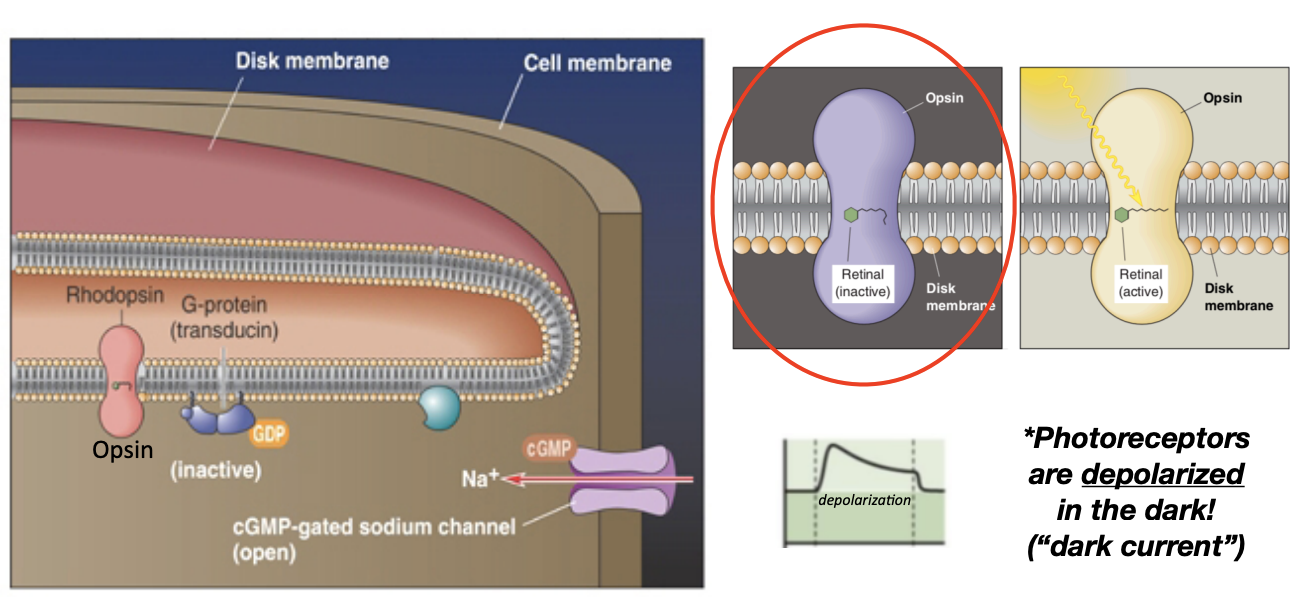
Photoreceptors only release glutamate in the dark
Photoreceptors are more active in the dark, shown by depolarization and glutamate release in darkness (“dark current”)
On pathway
cells are depolarized by light due to mGluR6 (GPCR) activation (“sign inverting”)— report information about “light” to the brain
Off pathway
cells are hyperpolarized by light due to ionotropic AMPARs (“sign conserving”)—report information about “darkness” to the brain
Light pathway
ganglion cells → bipolar cells → photoreceptors