2. Anatomy of the Brain and Spinal Cord
1/36
There's no tags or description
Looks like no tags are added yet.
Name | Mastery | Learn | Test | Matching | Spaced | Call with Kai |
|---|
No study sessions yet.
37 Terms
What is the Meninges?
3 layers of tissue provide
protection to the brain and spinal cord
What is cerebrospinal fliud?
Where is CSF derived from?
How does it Leave?
Fluid = cerebroventricular/cerebrospinal fluid (CSF)
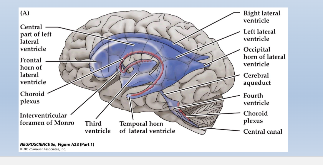
Derived from choroid plexus – a specialized vascular tissue within walls of ventricles; filters capillary blood and secretes the CSF product into the ventricles.
CSF leaves ventricles through several foramina (openings/passages) into the subarachnoid space surrounding the brain/spinal surface
CSF then drains out of subarachnoid space through arachnoid villi/granulations, into subdural sinuses, and then back into the general venous blood circulation
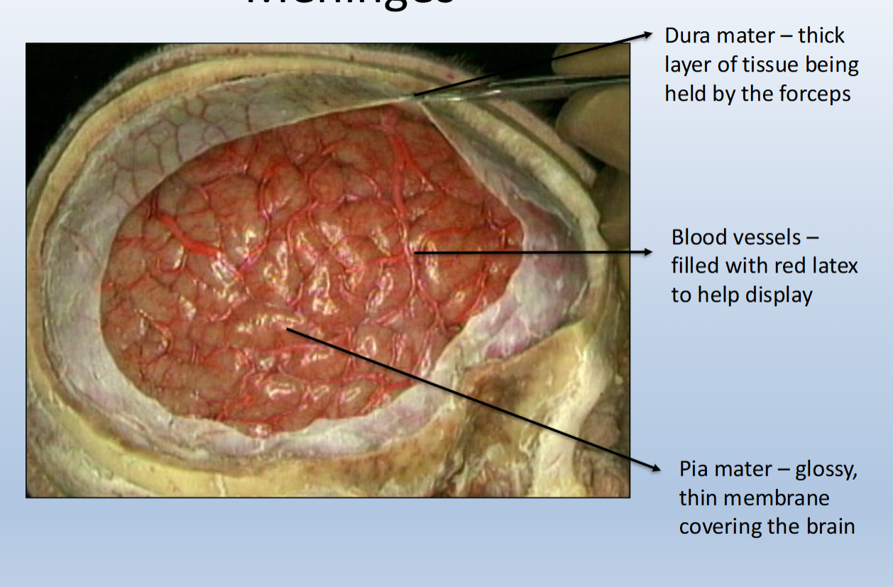
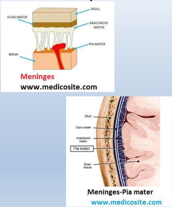
What are the different layers of the Meninges
Dura mater = “tough mother”
Outermost layer, it is tough and leathery
Arachnoid mater = “spider-like”
This is the middle layer, it is fairly delicate and impermeable
Arachnoid mater is separated from the dura by the subdural space
Arachnoid mater is separated from the pia mater by the subarachnoid space, which is filled with cerebrospinal fluid.
Pia mater = “tender mother”
The innermost layer, it adheres to the surface of the brain, covering the gyri and descending into the sulci.
It appears glossy, is so thin it is almost invisible to the naked eye
What is Meningitis?
Meningitis = infection/inflammation of the meninges
• viral meningitis
• bacterial meningitis (more serious, often fatal, immunization available)
What is the ventricular system of the brain?
The Ventricular system is a series of interconnected, fluid-filled spaces within the core of the CNS (brain and spinal cord)
What is the purpose of Cerebrospinal fluid?
1) Buoyancy: The mass of the human brain is about 1.5 kg (~ 3 pounds) but the net weight when suspended in CSF is equivalent 25-50 grams.
This is called“neutral buoyancy”
– the brain can be dense without being damaged by its own
weight.
2) Protection: CSF protects the brain tissue from injury by providing a fluid buffer that acts as a shock absorber
T or F, the ventricular system only protects the brain?
Ventricular system extends down to
spinal cord: protects the entire central
nervous system (not just the brain!)
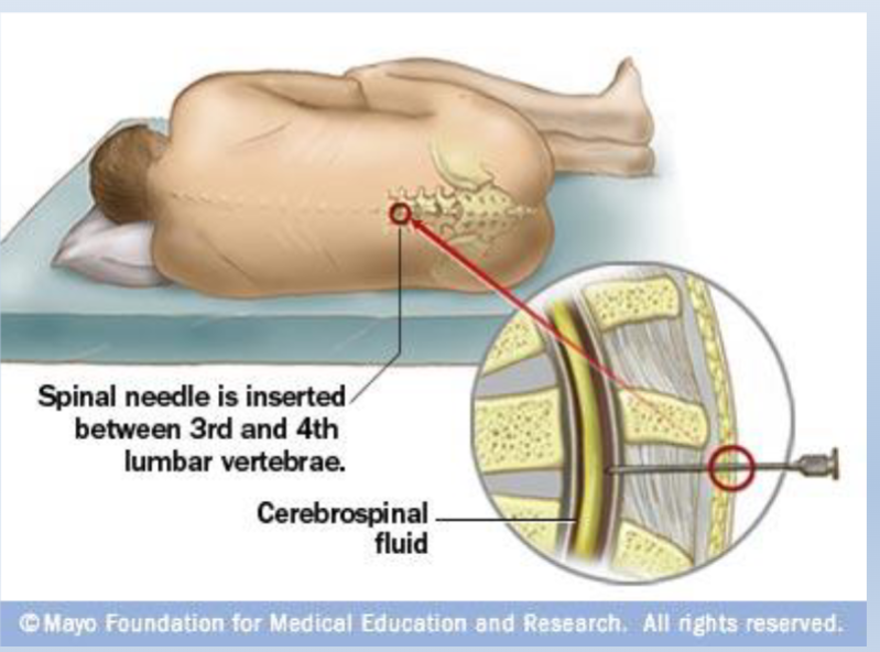
Clinical Applications: Ventricular System
Spinal tap/epidural
• CSF can be collected for testing (i.e. meningitis)
• Drugs can be delivered into the CSF (intrathecal administration)
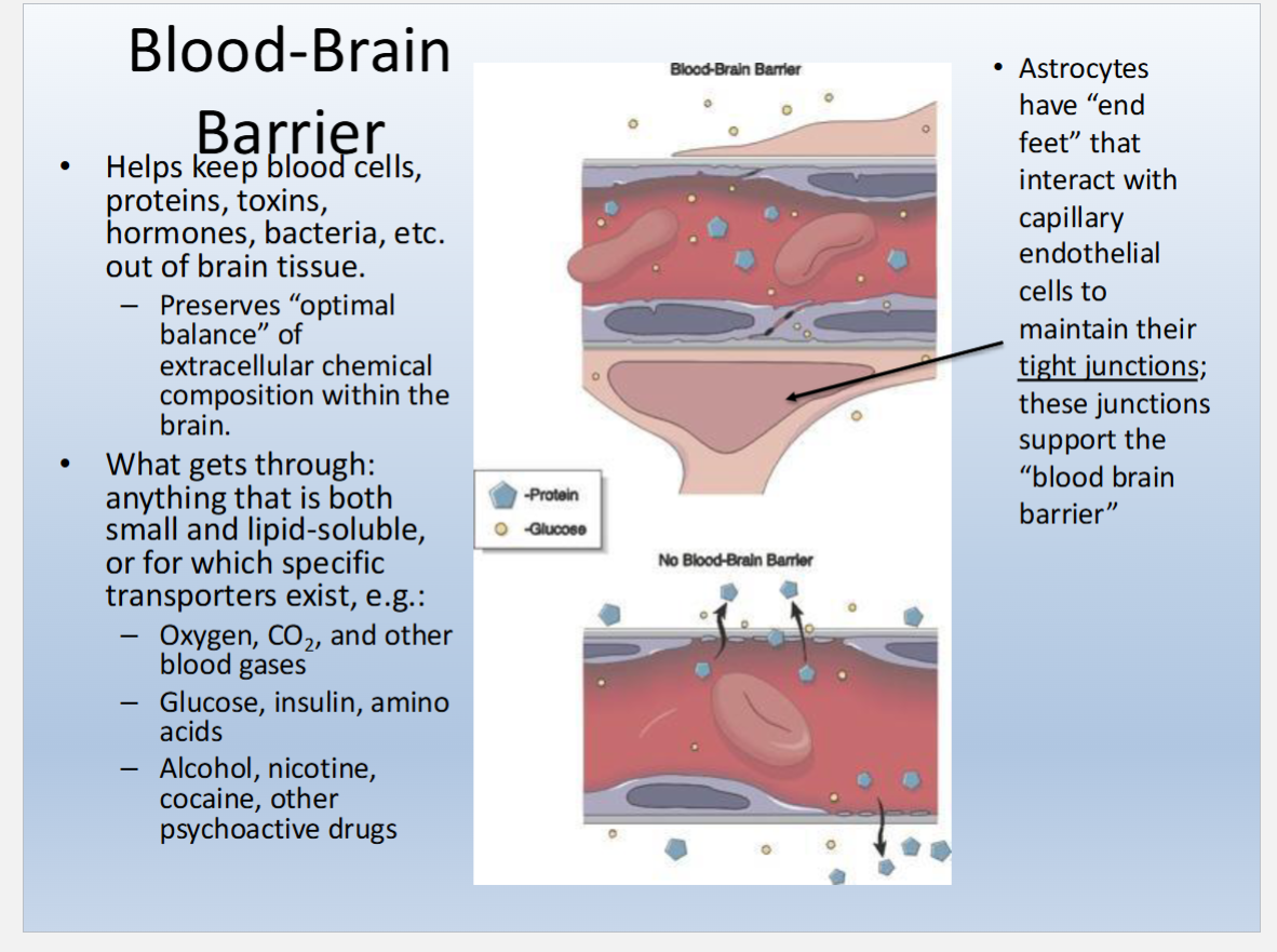
Describe the Blood Brain Barrier (BBB)
protects the brain from substances in the blood
Do not confuse with the role of the meninges
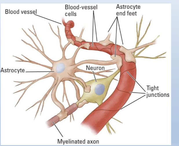
BBBarrier is formed by the tight junctions between capillary endothelial cells within the brain & spinal cord
These tight junctions are maintained by astrocytes via their “end feet”, which contact vascular endothelial cells
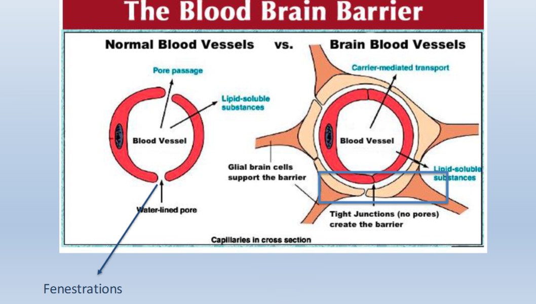
T or F? Brain capillaries are fenestrated?
Brain capillaries are NON-FENESTRATED (have tights junctions that form a barrier)
Capillaries in the rest of the body’s tissues are FENESTRATED (no tight junctions/no barriers)
What is kept in and out by the blood brain barrier?
____ organs are not protected by the BBB
Circumventricular
• A few small, special brain regions do
contain fenestrated capillaries . . .
specialized for detecting toxins,
hormones, etc.
– Circumventricular organs (on brain midline, adjacent to ventricles)
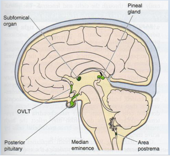
What functions might these structures (Circumventricular organs) serve?
• Receptors detect osmolality of extracellular fluid –
triggers thirst
• Receptors detect the presence of toxins in the blood

Axes and Directional Terminology

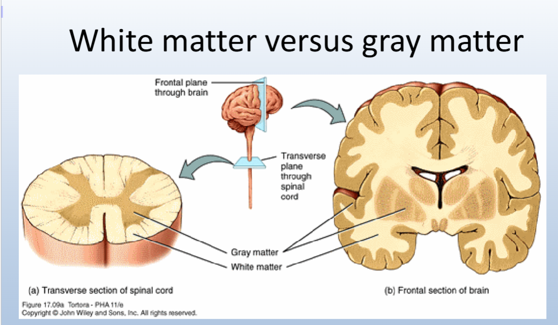
White vs Gray Matter
• White matter = myelinated axons.
• Gray matter – consists mostly of cell bodies and dendrites
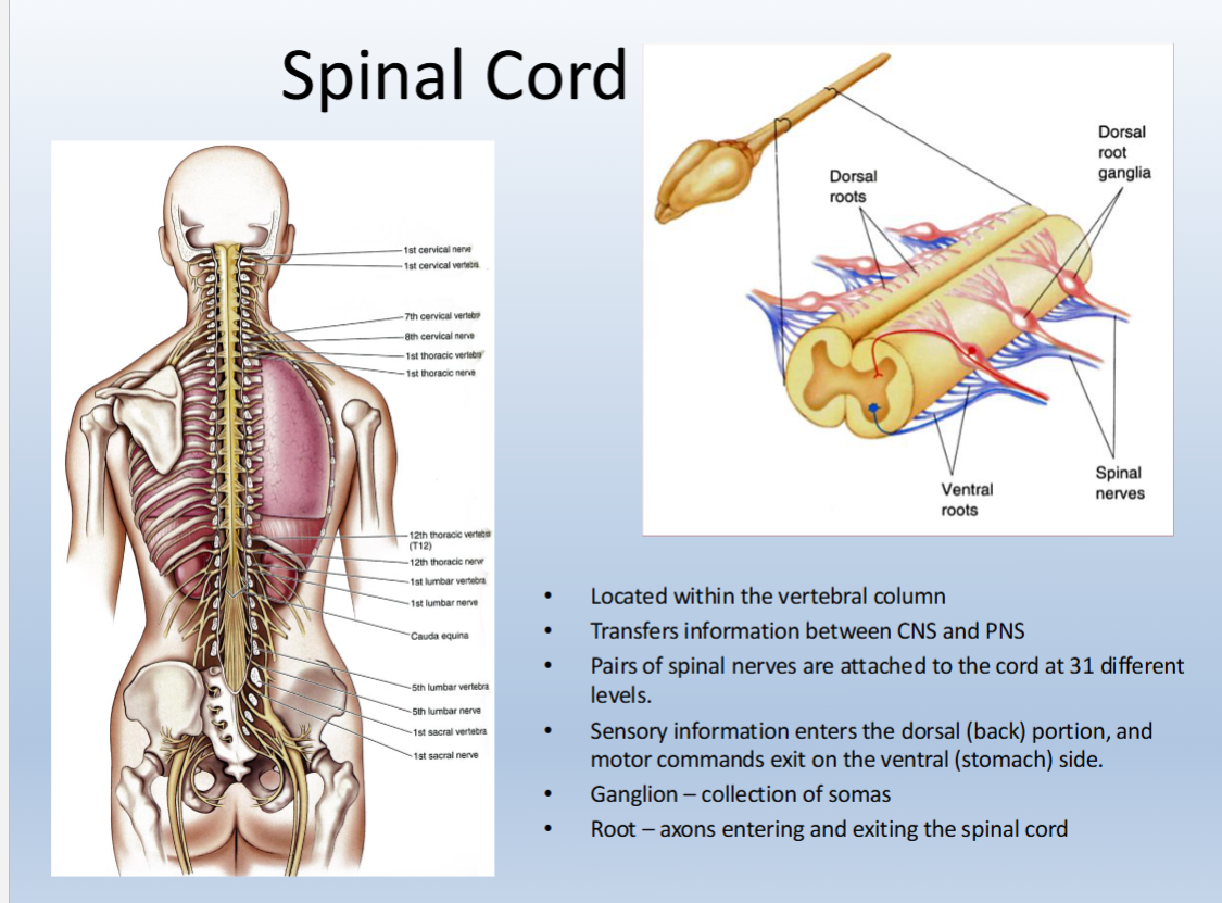
What are the features of the Spinal Cord?
• Located within the vertebral column
• Transfers information between CNS and PNS
• Pairs of spinal nerves are attached to the cord at 31 different
levels.
• Sensory information enters the dorsal (back) portion, and
motor commands exit on the ventral (stomach) side.
• Ganglion – collection of somas
• Root – axons entering and exiting the spinal cord
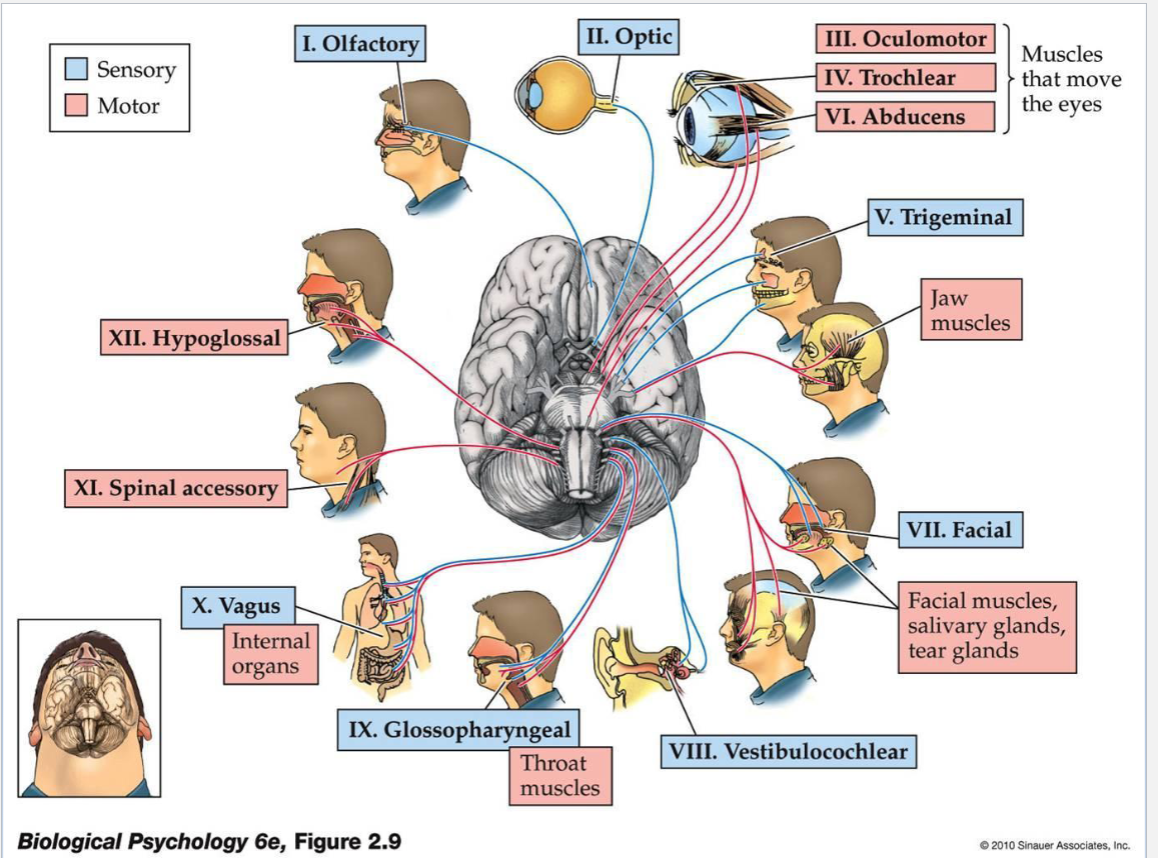
What are the cranial nerves?
• 12 pairs of nerves
• Emerge from the brain, rather than from segments
of the spinal cord
• Send motor commands to and receive sensory
information from the head and neck
Cranial nerves can be identified by:
• Rostro-caudal position (I-XII)
• Information type (sensory vs. motor)
• Function
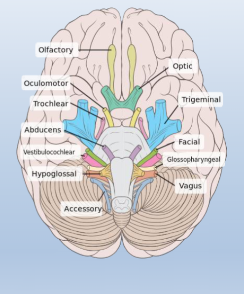
Name all the Cranial Nerves and Distinguish between Sensory and Motor
Sensory
Olfactory
Optic
Trigeminal
Facial
Vestibulocochlear
Glossopharyngeal
Vagus
Motor
Oculomotor
Trochlear
Abducens
Spinal Accessory
Hypoglossal
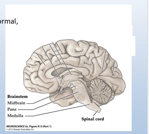
Divisions of the brainstem and their functions
Medulla:
• Includes neurons that maintain normal, rhythmic breathing
Pons region:
• Pons (structure): includes axons that allow the cerebellum to communicate with the brainstem and the cerebral cortex
• Fourth ventricle is on the dorsal side of the pons region
Midbrain:
• Superior and inferior colliculi: localization of visual and auditory
stimuli
All levels of the brainstem contain
sensory and motor axons
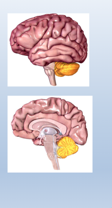
Cerebellum
Roles of the cerebellum:
Motor planning:
• Aids the motor cortices in planning complex movements
Motor learning:
• Error correction when learning movements: compares intended
movement with actual movement and corrects errors that might occur
• Synapses change with experience
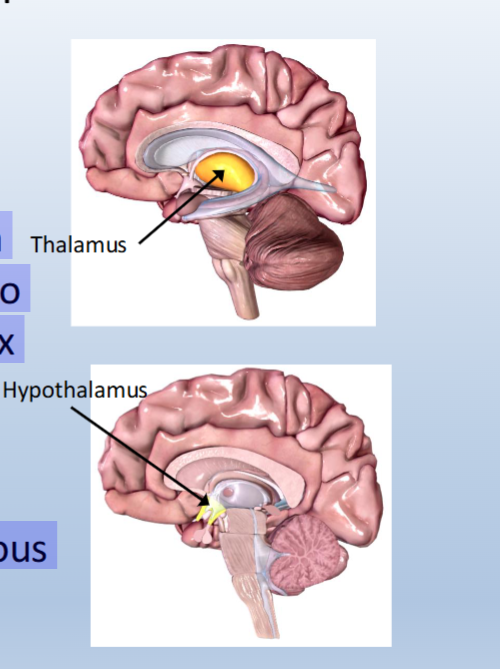
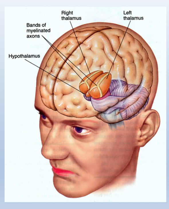
Diencephalon
Components:
Thalamus:
• Located rostral to the midbrain
• “Relay” for information going to and coming from the neocortex
Hypothalamus:
• Located below the thalamus
• Regulates the autonomic nervous system
• Regulates hormone release
Gross Structures of the cerebral cortex (neocortex)
What are the main overaching functions of the cerebral cortex?
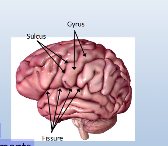
Gross structures:
• Sulci (singular: sulcus): grooves
• Fissures: deep sulci
• Gyri (singular: gyrus): rounded regions between sulci
Functions:
• Processing of sensory input
• Initiation/planning of movements
• “Higher-order” functions including memory, cognition, language
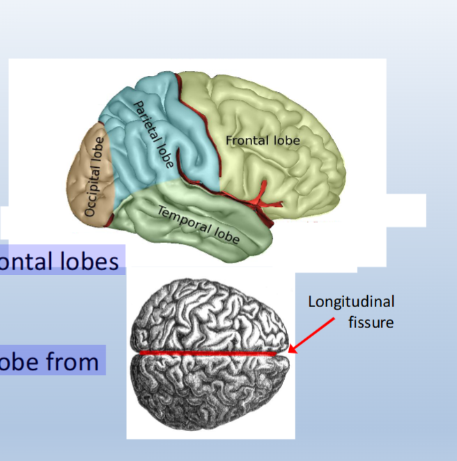
Distinct features of cerebral cortex + lobes
• Occipital lobe
• Parietal lobe
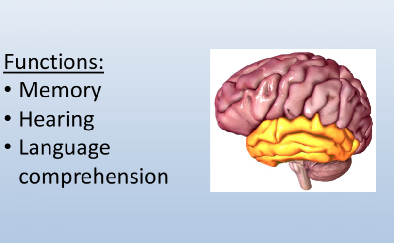
• Temporal lobe
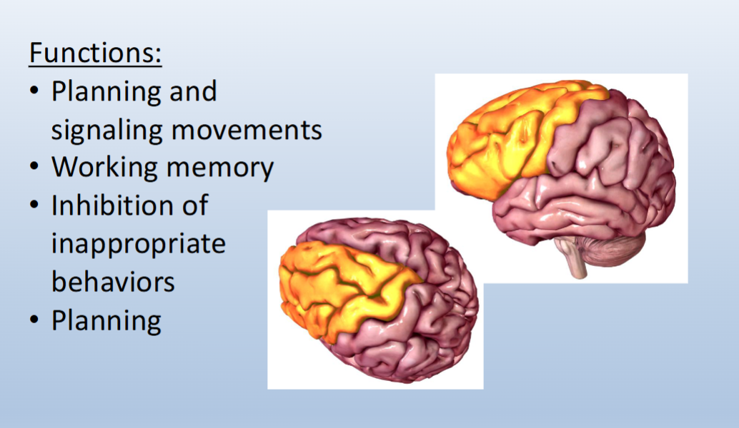
• Frontal lobe
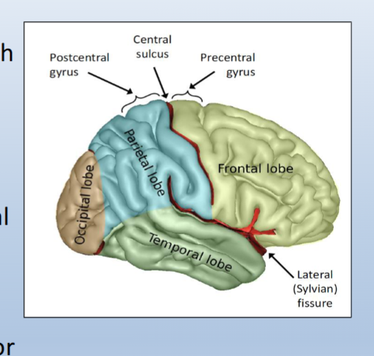
Central sulcus:
• Separates parietal and frontal lobes
Lateral fissure:
• Separates the temporal lobe from those surrounding it
Longitudinal fissure:
• Separates the two hemispheres of the brain
Pre and Post central gyri of cerebral cortex
Postcentral gyrus:
• Directly caudal to the central sulcus
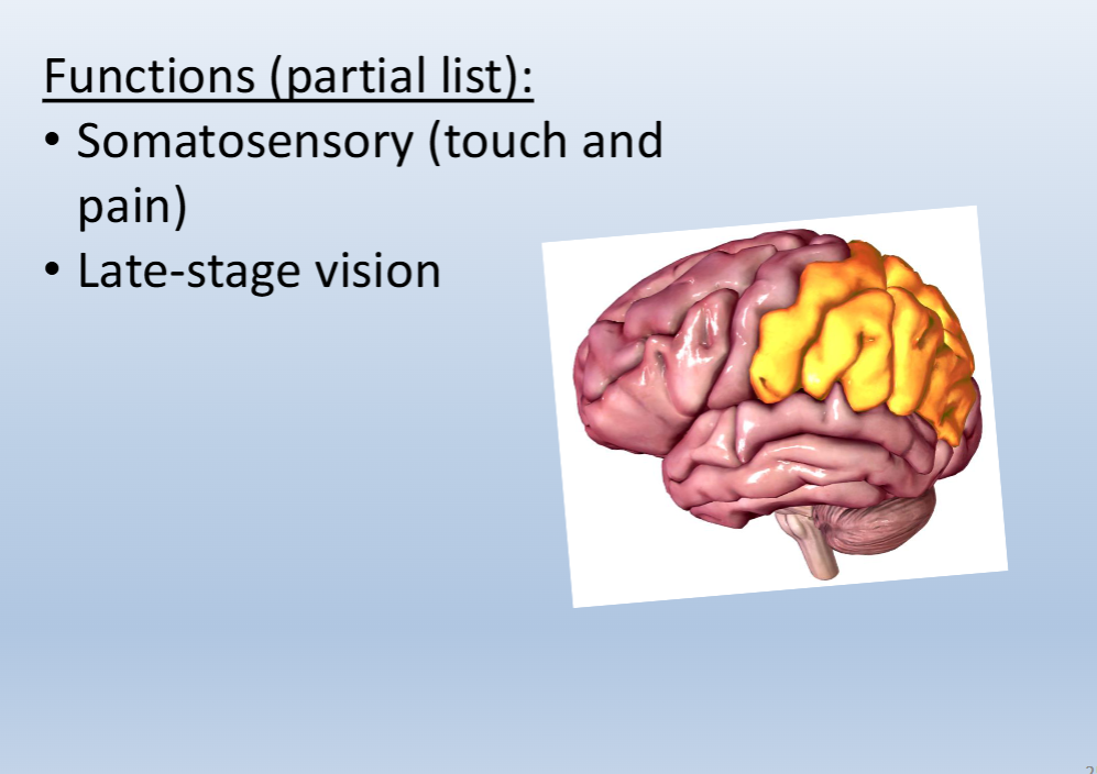
• Contains the primary somatosensory cortex, which processes touch and pain information
Precentral gyrus:
• Directly rostral to the central sulcus
• Contains the primary motor cortex, which helps plan movements and sends motor (movement) signals to the spinal cord
What is the function of the occipital lobe

What is the function of the parietal lobe?
What is the function of the temporal lobe
What is the function of the frontal lobe?
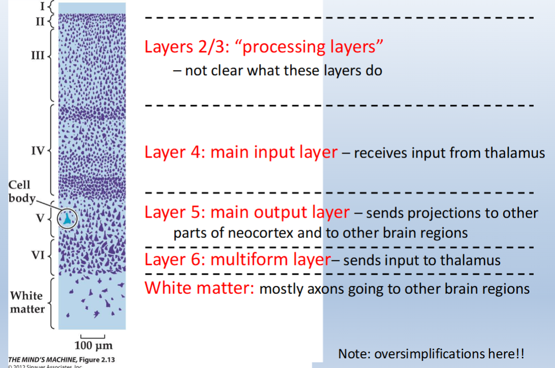
Layers of neocortex: function of each layer
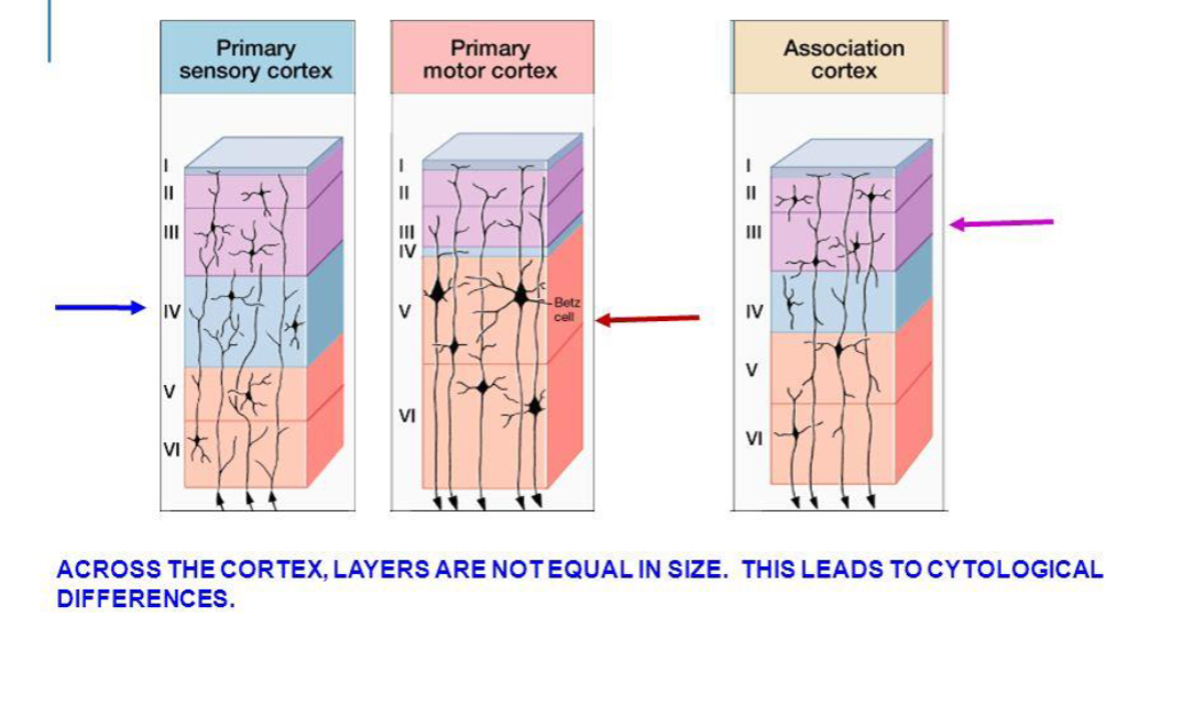
T or F, the layers of the cortex are the same size throughout the neocortex?
False
What is the limbic system and what is it responsible for?
• A group of interconnected structures that are related to
emotional behavior and emotional interpretation of the world.
• Sexual behavior
• Involved in the formation of memory, contains primary reward and punishment centers.
• Site of action of drugs which produce euphoria (direct and
indirect)
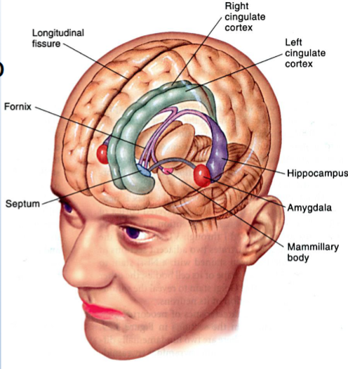
What is the hypothalamus?
Part of Limbic System
• Regulates many
motivated functions
(e.g., eating and
drinking), sleep/wake
cycle
• Controls activity of the
pituitary gland (master
gland that interacts with
the hypothalamus to
regulate many functions
via the release of
hormones)
What is the Hippocampus?
• Involved in memory
consolidation and provide
the organism’s context
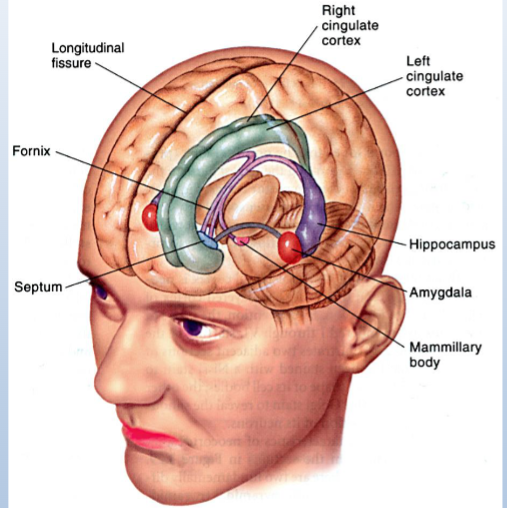
What is the Amygdala
• Coordinates autonomic
responses in with
emotional states

How does the cerebral cortex impact limbic system activity?
• Interacts with subcortical structures
to guide behavior

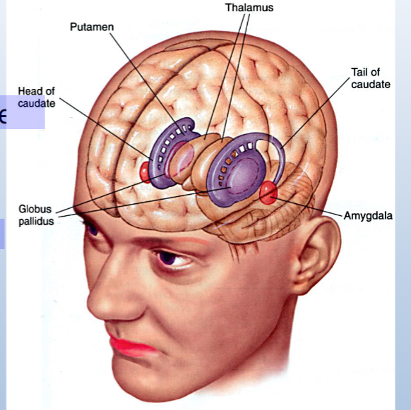
What is the basal ganglia?
Group of interconnected structures that control
voluntary, smooth movement
Structures include:
striatum (caudate/putamen)
globus pallidus
substantia nigra
Actions of stimulants in the basal ganglia increases motor
activity
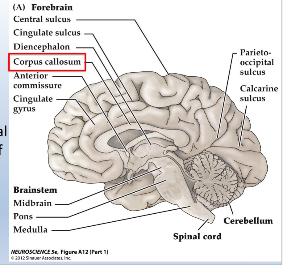
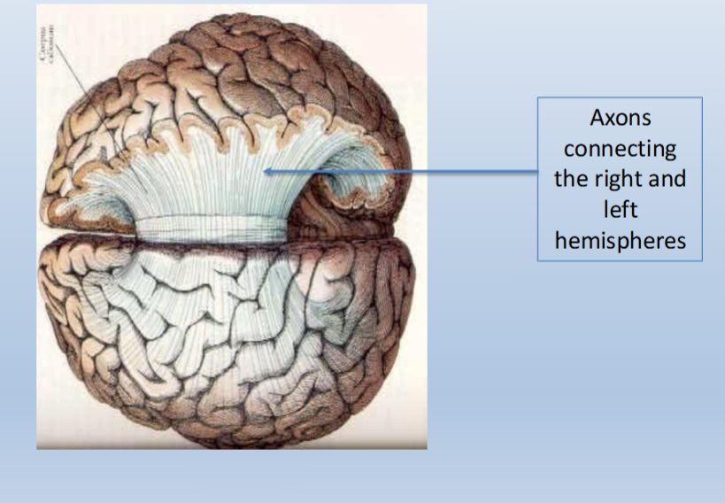
What is the corpus callosum
- Long-range neurons that connect the two halves of the brain