5. motor functions + spinal reflexes
1/46
There's no tags or description
Looks like no tags are added yet.
Name | Mastery | Learn | Test | Matching | Spaced |
|---|
No study sessions yet.
47 Terms
Voluntary movement requires:
complex interaction of the corticospinal (pyramidal) tracts, basal ganglia, and cerebellum to
ensure smooth, purposeful movement without extraneous muscular contractions
Skeletal motor axis
Projection from motor cortex to motor neuron
Receives extensive input from thalamus, brainstem and cerebellum
Motor Cortex
Divided into 3 sub areas
Primary motor cortex
Premotor area
Supplemental motor area
Motor Cortex: Primary motor cortex
Unequal topographic representation (some areas receive more cortical space and attention than others)
Fine motor movement elicited by stimulation
Motor Cortex: Premotor area
Stimulation results in movement of muscle groups (patterns) to perform a specific task
Works in concert with other motor areas
Motor Cortex: Supplemental motor area
Functions in concert with premotor area to provide attitudinal, fixation, or positional movement for the body
Provides background for fine motor control of the arms and hands by premotor and primary motor cortex
Transmission of Cortical Motor Signals: Direct pathway
Corticospinal tract (pyramidal)
Directs discrete detailed movement (fine-skilled motor behavior)
Transmission of Cortical Motor Signals: Indirect pathway (extrpyramidal)
Modulate signal from corticospinal tract to motor neuron
Integrate sensory and posture information
Signals through basal ganglia, cerebellum, and brainstem nuclei
Trunk musculature
Upright posture
Locomotion
Orienting responses
Giant pyramidal cells (Betz cells)
originate in primary motor cortex and give rise to large fibers with fast transmission rates
Cross in pyramid of medulla and descend spinal cord in corticospinal tracts
Synapse with interneurons or directly with motor neurons
antigravity muscles
muscles of the spinal column and the extensor muscles of the legs support the body against gravity
These muscles are under the influence of brainstem nuclei
Pontine reticular nuclei VS Medullary reticular nuclei
Pontine reticular nuclei excite the antigravity muscles
Medullary reticular nuclei inhibit the antigravity muscles
Cerebellum (little brain)
Responsible for coordinating and sequencing muscle activity
Compares actual motor movements with intended movements
Makes corrective changes
Cerebellum: Afferent signals from motor and cortex:
transmits information about intended motion
Cerebellum: Afferent signals from muscle spindles and Golgi tendon organs (proprioception)
Transmits information about status of muscle contraction/tension and limb position
Cerebellum: Efferent signals
Pathways involved in equilibrium control and coordinating opposing influences on motor neurons
Basal Ganglia
Four nuclei involved in control of complex patterns of motor activity
Receives input from primary, premotor, supplemental and prefrontal cortex
Inhibitory output to cortex
Suppression of conflicting motor activity
functions of basal ganglia
Postural stability: with damage, the head falls over chin and the person walks bent over at the waist
Motor programs: converts an overall goal into programs which determine the specific movement
Parkinson disease – inability to initiate movements
“Extrapyramidal symptoms”
“Extrapyramidal symptoms”
involuntary movements
Lack of smoothness in their movements
Tremor
Difficulty in the initiation of a movement
Difficulty maintaining an upright posture
Many of the symptoms of Parkinson’s disease are classic extrapyramidal symptoms
summary of motor control
Cortical level
Issues commands to set into motion the patterns available in the spinal cord
Control the intensity and modifies the timing
Brainstem level
Maintains equilibrium by adjusting axial tone
Cerebellum
Function with all levels of control to adjust cord motor activity, equilibrium, and planning of motor activity
Basal ganglia
Functions to assist cortex in executing subconscious but learned patterns of movement, and to plan sequential patterns to accomplish a purposeful task
Spinal cord level
Preprogramming of patterns of movement of all muscles (i.e., withdrawal reflex, walking
movements, etc.)
Neuronal circuits
for walking and various reflexes
contained within the spinal cord
Higher brain centers activate and command these circuits
walking + maintaining equilibrium
Motor neuron cell body receives inhibitory and excitatory inputs from:
1. Afferent neurons
2. Spinal cord interneurons
3. Cortex via descending tracts
Motor neuron output at synapse is _________ to the muscle fiber
ALWAYS excitatory
Sensory fibers
enter cord and are transmitted to higher centers, or they synapse locally to elicit motor reflexes
Motor neurons
located in the anterior portion of the cord
50 - 100% bigger than other neurons
Interneurons
30x more abundant than anterior motor neurons
Small and highly excitable
Most signals from brain terminate on interneurons
Comprise neural circuitry for motor reflexes
Propriospinal fibers
Travel up and down cord for 1 - 2 segments
Provide pathways for multi-segmental reflexes
Important for proprioception
Motor unit
single motor neuron and all associated muscle fibers
Alpha motor neurons
Large type Aα fibers (~14 microns)
Stimulation can excite 3 to >100 extrafusal muscle fibers (Motor unit)
Gamma motor neurons
Smaller type Aγ fibers (~5 microns)
Stimulation excites intrafusal fibers in muscle spindle
Maintains tone
how are α-motor neurons and γ-motor neurons activated during a muscle contraction?
co-activated
Extrafusal fibers
Make up bulk of muscle
Stimulated by alpha motor neurons
Provide force for muscle contraction
Intrafusal fibers
Smaller than extrafusal fibers
Encapsulated in sheaths to form muscle spindles
Run in parallel with extrafusal fibers, but much shorter (~10 mm)
Sensory Receptors of Muscle
Muscle Spindle + Golgi Tendon Organ
Signals from muscle sensory receptors are (mainly) for intrinsic muscle control.
mainly occur subconsciously.
transmit information to cerebrum and cerebellum (as well as to spinal cord).
Sensory Receptors of Muscle: Muscle Spindle
Located in muscle belly
Senses muscle length and rate of change in length
Detects both static and dynamic changes in muscle length
Consist of intrafusal muscle fibers (3 to 12 per spindle) in parallel with extrafusal muscle
fibers
Intrafusal fibers (3 to 12 per spindle)
Do not contribute to muscle tension (Functions as sensory receptor only)
Innervated by gamma motor neurons (type Aγ)
Two types of intrafusal fibers: Bag + Chain
Stretch Reflex (myotatic reflex)
Sudden stretch of muscle stimulates receptors in muscle spindle
Afferent neuron synapses with α-motor neuron and inhibitory interneuron
Causes contraction of stretched muscle and inhibition of opposing muscle
Opposes further stretch of muscle
MONOSYNAPTIC REFLEX ARCH
Myotatic (stretch) reflex in the jaw
Helps determine resting position of mandible
Increases tone of elevator muscles to counteract effects of gravity
Sensory Receptors of Muscle: Golgi Tendon Organ
Low-threshold mechanoreceptors located in tendon
Senses tendon tension and rate of change in tension
Increased muscle tension compresses nerve endings, opens stretch-sensitive ion channels
Function is to equalize force among muscle fibers
Only affects an individual muscle (adjacent muscles are not affected)
Autogenic inhibition reflex
a sudden relaxation of muscle at very high muscle tensions (protects against muscle tear)
However, Golgi tendon organs signal muscle force through the entire physiological range,
not only at high levels of tension
Inverse stretch reflex (Golgi tendon reflex)
Protective: prevent muscle damage
DISYNAPTIC reflex arc
Golgi tendon organ: tension receptor (NOT length)
In series with the extrafusal muscle fibers
Stimulus: excessive contraction of the muscle
Sensory afferent synapses with an inhibitory interneuron
Hyperpolarizes the α motor neuron to the extensor muscle and causes muscle to relax
Transmission of muscle length and tension information to brain
Signal from Muscle Spindle and Golgi tendon are also transmitted to higher centers (not just the spinal cord)
This informs brain of instantaneous changes in muscle tension and length
Information is transmitted at 120 m/sec
Important for feedback control of motor activity
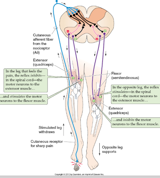
Flexor Withdrawal and Crossed Extensor Reflex
Withdraws a limb from a painful stimulus
Activated by Nociceptors (pain afferent)
Polysynaptic reflex arch
Reciprocal innervation: to obtain movement around a joint, need to contract flexors and inhibit extensors
Painful stimulus causes combined reflex:
Withdrawal
Ipsilateral (same side)
Contract flexors
Inhibit extensors
Crossed extensor
Contralateral (opposite side)
Inhibit flexors
Contract extensors
Withdrawal reflex in the jaw
Withdrawal reflex in the jaw
can be initiated by any painful stimulus in the oral cavity
Also called jaw-opening reflex or nociceptive reflex
Noxious stimulus
e.g.: biting down on popcorn kernel
causes inhibition of elevator muscles and stimulates jaw-opening muscle
Other Reflexes for Posture and Locomotion
Pressure on bottom of feet causes extensor reflex
more complex than flexor-crossed extensor reflex
Basic walking reflexes reside in spinal cord