Ch 15 - Intracellular Compartments and Protein Transport
1/136
Earn XP
Description and Tags
- membrane-enclosed organelles - protein sorting - vesicular transport - secretory pathways - endocytic pathways
Name | Mastery | Learn | Test | Matching | Spaced | Call with Kai |
|---|
No analytics yet
Send a link to your students to track their progress
137 Terms
membrane-bound organelles
surrounded by the cytosol, which is enclosed by the plasma membrane
occupy nearly 50% of cell volume
allow spatial separation of different cellular functions
cytosol function
contains many metabolic pathways; protein synthesis; the cytoskeleton
nucleus function
contains main genome; DNA and RNA synthesis
endoplasmic reticulum (ER) function
synthesis of most lipids; synthesis of proteins for distribution to many organelles and to the plasma membrane
golgi apparatus function
modification, sorting, and packaging of proteins and lipids for either secretion or delivery to another organelle
lysosomes function
intracellular degradation
endosomes function
sorting of endocytosed material
mitochondria function
ATP synthesis by oxidative phosphorylation
chloroplasts (in photosynthetic cells) function
ATP synthesis and carbon fixation by photosynthesis
peroxisomes function
oxidative breakdown of toxic molecules
cytosol percentage of total cell volume (highest)
55%
likely originated by invagination of the plasma membrane
nuclear envelope, membranes of ER, golgi apparatus, endosomes and lysosomes
vesicular traffic
how organelles (w/ exception of nucleus) communicate extensively with one another and with the outside of the cell
likely evolved from bacteria that were engulfed by primitive eukaryotic cells
remain isolated from the vesicular traffic
mitochondria and chloroplasts
signal sequence of protein
directs the protein to a particular organelle
cytosol
where synthesis of virtually all proteins begins
remain in the cytosol
proteins that lack a signal sequence
into the nucleus, mitochondria, chloroplasts, peroxisomes and the ER
where different signal sequences direct proteins
depends on the organelle
the mechanism by which a protein is transported into an organelle
first mechanism of transport
transport of folded proteins into the nucleus through nuclear pores
second mechanism of transport
transport of unfolded proteins into the ER, mitochondria or chloroplasts across their membranes by protein translocators
third mechanism of transport
transport of proteins in transport vesicles that pinch off from the ER and fuse with a compartment of the endomembrane system
common themes of mechanisms of protein import into organelles
sequence-specific signal on the “cargo” protein
signal sequence is recognized by receptor proteins
directional movement by transport machinery requires energy
nuclear import
large pore, competent for fully folded proteins/complexes. GTP hydrolysis provides energy
mitochondria/chloroplasts
narrow translocation channel; proteins are unfolded and pulled into organelles by chaperone proteins (which hydrolyze ATP for energy)
ER
proteins enter as they are being synthesized
signal sequences
necessary and sufficient to direct a protein to a particular destination
typically 15-60 amino acids long; exact sequence can vary
hydrophobicity or the order of charged amino acids are more important than the exact sequence
often (but not always) removed from the protein once it has been sorted
deleting a signal sequence from an ER
converts it into a cytosolic protein
adding an ER signal sequence to a cytosolic protein
directs it to the ER
the nuclear envelope
surrounds the nucleus, and consists of an inner membrane and an outer membrane
outer membrane = continuous w/ ER
inner membrane = contains proteins that act as binding sites for chromosomes and the nuclear lamina
what transport across the nuclear envelope occurs through
nuclear pores
the nuclear pore complex
acts as a selective gate
large structure composed of ~30 different proteins
proteins that line it are unstructured and form a mesh that fills the pore, preventing passage of large molecules
how proteins enter the nucleus
in their mature, fully folded state
nuclear localization signal (NLS)
signal sequence that directs transport into the nucleus
nuclear import receptor
cytosolic protein that NLS is recognized and bound by
interacts w/ fibrils that extend from nuclear pore complexes
disrupts the protein mesh that fills the pore and opens a passageway into the nucleus
Ran
a small monomeric GTPase that exists in two conformations: one bound to GTP and one bound to GDP
Ran-GTP
is present in high concentrations in the nucleus
Ran-GDP
is present in high concentrations in the cytosol
GTP hydrolysis
facilitates nuclear transport
in the nucleus, Ran-GTP
binds a nuclear import receptor, allowing the prospective nuclear protein to be released
the import receptor, bound to Ran-GTP
returns to the cytosol
hydrolysis of GTP causes
Ran-GDP to release the import receptor, freeing it to bind a new prospective nuclear protein
proteins must unfold
in order to enter mitochondria or chloroplasts
step 1
protein synthesis and folding in the cytosol
step 2
binding of signal sequence to an import receptor on mitochondrial surface
step 3
simultaneous transport across the outer and inner membranes by protein translocators. the protein is unfolded during the transfer process
step 4
cleavage of the signal sequence
mitochondrial chaperone proteins
help pull in and refold proteins (use energy from ATP hydrolysis)
proteins imported into peroxisomes
do not need to unfold, are imported via a related mechanism and also by vesicular transport
ER is the entry point for proteins destined for …
the ER, golgi apparatus, endosomes, lysosomes, and proteins destined for the cell surface
protein will not re-enter the cytosol once it is inside ___________ or embedded in the ______________
the ER lumen, ER membrane
transport vesicles
how proteins are transported to their destination
hydrophobic ER signal sequence
direct proteins that enter the ER while being synthesized
soluble proteins
translocated across the membrane into the ER lumen
transmembrane proteins
partly translocated across the membrane and remain embedded in it
create the rough ER
membrane-bound ribosomes
ribosome synthesizing the protein
attaches to the ER membrane to initiate transfer
structurally identical
membrane-bound and free (cytosolic) ribosomes
polyribosome
when many ribosomes bind the same mRNA molecule
signal recognition particle (SRP)
binds both the ribosome and the ER signal sequence as it emerges from the ribosome
protein synthesis slows down until this binds to its receptor in the ER membrane
once bound, it is released, the ribosome is passed to a protein translocator and protein synthesis resumes
soluble proteins made in the ER
released into the ER lumen
cleaved signal
released from the protein translocator into the lipid bilayer and degraded
N-terminal ER signal sequence
initiates transfer into the ER
stop-transfer sequence
halts the transfer
located further along the polypeptide chain
n-terminal signal sequence is cleaved
stop-transfer sequence remains in the bilayer, forming an alpha-helix that spans the membrane
the protein has a defined and permanent orientation
N-terminus in the ER lumen & C-terminus in the cytosol
start-transfer sequence
an internal signal sequence in some proteins
initiates protein transfer into the ER
vesicular transport
allows material to exit the cell (exocytosis) and enter the cell (endocytosis)
continual budding and fusion of transport vesicles enables transport from the ER to the golgi apparatus, and from the golgi to other compartments (this is energetically unfavorable)
is highly selective
secretory pathways
ER → golgi apparatus → cell surface
at golgi, a side branch leads through endosomes to lysosomes
Quality control mechanism: proteins are checked for proper folding and assembly so that only correctly build proteins make it to the cell surface
misfolded proteins and incorrect assemblies are degraded inside the cell
endocytic pathway
plasma membrane → endosomes → lysosomes
allows ingestion and degradation of extracellular molecules
vesicle budding
driven by the assembly of a protein coat
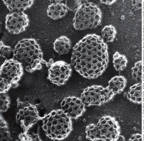
clathrin-coated pit
clathrin molecules assemble into a basketlike network on the cytosolic side of the membrane
clathrin
protein that makes up protein coat
step 1 of mechanism of budding of a clathrin-coated vesicle
cargo receptors bind molecules selected for transport
step 2 of mechanism of budding of a clathrin-coated vesicle
adaptins capture cargo receptors and bind clathrin. clathrin assembles into a basketlike array, forming a clathrin-coated pit
step 3 of mechanism of budding of a clathrin-coated vesicle
dynamin (a GTPase) assembles as a ring around the neck of a clathrin-coated pit, hydrolyzes GTP to help pinch off the vesicle
step 4 of mechanism of budding of a clathrin-coated vesicle
following budding, the vesicle sheds its protein coat (ie clathrin and adaptins), allowing direct interaction with the membrane of its target compartment for fusion. uncoating of clathrin requires energy from ATP hydrolysis
Rab proteins (GTPases) on the vesicle surface
are recognized by tethering proteins on the target membrane
matching Rab and tethering proteins
ensures that transport vesicles fuse only with the correct membrane
vesicle docking depends on :
tethers, Rab proteins and SNAREs
SNARE proteins
drive vesicle fusion
SNARES on the vesicle
(v-SNAREs) bind complementary t-SNAREs)
SNAREs on the target membrane
t-SNAREs
during vesicle fusion,
complementary SNAREs wind around one another, pulling the vesicle’s membrane close to the target membrane and displacing water molecules
mediated by transport vesicles
movement between compartments
energetically unfavorable processes
vesicle budding, scission and fusion
budding
driven by protein coat assembly (clathrin, COP)
scission
(pinching off from membrane) is driven by assembly and GTPase activity of Dynamin
fusion
driven by v-SNARE/t-SNARE winding
cargo selection
involves specific cargo protein, a receptor for the cargo, and adaptins that link the receptor to the clathrin coat, and adaptins that link the receptors to the clathrin coat
provide selective vesicle docking
rabs/tethering proteins
covalently modified in the ER
most proteins
disulfide bond formation
catalyzed by an enzyme in the ER lumen
disulfide bonds
help stabilize the structure of secreted proteins
glycosylate in the ER
many proteins
a branched oligosaccaride containing 14 sugars
transferred from a lipid (called dolichol) to the side chain of an asparagine amino acid
N-linked
oligosaccharides linked to an asparagine side chain
subsequent modification of the oligosaccharide
begins in the ER and continues to the golgi
glycosylation
helps protect a protein from degradation by preventing binding of proteases
can help mediate binding to chaperone proteins
this ensures that the protein will be retained in the ER until it is properly folded → quality control mechanism
oligosaccharides
serves as a transport signal for packaging the protein into an appropriate transport vesicle
on cell surface, can function in cell-cell recognition