Nursing Care of a Family when a Child has Infectious Disorder
1/131
There's no tags or description
Looks like no tags are added yet.
Name | Mastery | Learn | Test | Matching | Spaced |
|---|
No study sessions yet.
132 Terms
infectious diseases
remain a leading cause of morbidity in children.
vaccination programs
have been the most effective in reducing infectious diseases in children
Pathogens
any organism that causes disease
viruses, bacteria, rickettsia, helminths and fungi.
5 types of microorganism pathogens
Incubation period
Prodromal period
Illness
Convalescent period
Stages of infectious disease
Incubation period
the time between the invasion of an organism and the onset of symptoms of infection. During this time, microorganisms grow and multiply the time between the invasion of an organism and the onset of symptoms of infection.
the virulence of the microorganism, the mechanism of spread, and the host
Incubation period vary widely depending on
Prodromal period
time between the beginning of nonspecific symptoms such as malaise, low-grade fever, fatigue, and arthralgia to the onset of disease specific symptoms like rash, diarrhea, and vomiting. Infectious diseases spread readily through communities from a person with the disease to any susceptible individual. This stage is generally short, ranging from hours to a few days.
Illness
stage during which specific symptoms occur
Exanthem
refers to a widespread rash, usually in children
Enanthem
rash on mucous membranes arising from a disease
Convalescent period
Interval between when symptoms first begin to fade and when the child returns to a healthy baseline. The return to baseline will vary from child to child depending on the host, other underlying illnesses and the type and severity of infection.
Chain of Infection
the method in which organisms are spread and enter a new individual to cause disease
Reservoir
container or place in which an organism grows and reproduces
Fomites
are inanimate objects that can also transmit infections from one person to another without direct contact with a human vector
Portal of exit
the route by which an organism leaves an infected child's body to be spread to others. Organism can be carried out of the body by upper respiratory excretions, feces, vomitus, saliva, urine, vaginal secretions, blood or lesion secretions.
Mode of transmission
refers to whether the infection is spread by direct or indirect contact and vectors.
Direct contact
spread by mucous membrane to mucous membrane
Indirect contact
spreads indirectly by fomites- inanimate objects. The most common means is the spread of mouth and nose secretion (droplet).
Droplet
Occurs when a person is in close contact (within 1 m) with someone who has respiratory symptoms. It may also occur through fomites in the immediate environment around the infected person.
Airborne
refers to the presence of microbes within droplet nuclei, which are generally considered to be particles <5ʯm in diameter, can remain in the air for long periods of time and be transmitted to others over distances greater than 1m
Vectors
insects, rats or other vermin- who may not be ill but are carriers of a human disease
Portal of entry
refers to the opening through which a pathogen can enter a child's body such as by inhalation, ingestion, or breaks in the skin from bites, abrasions, or burns.
Age
Gender
Virulence
Body Defenses
Certain characteristics make some individuals more prone to infection than others
Macules, Papules, Plaques, Vesicles, Bullae, Pustules, Urticaria, Scales, Crusts, Erosions, Ulcers, Petechiae, Purpura, Nodules, Atrophy, Scars, Telangiectases
Types of Lesions
Macules
•flat, nonpalpable lesions usually < 10 mm in diameter. represent a change in color and are not raised or depressed compared to the skin surface.
patch
large macule greater than 1 cm in diameter
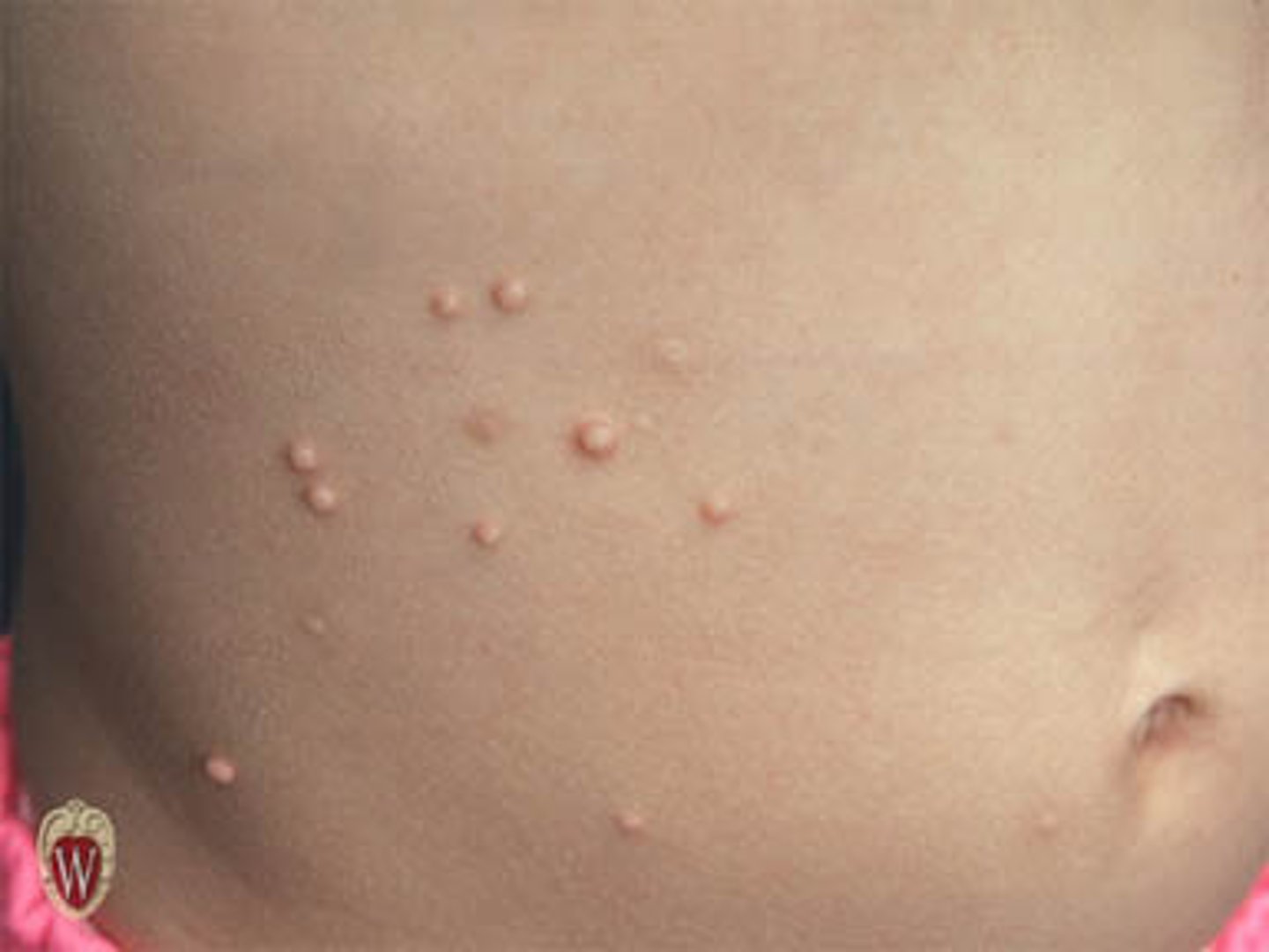
Papules
elevated lesions usually < 10 mm in diameter that can be felt or palpated.
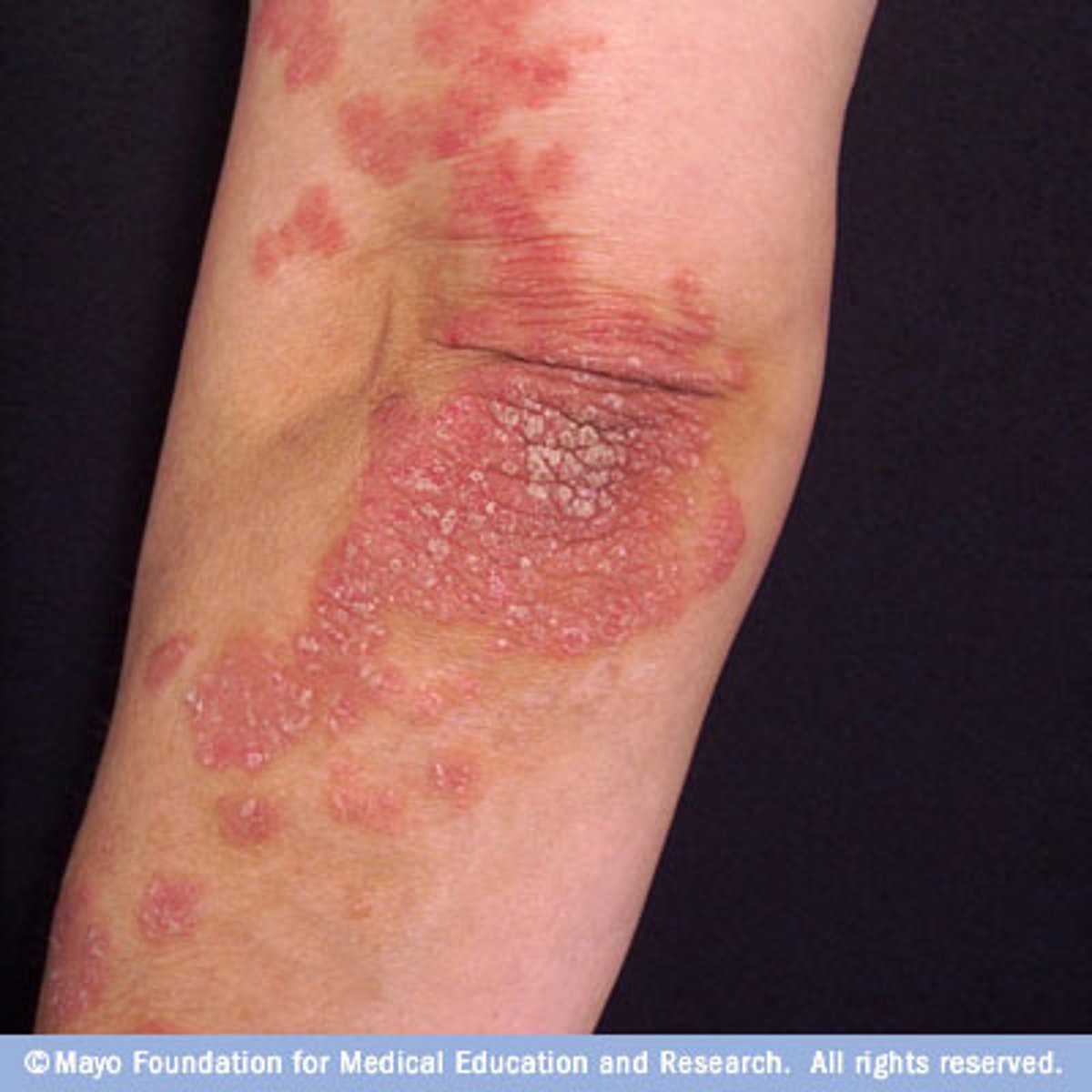
Plaques
palpable lesions > 10 mm in diameter that are elevated or depressed compared to the skin surface. may be flat topped or rounded. Lesions of psoriasis and granuloma annulare commonly form this.
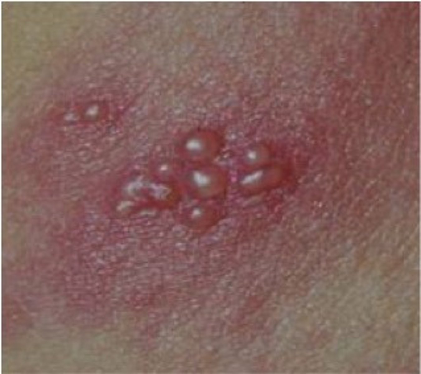
Vesicles
small, fluid-filled blisters < 10 mm in diameter. Are characteristic of herpes infections, acute allergic contact dermatitis, and some autoimmune blistering disorders
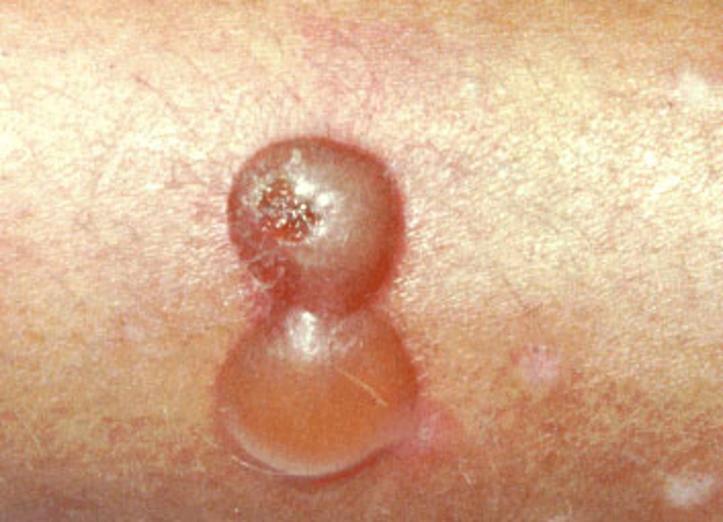
Bullae
Fluid-filled blisters > 10 mm in diameter. These may be caused by burns, bites, irritant contact dermatitis or allergic contact dermatitis, and drug reactions.
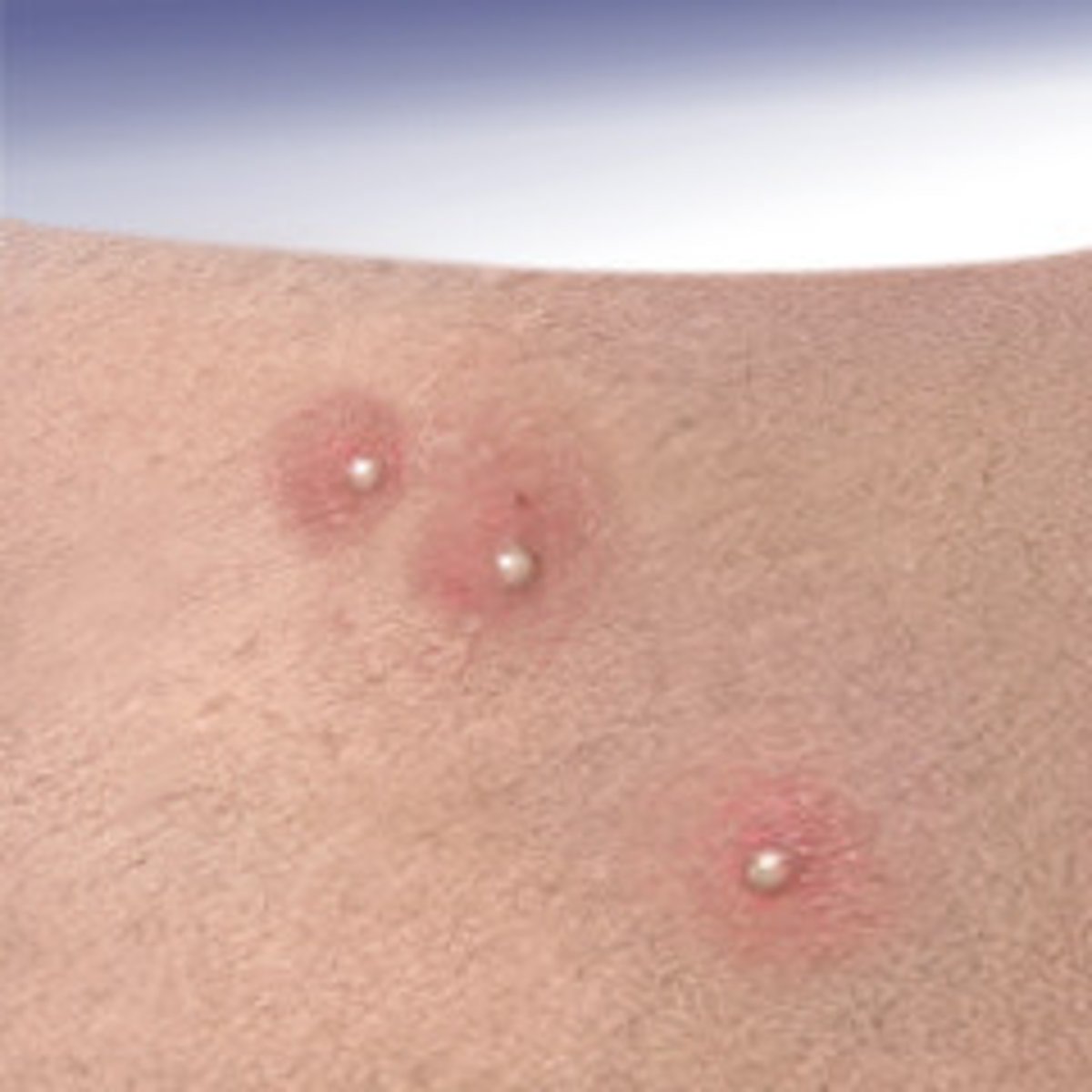
Pustules
vesicles that contain pus. They are elevated, usually yellow-topped lesions that contain pus
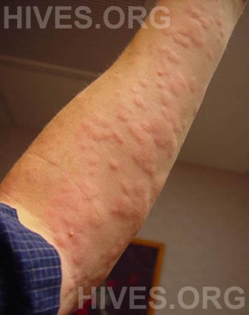
Urticaria (wheals or hives)
characterized by elevated lesions caused by localized edema. are pruritic and erythematous. are a common manifestation of hypersensitivity to medications, stings or bites, autoimmunity, and, less commonly, physical stimuli including temperature, pressure, and sunlight. Typically lasts < 24 hours.
Scales
heaped-up accumulations of horny epithelium. is a characteristic feature of many dermatophytoses, including tinea capitis, resulting in the large bald patches.
Crusts (scabs)
consist of dried serum, blood, or pus. Can occur in inflammatory or infectious skin diseases
Erosions
are open areas of skin that result from loss of part or all of the epidermis. can be traumatic or can occur with various inflammatory or infectious skin diseases
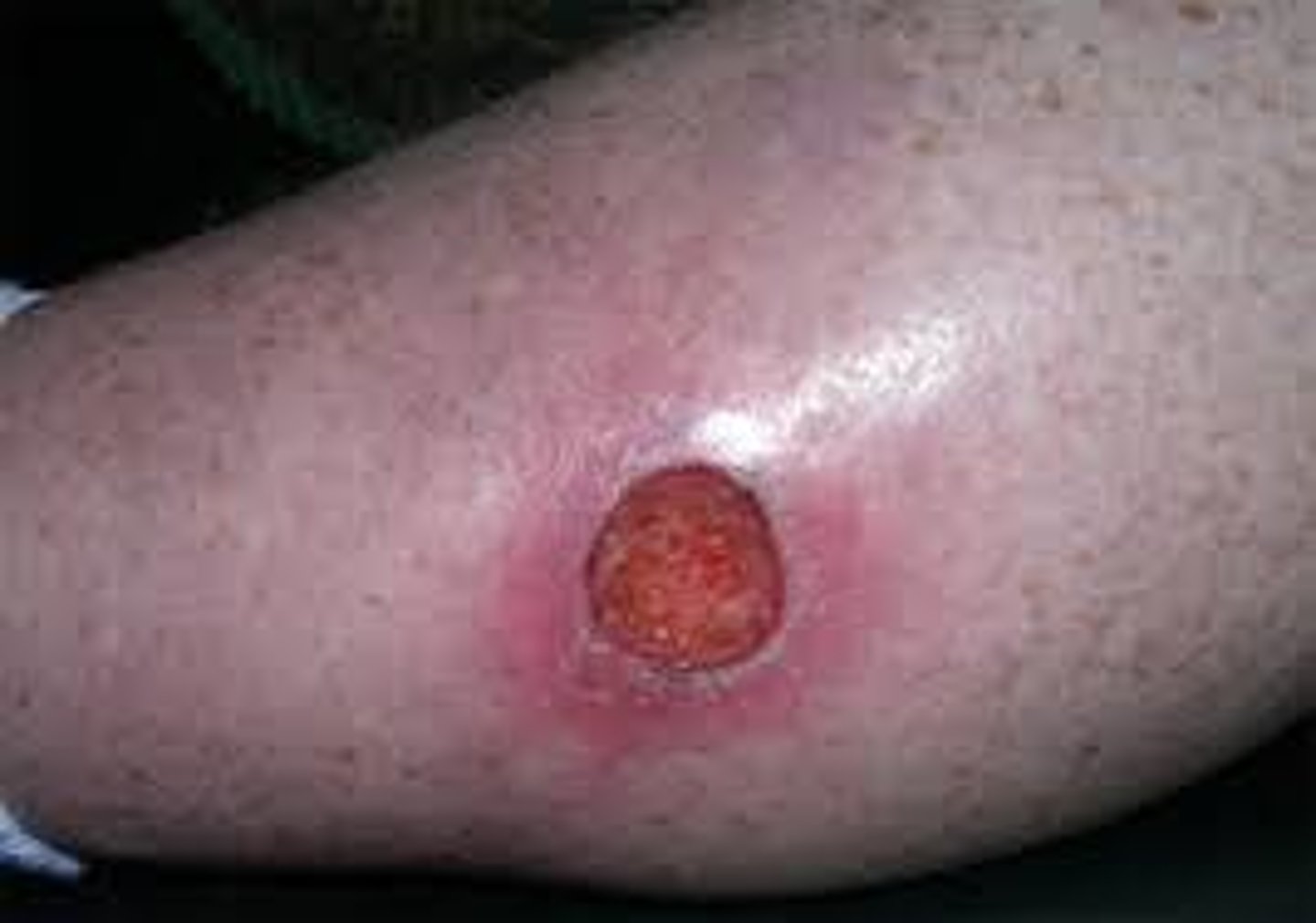
excoriation
a linear erosion caused by scratching, rubbing, or picking.
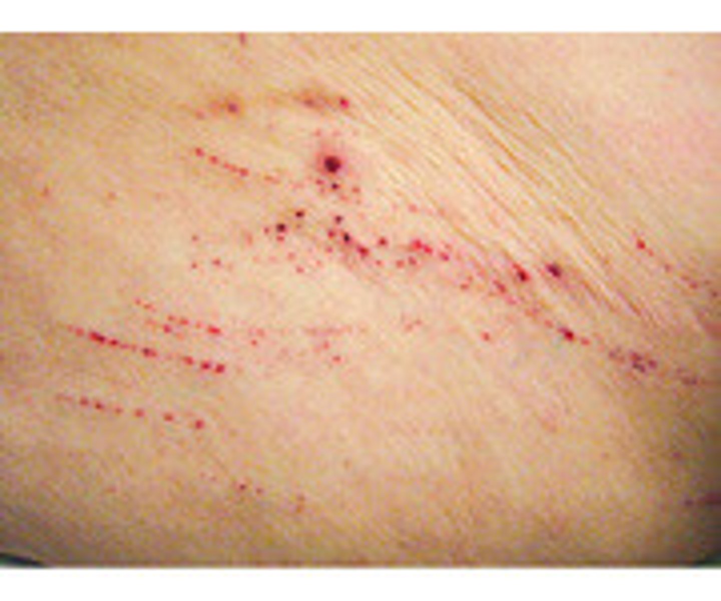
Petechiae
non-blanchable punctate foci of hemorrhage. Causes include platelet abnormalities (eg, thrombocytopenia, platelet dysfunction), vasculitis, and infections
Purpura
larger area of hemorrhage that may be palpable. does not blanch. may indicate a coagulopathy. Large areas may be called ecchymoses or, colloquially, bruises
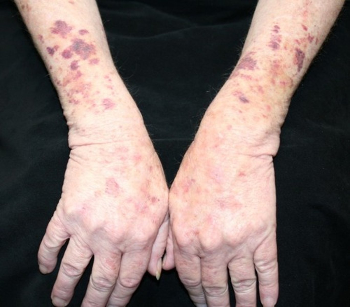
Leukocytoclastic vasculitis
Palpable purpura is considered the hallmark of
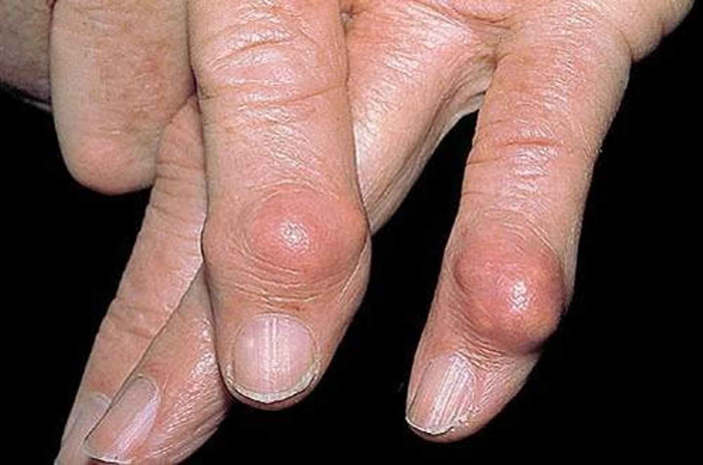
Nodules
are firm papules or lesions that extend into the dermis or subcutaneous tissue
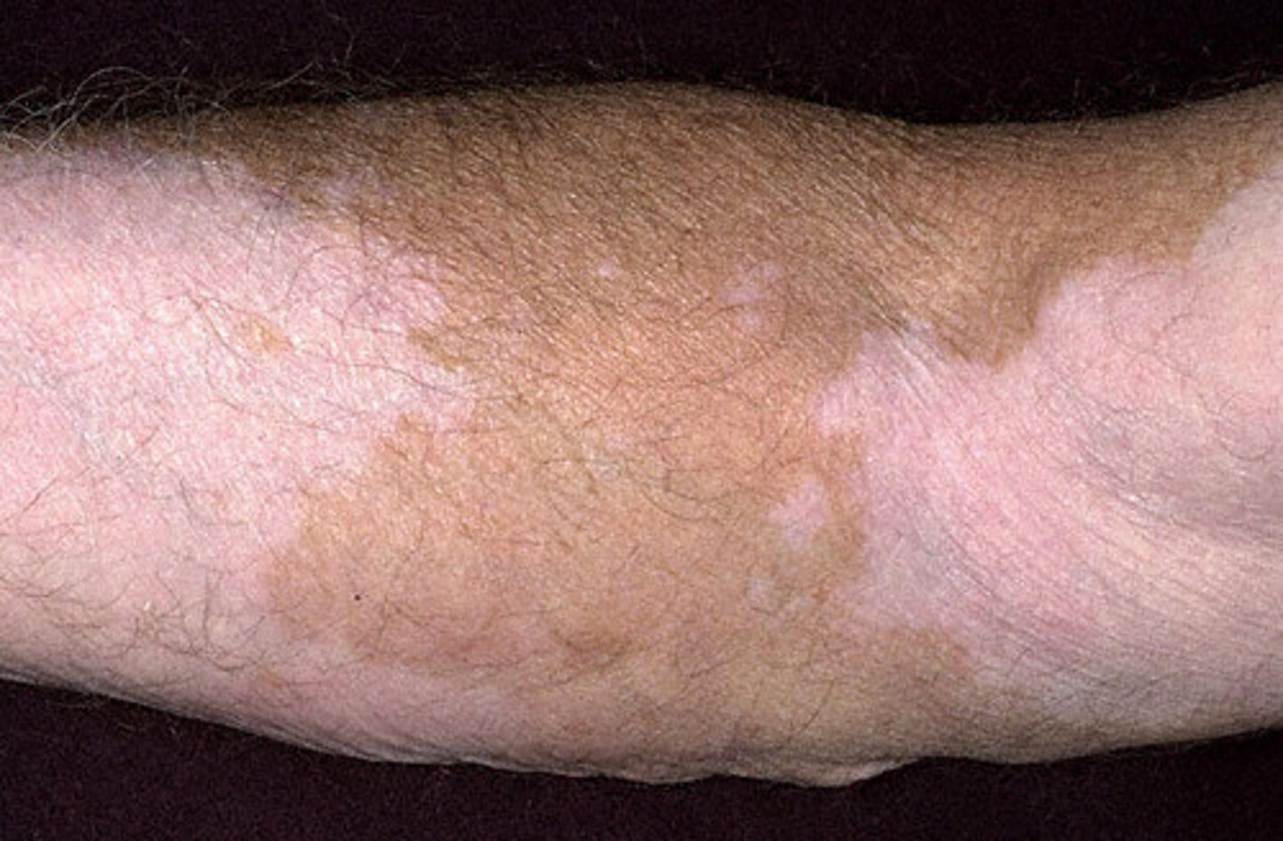
Atrophy
thinning of the skin, which may appear dry and wrinkled, resembling cigarette paper. may be caused by chronic sun exposure, aging, and some inflammatory and neoplastic skin diseases. also may result from long-term use of potent topical corticosteroids
Scars
are areas of fibrosis that replace normal skin after injury. Some scars become hypertrophic or thickened and raised
Keloids
are hypertrophic scars that extend beyond the original wound margin.
telangiectases (vascular lesions)
Foci of small, permanently dilated blood vessels that may occur in areas of sun damage. Are small, dilated blood vessels; they are most often idiopathic.
Causative agent: Human herpesvirus 6 (HHV-6)
Incubation period: 9 to 10 days
Period of communicability: during febrile period
Mode of transmission: unknown
Immunity: contracting the disease offers lasting natural immunity; no vaccine is available
Details of Exanthem Subitum (Roseola Infantum)
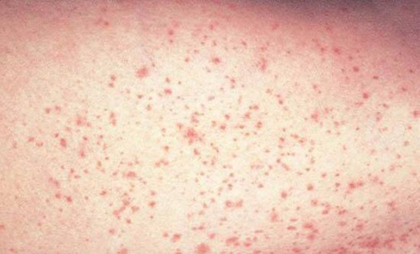
•First symptoms is fever 101-105 degree Fahrenheit or 38.3-40.6 degree Celsius
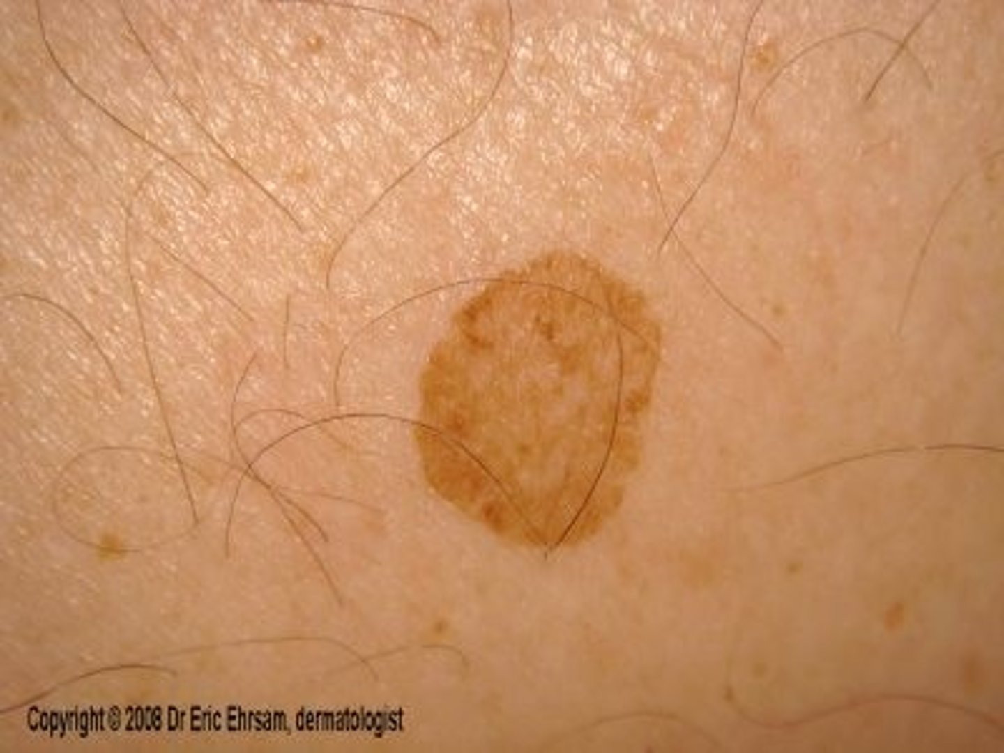
•Rash of discrete, rose-pink macules approximately 2 to 3 mm in size
Hallmark appearance of a rash appearing immediately after the sharp decline in fever
•Lesions occur mostly prominently on the trunk, fade on pressing and last 1-2 days
•Coryza (upper respiratory symptoms), conjunctivitis, or cough
Assessment of Exanthem Subitum (Roseola Infantum)
10% to 15% of children.
Roseola is a cause of febrile seizure in up to
encephalitis, encephalopathy and bulging fontanels
Complications of Exanthem Subitum
Acetaminophen (Tynenol) or ibuprofen (Motrin) if the child is over 6 months - fever. Follow standard infection precautions.
Therapeutic management of Exanthem Subitum
•Causative agent: rubella virus
•Incubation period: generally 14 days within a range of 12-23 days
•Period of communicability: 7 days before to approximately 7 days after the rash appears
•Mode of transmission: direct and indirect contact with droplets
•Immunity: contacting the disease offers lasting natural immunity
•Active artificial immunity: attenuated live virus vaccine (MMR)
•Passive artificial immunity: Immune serum globulin is considered for pregnant women exposed to the virus
Details of German Measles
German Measles
Tigdas Hangin (Tagalog), Dupang (Cebuano) was first described in 1691 by a German physician Daniel Sennert calling it röteln, or rubella, for the red-coloured rash that accompanies the illness
isolated in 1962, and a vaccine was made available in 1969
When was the rubella first isolated and then a vaccine made available
Norman McAlister Gregg
Australian ophthalmologist that discovered that prenatal infection with the virus was responsible for congenital malformations in children in 1941
togavirus, genus Rubivirus
Rubella virus is classified as a
group A arboviruses, such as eastern and western equine encephalitis viruses
Rubella virus is most closely related to
lipid solvents, trypsin, formalin, ultraviolet light, low pH, heat, and amantadine.
Rubella virus is relatively unstable and is inactivated by
• Rubella
• Rosy disease
• Three-day measles
• Rotein
names for German Measles
Pathognomonic Sign
a sign whose presence means that a particular disease is present beyond any doubt
Forscheimer's spots
fleeting enanthem seen as small, red spots (petechiae) on the soft palate and may precede or accompany the skin rash of rubella (German measles)
•A mild maculo papular rash (pink) (non itchy) that starts on the face and spreads to the neck, the chest, and the rest of the body.
•Swollen glands (lymph nodes), especially behind the ear and at the back of the head.
•Adults, especially women, also may have joint pain. Older children and teens also may have eye pain, a sore throat, and body aches. Young children may have only a rash.
Illness stage of Rubella (1-2 days of illness)
•Disappearance of the rash
•Enlargement of lymph nodes subsides
Convalescence of Rubella (3rd day of illness)
congenital rubella syndrome
Defects of the eye, heart, brain and large arteries are most common
cataract, deafness and cardiac abnormalities
Classical Triad of Rubella
Deafness
the most common and often the sole manifestation of congenital rubella infection, especially after the fourth month of gestation
cataracts, glaucoma, retinopathy, and microphthalmia may occur
Eye defects in Rubella
patent ductus arteriosus, ventricular septal defect, pulmonic stenosis, and coarctation of the aorta are possible
Cardiac defects in Rubella
microcephaly and mental retardation
Neurologic abnormalities in Rubella
bone lesions, splenomegaly, hepatitis, and thrombocytopenia with purpura may occur.
Other abnormalities in Rubella
fetal death, spontaneous abortion, or preterm delivery
Most severe effects of Rubella in utero
early gestation. As many as 85% of infants infected in the first trimester of pregnancy will be found to be affected if followed after birth
Infection with rubella virus is most severe in
2 to 4 years
Manifestations of CRS may be delayed from
Diabetes mellitus appearing in later childhood. Progressive encephalopathy resembling subacute sclerosing panencephalitis. Have a higher than expected incidence of autism.
Complications of CRS in later life
20th week of gestation. The overall risk of defects during the third trimester is probably no greater than that associated with uncomplicated pregnancies.
defects are rare when infection occurs after what week of gestation
•Vaccine (MMR or Measles Mumps Rubella) should be given on or after the child's first birthday; the recommended age range is from 12-15 months. The 2nd dose is usually given when the child is 4-6 years old, or before he or she enters kindergarten or first grade.
•Gammaglobulin (after exposure for 72 hours if pregnant)
Treatment/ Prevention of Rubella
Measles
Tigdas (Tagalog), Tipdas (Cebuano) is a highly contagious respiratory infection that's caused by a virus. It causes a total-body skin rash and flu-like symptoms, including a fever, cough, and runny nose. It is caused by a single-stranded, enveloped RNA virus with 1 serotype
Morbillivirus in the Paramyxoviridae family.
Measles is classified as a member of the genus
Humans
are the only natural hosts of measles virus and mumps virus.
•Rubeola
•Morbilli
•Hard Measles
•10-day measles
•Red measles
•Little red disease
Other names for measles
•Fever
•Dry cough
•Runny nose
•Sore throat
•Inflamed eyes (conjunctivitis)
•A skin rash made up of large, flat blotches that often flow into one another
•Koplik's spots
Signs and symptoms of measles typically include:
infected droplets spray into the air (airborne), where other people can inhale them
Mode of transmission of Measles
•High grade fever (40-41 degree Celsius)
•3 C's- cough, colds/coryza, conjunctivitis
Prodromal stage of Measles (3-4 days)
Koplik's spot
Pathognomonic sign of measles
•Appearance of maculopapular rashes (non-itchy)
•Reddish blotchy
•Behind the ears, eyes, entire face, neck and entire torso (head to toe)
Illness stage of measles (2-3 days)
•Disappearance of rashes (fine branny desquamation)
•On the road to recovery
Convalescence stage of measles
Ear infection (otitis media)
Bronchitis, laryngitis or croup
Pneumonia
Encephalitis
Pregnancy problems
Low platelet count (thrombocytopenia)
Complications of measles
Vaccination
the most cost-effective protection against measles.
first dose given when a child is 9 months old and the second dose given when a child is 12-months old.
When is the measles vaccine given
Post-exposure vaccination
Non-immunized people, including infants, may be given the measles vaccination within 72 hours of exposure to the measles virus to provide protection against the disease. If measles still develops, the illness usually has milder symptoms and lasts for a shorter time.
immune serum globulin
Pregnant women, infants and people with weakened immune systems who are exposed to the virus may receive an injection of what within six days of exposure to the virus, these antibodies can prevent measles or make symptoms less severe.
Chickenpox
Bulutong tubig (Tagalog), Hangga (Cebuano), also known as Varicella, is a highly contagious disease. an airborne disease which spreads easily from one person to the next through the coughs and sneezes (nasopharyngeal secretions) of an infected person. It may be spread from one to two days before the rash appears until all lesions have crusted over. It may also spread through contact with the blisters.
varicella zoster virus
Causative agent of Chickenpox
14 to 16 days
incubation period of chicken pox
about 1 or 2 days after the first symptoms start.
itchy chickenpox rash usually appears
Most people recover in about 2 weeks.
Recovery period of chickenpox
•Presence/ absence of high-grade fever
•Malaise
•Muscle pain
•Headache
Prodromal stage of Chickenpox
•Rashes appear (itchy) distribution: center to peripheries
•Abundantly found (covered areas such as scalp)
•Macule, papule, vesicle, pustule
Illness stage of chickenpox
•Crusting of rashes: falls off (peel off), dry
•On the road to recovery
Convalescence stage of chickenpox
first six months
During pregnancy the dangers to the fetus associated with a primary VZV infection are greater in
congenital varicella syndrome
If infection occurs during the first 28 weeks of gestation. Effects on the fetus can range in severity from underdeveloped toes and fingers to severe anal and bladder malformation. Possible problems include: