Anatomy Lab 2 Test 2
1/293
There's no tags or description
Looks like no tags are added yet.
Name | Mastery | Learn | Test | Matching | Spaced | Call with Kai |
|---|
No analytics yet
Send a link to your students to track their progress
294 Terms
The sound of a heartbeat comes from…
Turbulence of blood flow caused by closure of the valves
Listening to sounds in the body is called
Auscultation
The first heart sound is what valves closing
AV
The second heart sound is caused by what valves closing
Semilunar
Where do you Auscultate the aortic valve
2nd intercostal space to the right of the sternum
Where do you auscultate the pulmonic valve
The second intercostal space to the left of the sternum
Where do you auscultate the Tricuspid and right heart sounds
The fourth intercostal space to the left of the sternum
Where do you auscultate the Mitral and left heart sounds
The fifth intercostal space in the midlclavavicuar line
Electrical events in order for the heart
SA node → junctional fibers→AV node→AV bundle→ bundle branches→ Purkinje fibers
What happens during the P-R interval
Beginning of atrial excitation to beginning of ventricular excitation
What happens during the S-T segment
Entire ventricular myocardium depolarized
What happens during the Q-T interval
Beginning of the ventricular depolarization through ventricular repolarization
What happens during the P wave
Depolarization of the SA node and atria
What happens during the QRS complex
Ventricular depolarization and atrial repolarization
What happens during the T wave
Ventricular repolarization

How many heartbeats are shown in this tracing?
6
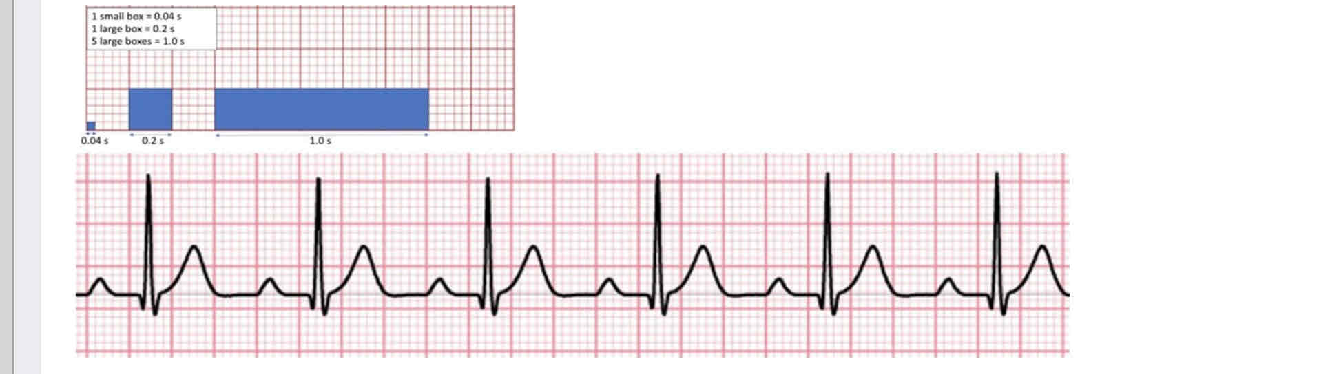
What is the beat period for this patient? Round to the nearest 100th
.76

What is the patients heart rate?
75 bpm

Is the patients heart rate considered bradycardia, tachycardia, or sinus rhythm
Sinus
How do you find heart rate on ecg?
Get average of three R intervals and divide 60 over the average
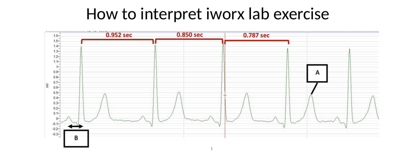
What is the beats per minute given this example
70 bpm
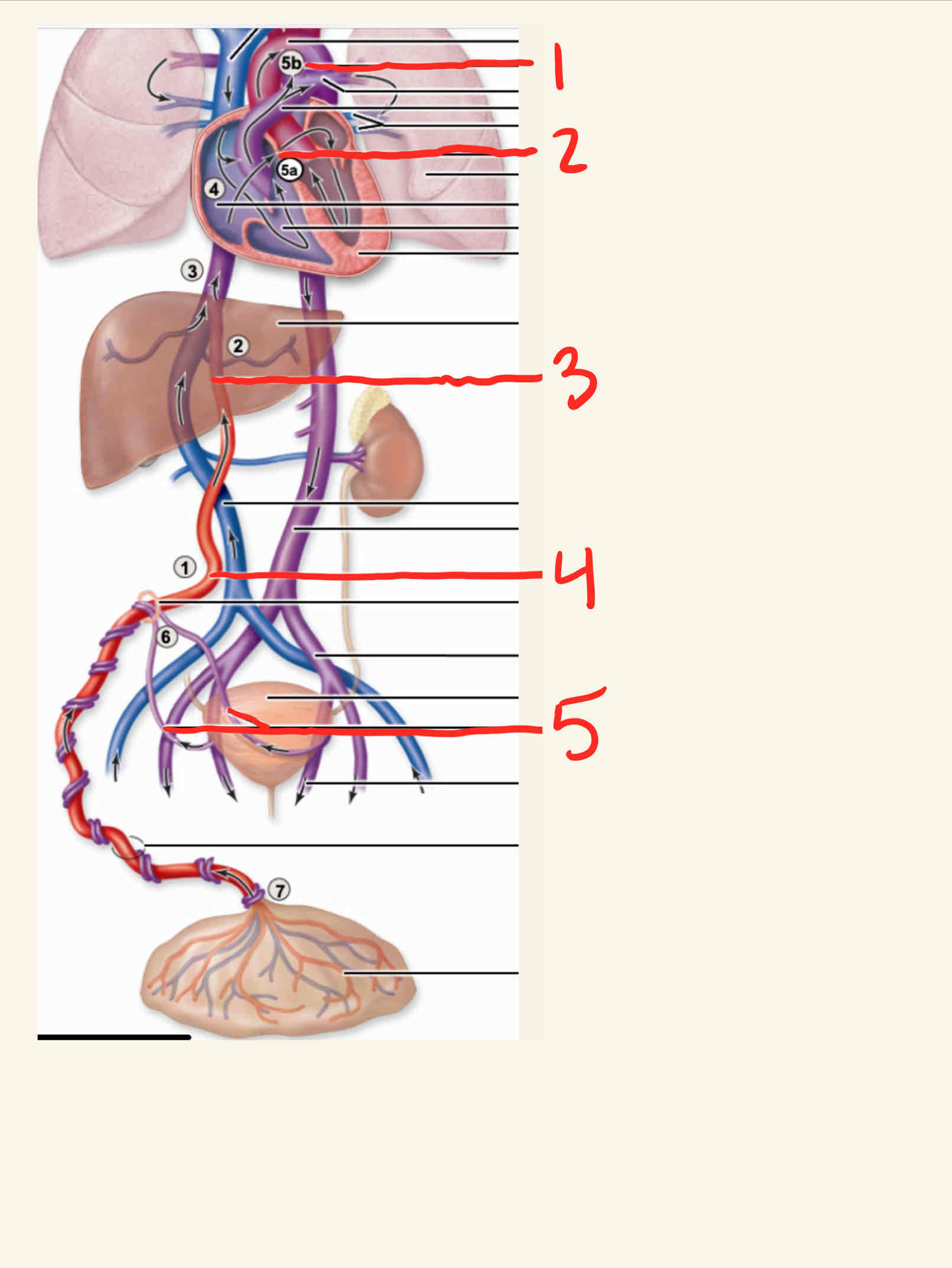
All of the body parts above the level of the diaphragm are drained by
Superior vena cava
All of the body parts below the level of the diaphragm are drained by
Inferior vena cava

What is the structure labeled 1
Artery

What is the structure labeled 2
Vein

What is the structure labeled 3
Tunica interna artery

What is the structure labeled 4
Tunica media artery

What is the structure labeled 5
Tunica externa artery

What is the structure labeled 6
Tunica interna vein

What is the structure labeled 7
Tunica media vein

What is the structure labeled 8
Tunica externa vein
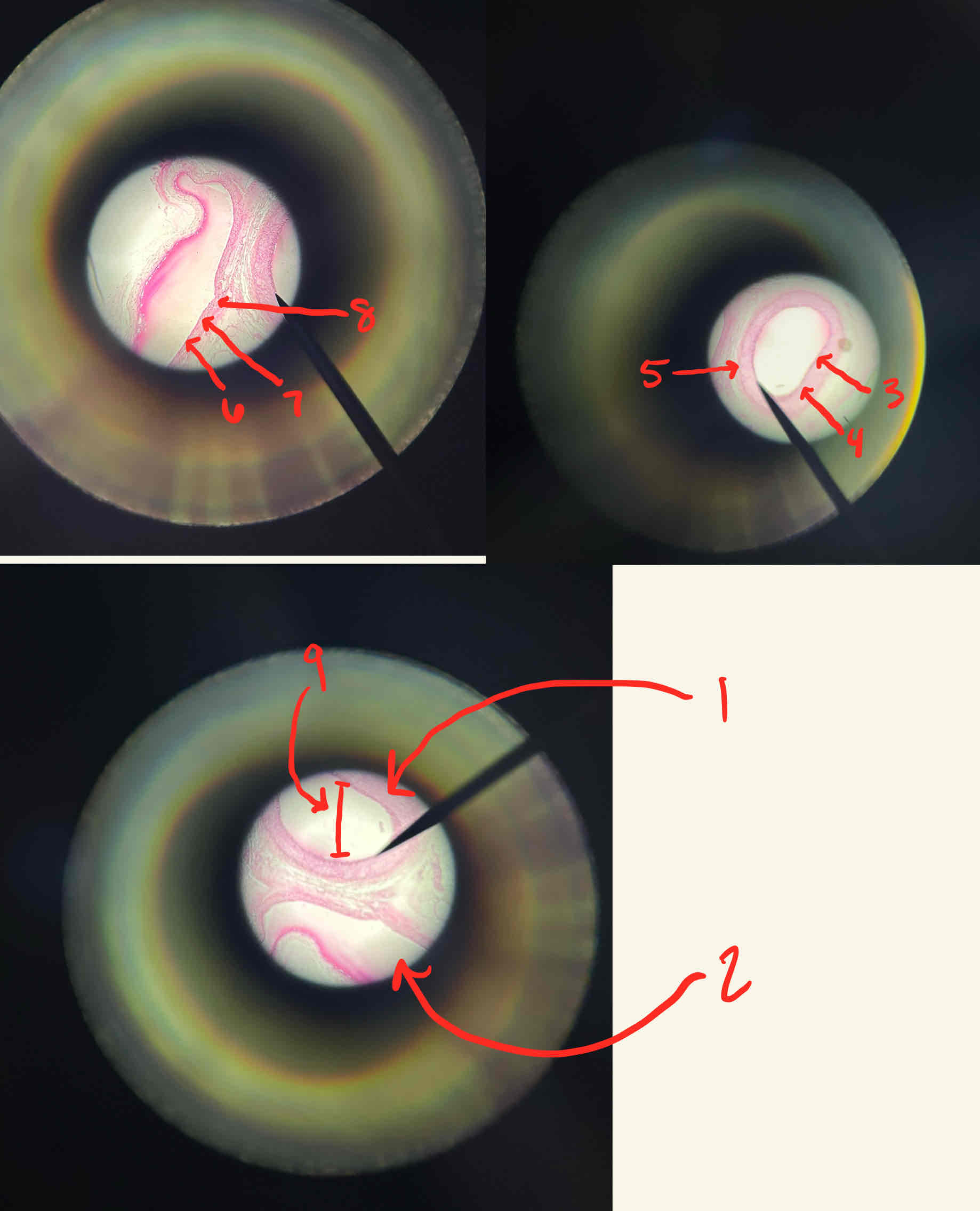
What is the structure labeled 9
Lumen

What is the structure labeled 1
Ductus arterious

What is the structure labeled 2
Foramen ovale

What is the structure labeled 3
Ductus venous
What is the structure labeled 4
Umbilical vein
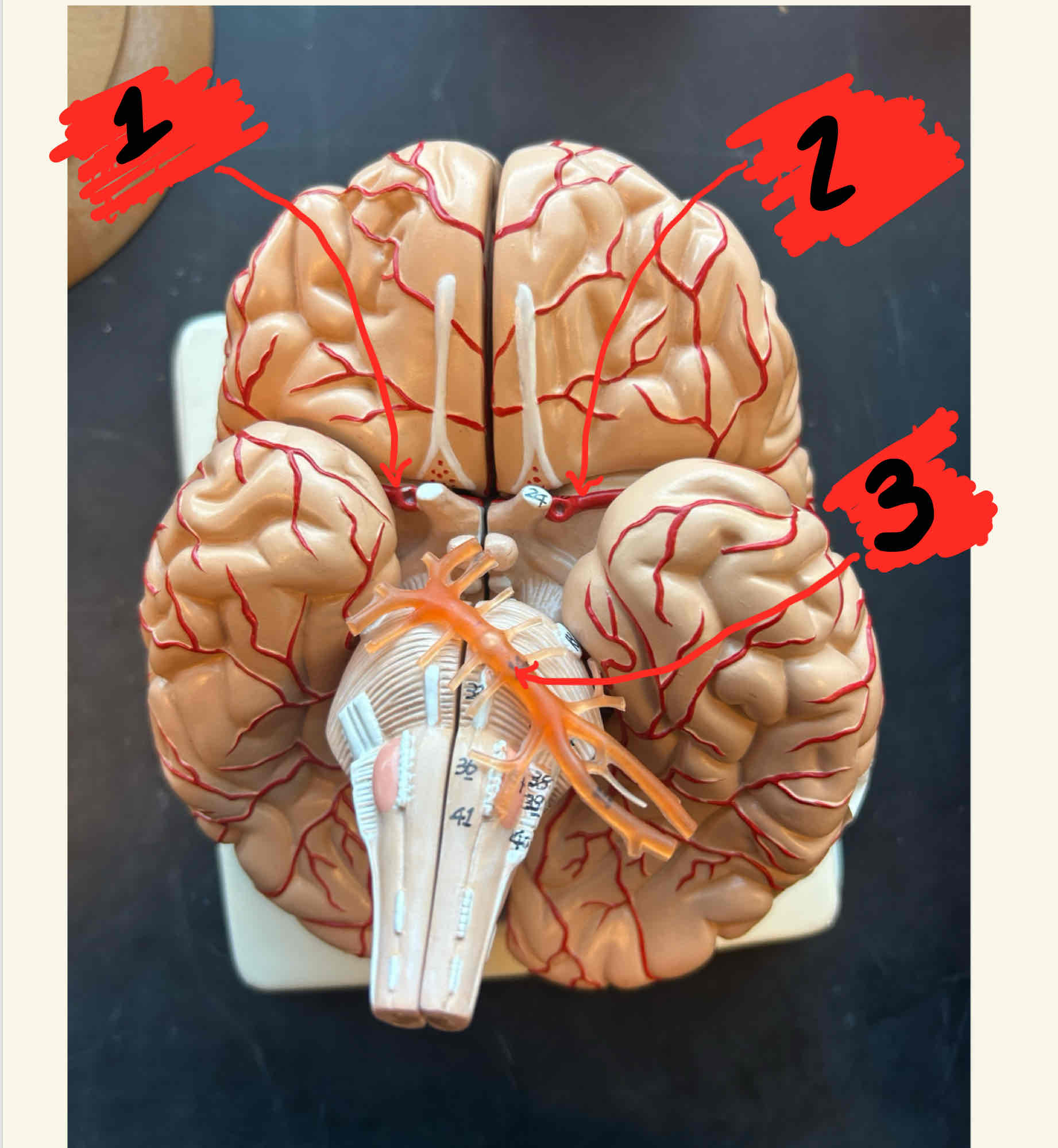
What is the structure labeled 2
Left internal corticoid

What is the structure labeled 3
Basilar artery
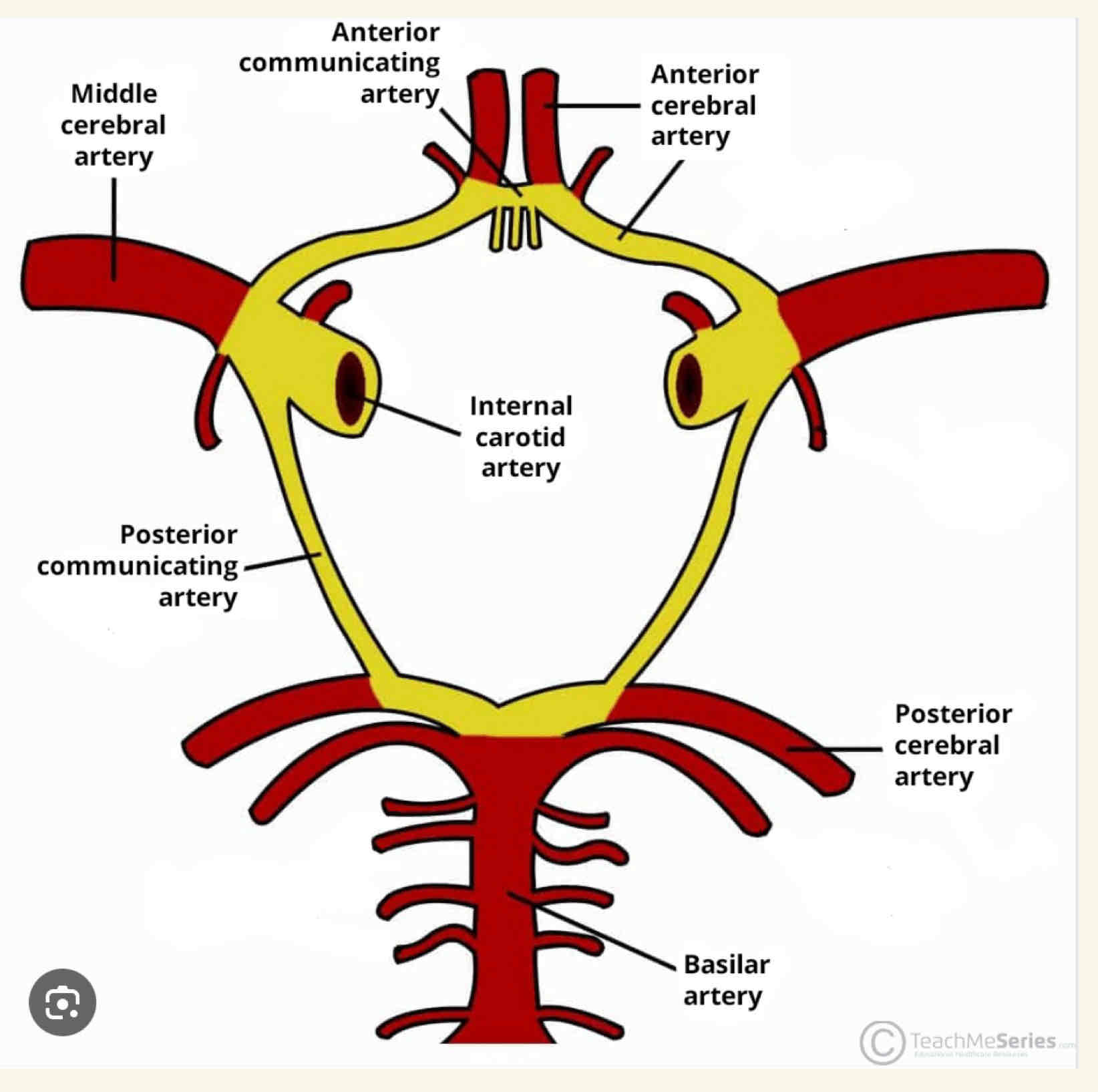
What is the structure highlighted
The circle of Willis

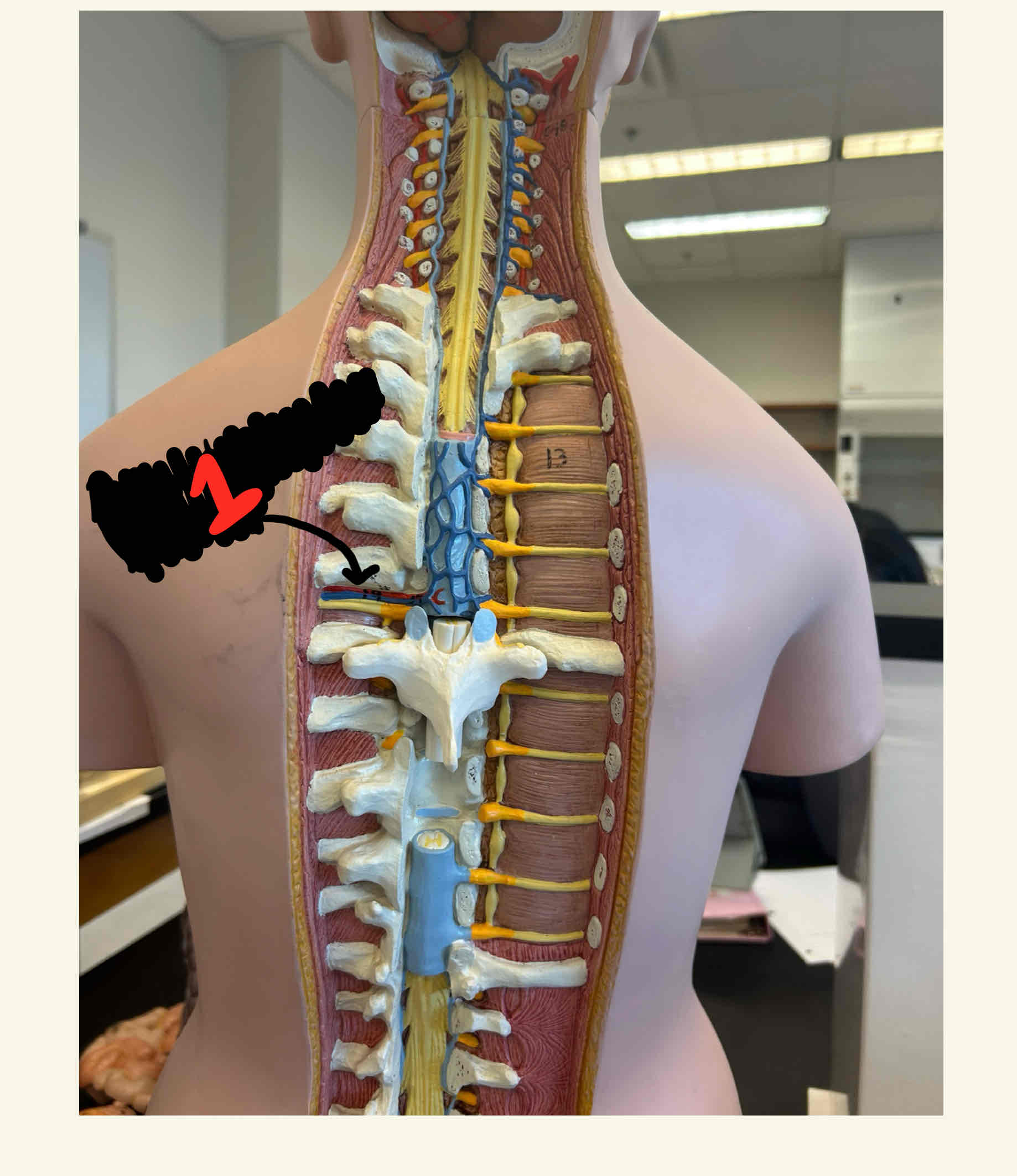
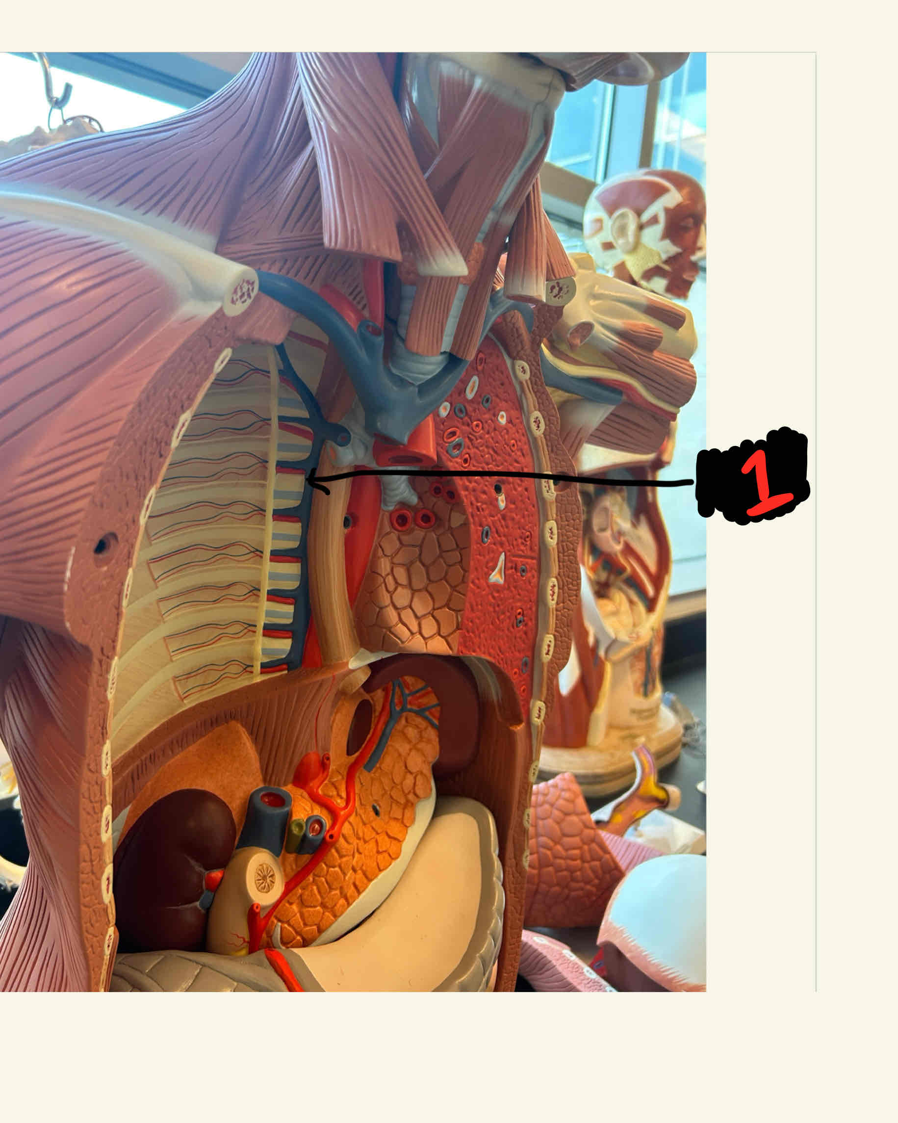
What is the structure labeled 1
Intercostal arteries
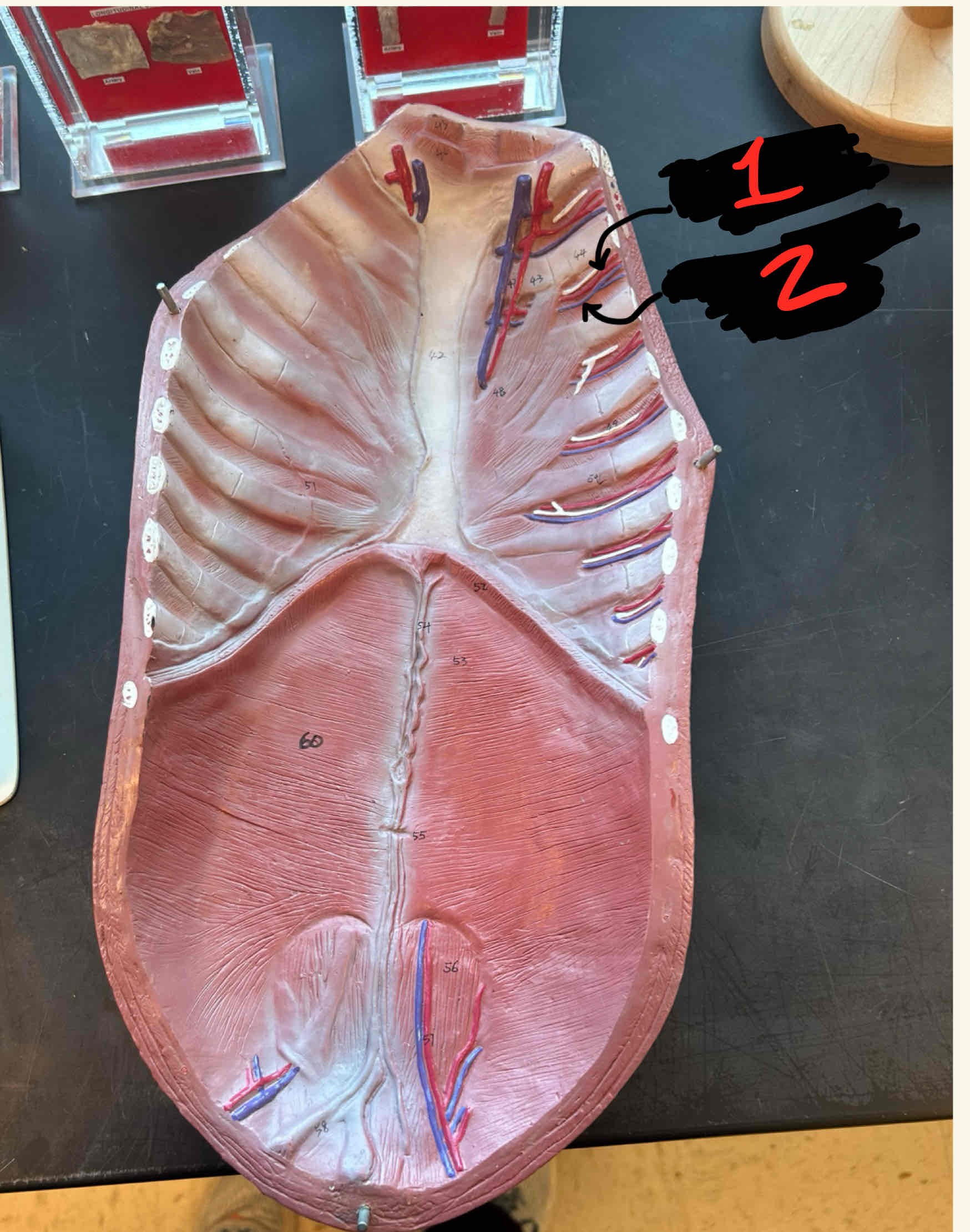
What is the structure labeled 2
Intercostal veins

What is the structure labeled 1
Internal carticoid

What is the structure labeled 2
External carticoid

What is the structure labeled 3
Brachiocephalic Trunk

What is the structure labeled 4
Pulmonic arteries

What is the structure labeled 5
Left carticoid

What is the structure labeled 6
Left subclavian

What is the structure labeled 7
Aortic arch

What is the structure labeled 8
pulmonary veins

What is the structure labeled 9
Descending aortic artery

What is the structure labeled 10
Superior vena cava

What is the structure labeled 11
Right subclavian
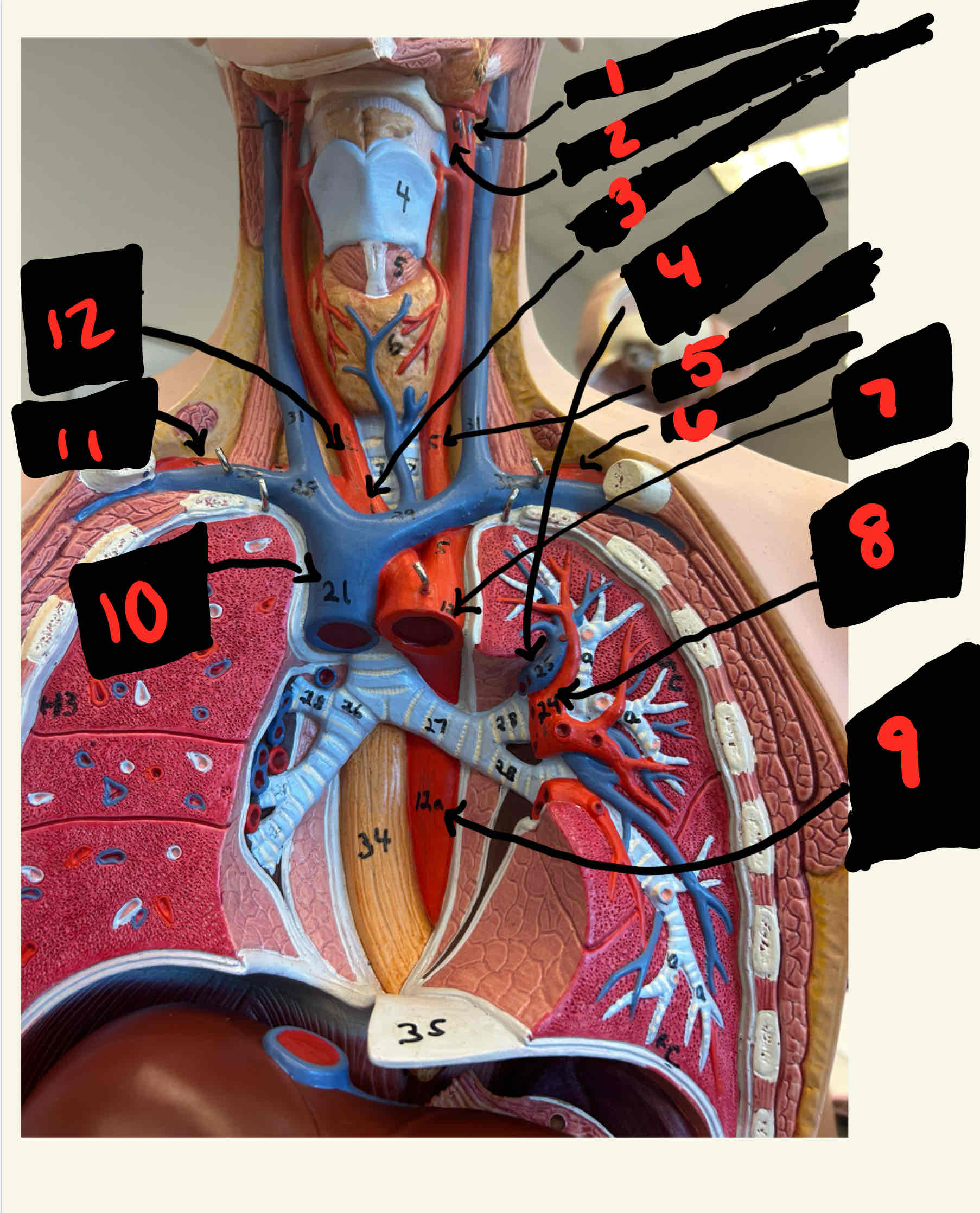
What is the structure labeled 12
Right carticoid artery

What is the structure labeled 1
Inferior mesenteric artery

What is the structure labeled 2
Gonadal artery

What is the structure labeled 3
left internal iliac artery

What is the structure labeled 4
left gonadal artery

What is the structure labeled 5
Lumbar artery

What is the structure labeled 6
Decending aortic artery
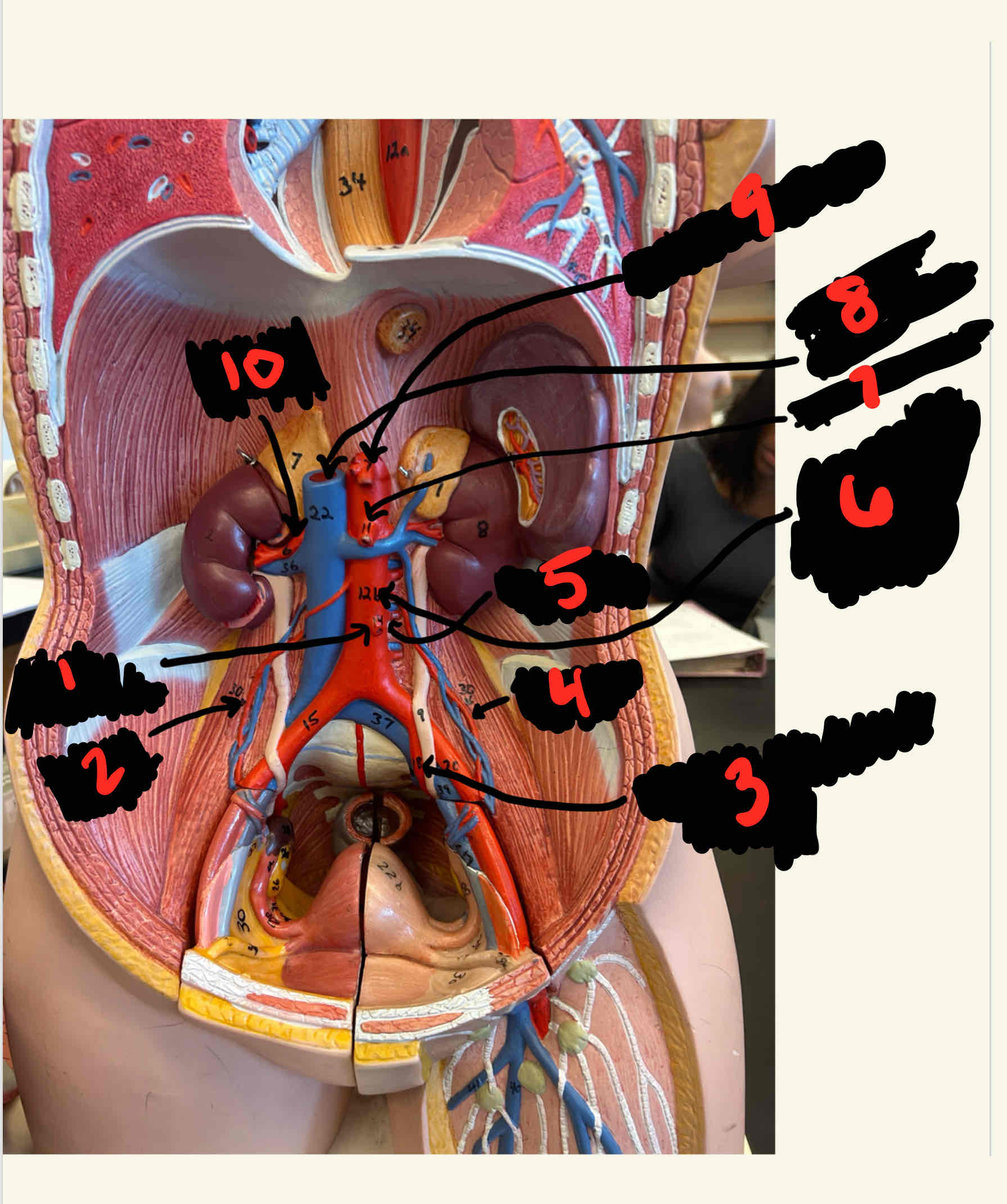
What is the structure labeled 7
Superior mesenteric

What is the structure labeled 8
Inferior Vena Cava

What is the structure labeled 9
Celiac Trunk

What is the structure labeled 10
Renal artery
What is the structure labeled 1
right vertebral artery

What is the structure labeled 1
Hepatic portal vein
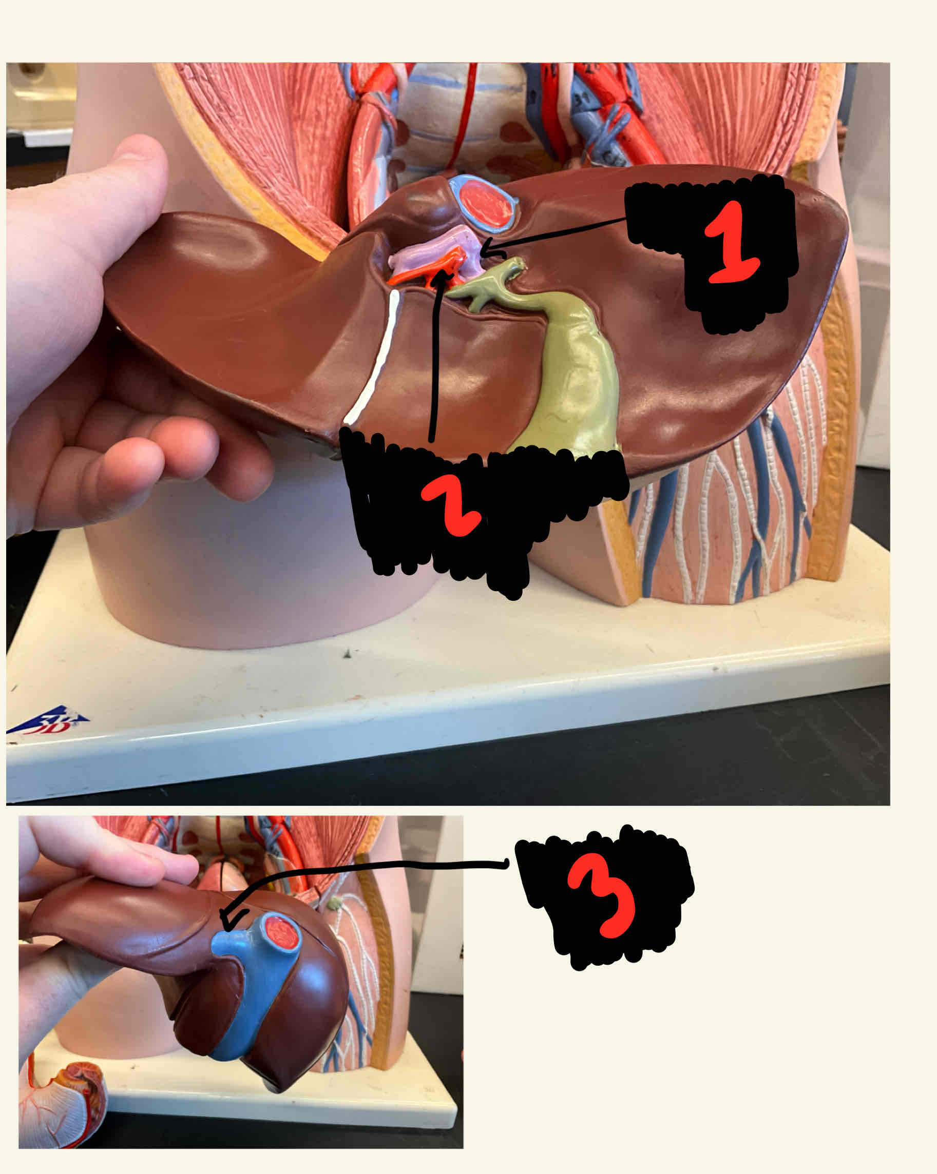
What is the structure labeled 2
Common hepatic artery

What is the structure labeled 3
Hepatic vein
What is the structure labeled 1
Gastric artery
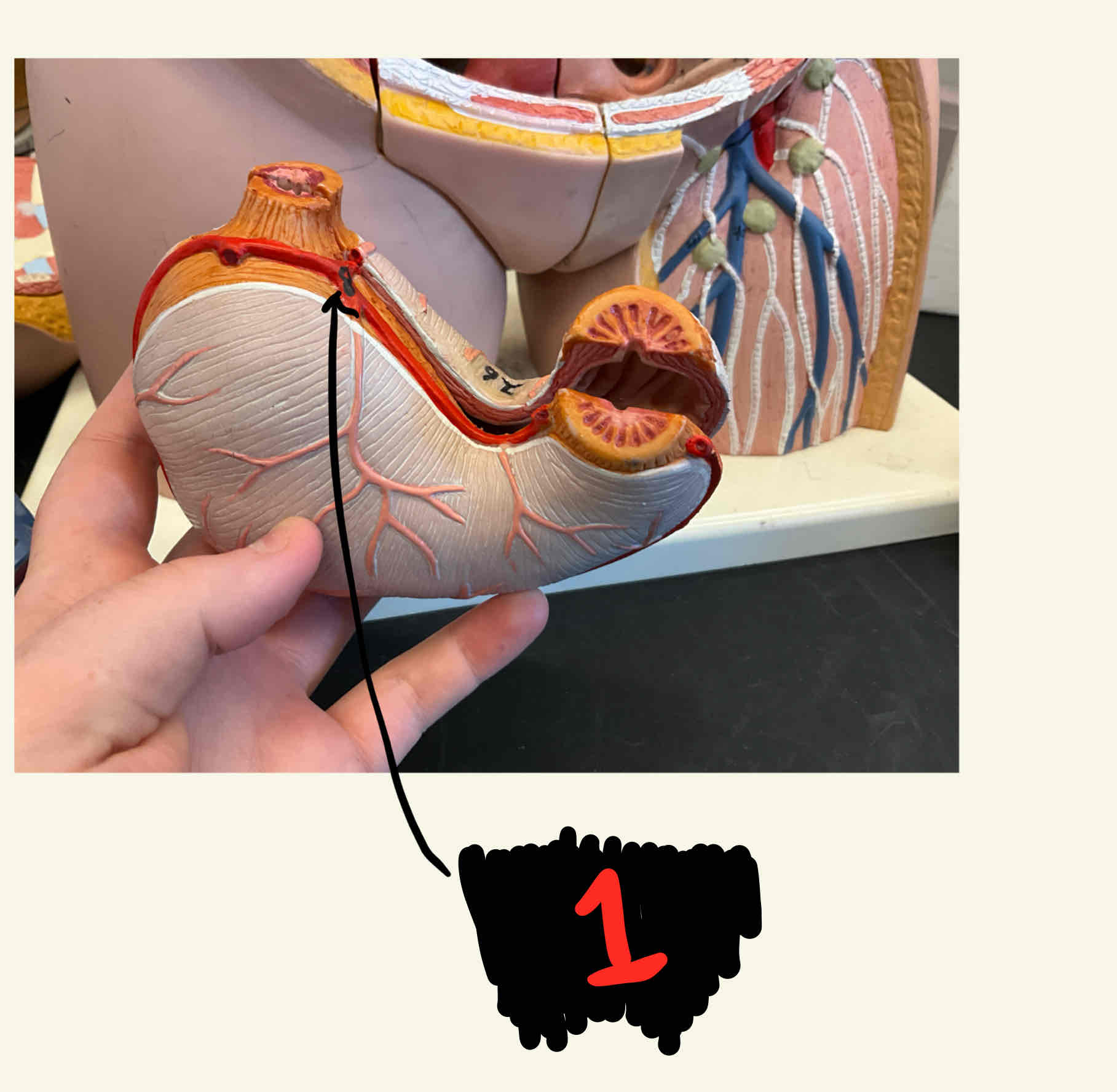
What is the structure labeled 1
Splenic artery
What is the structure labeled 1
Splenic vein

What is the structure labeled 2
Right subclavian
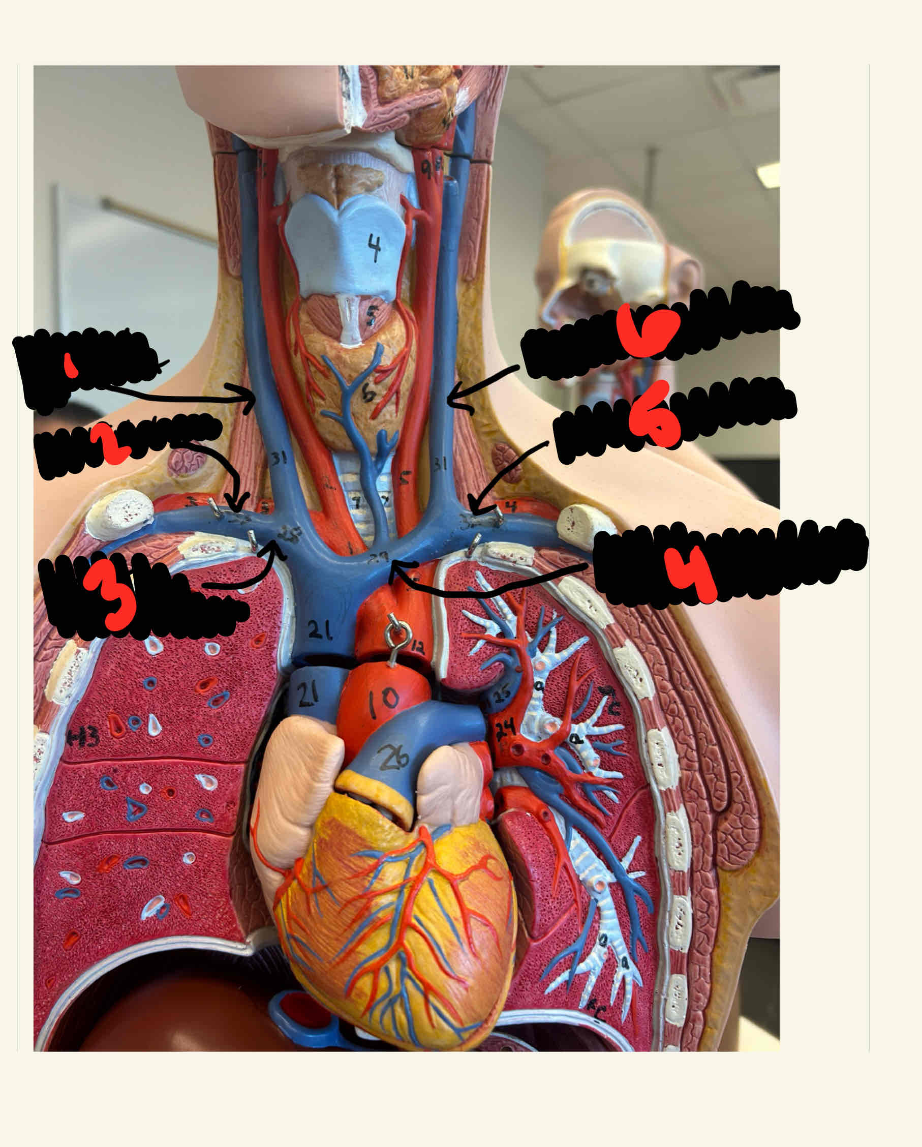
What is the structure labeled 3
Right braciocephalic

What is the structure labeled 4
Left brachiocephalic

What is the structure labeled 5
Left subclavian

What is the structure labeled 6
Internal left jugular
What is the structure labeled 1
Azygous
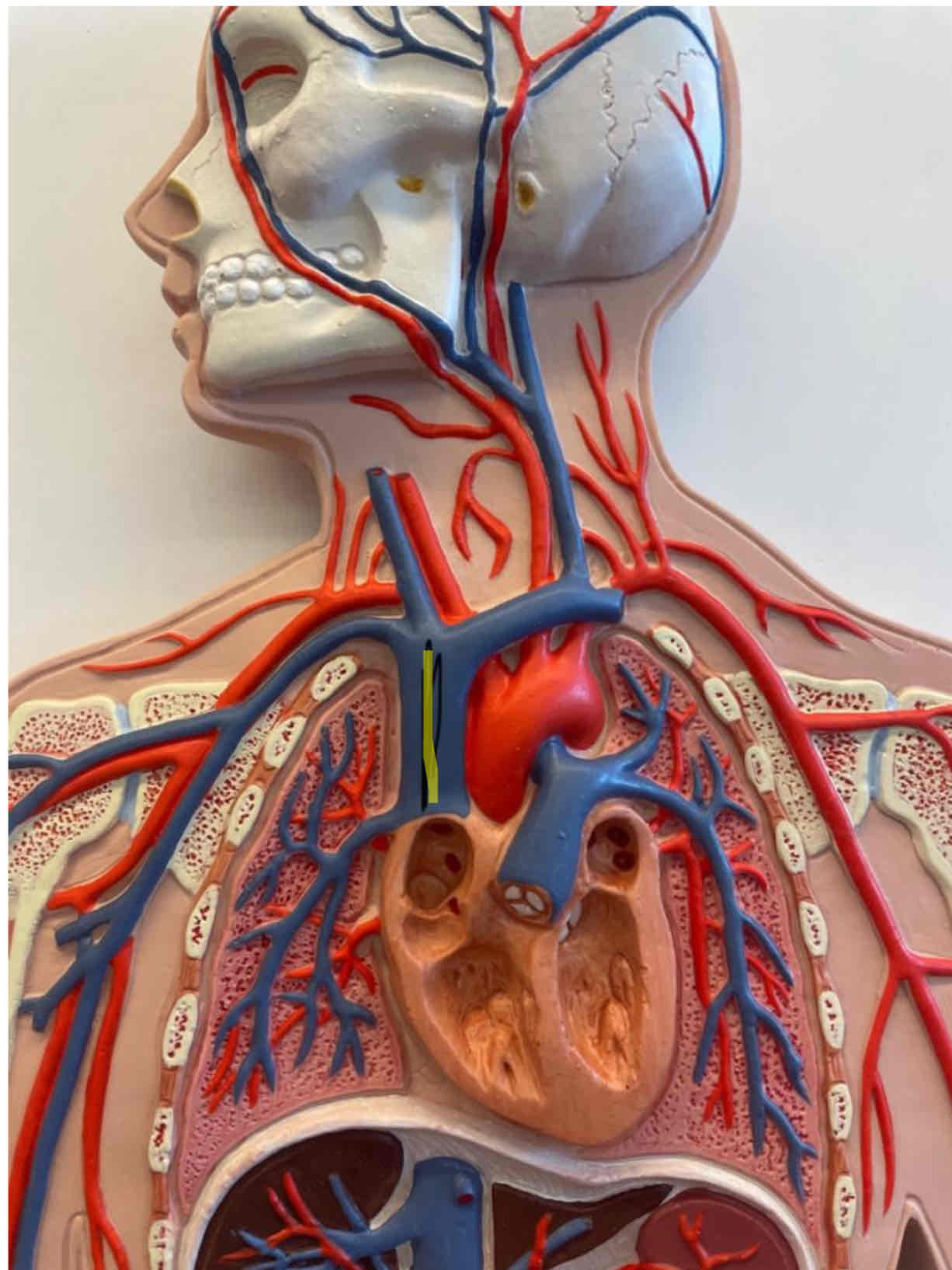
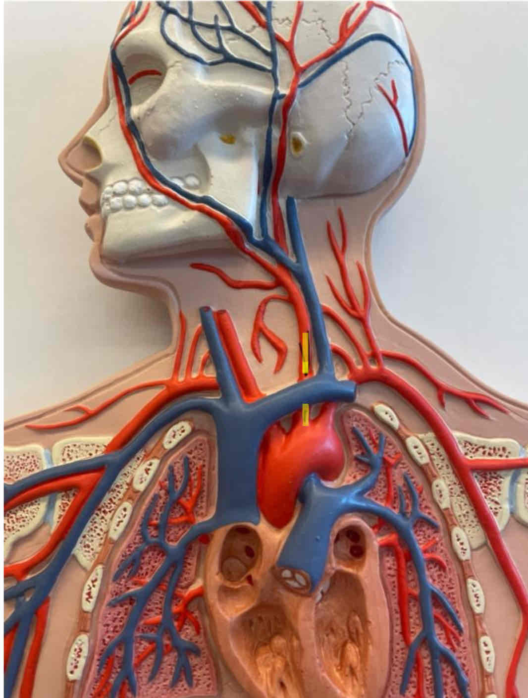
What is the highlighted area
Superior vena cava
full body model
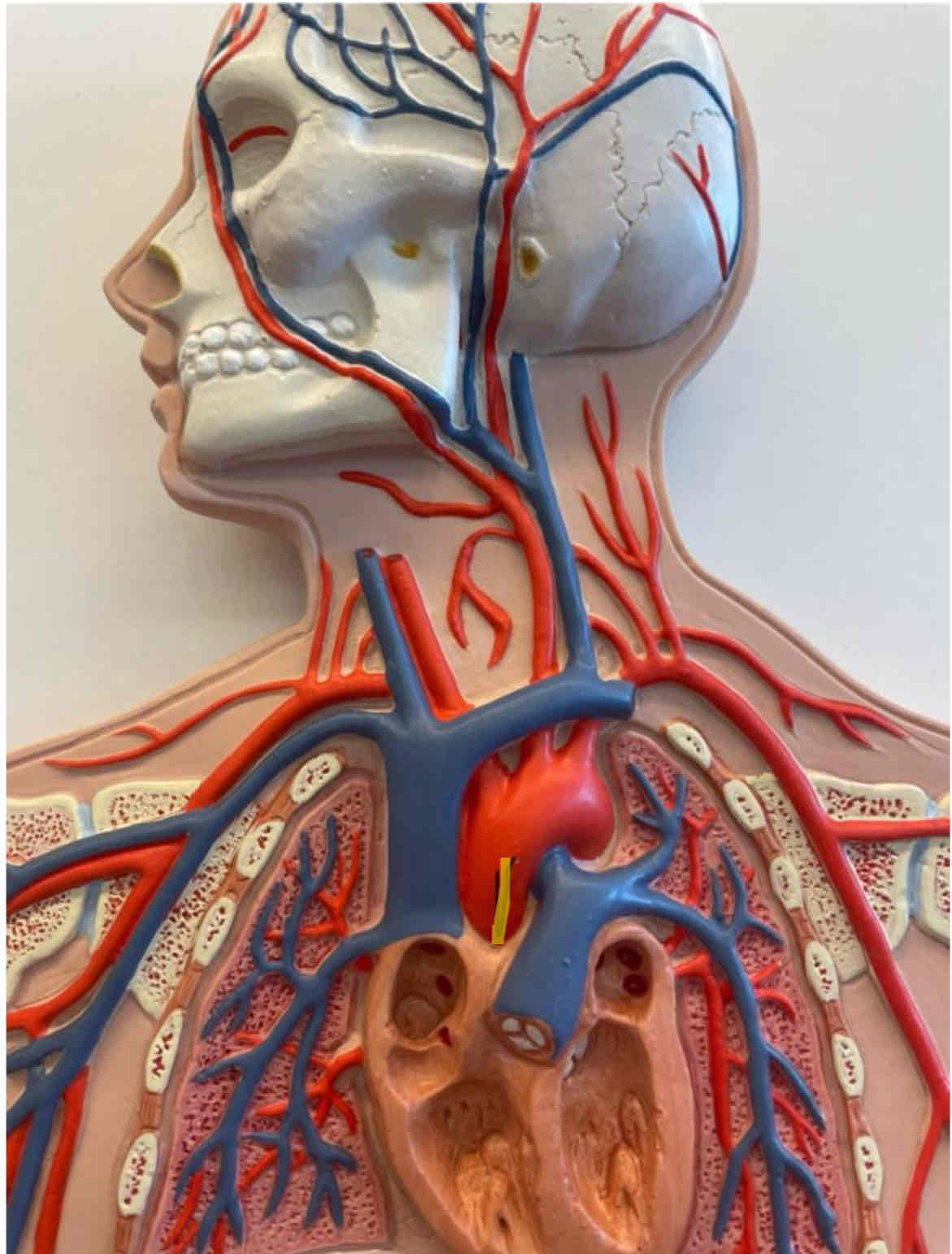
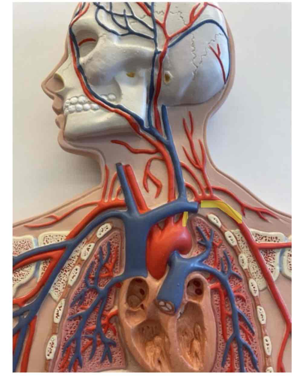
What is the highlighted area
Ascending aorta
full body model
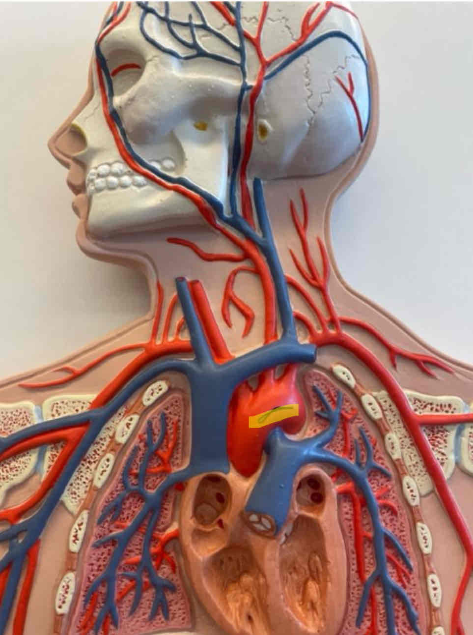
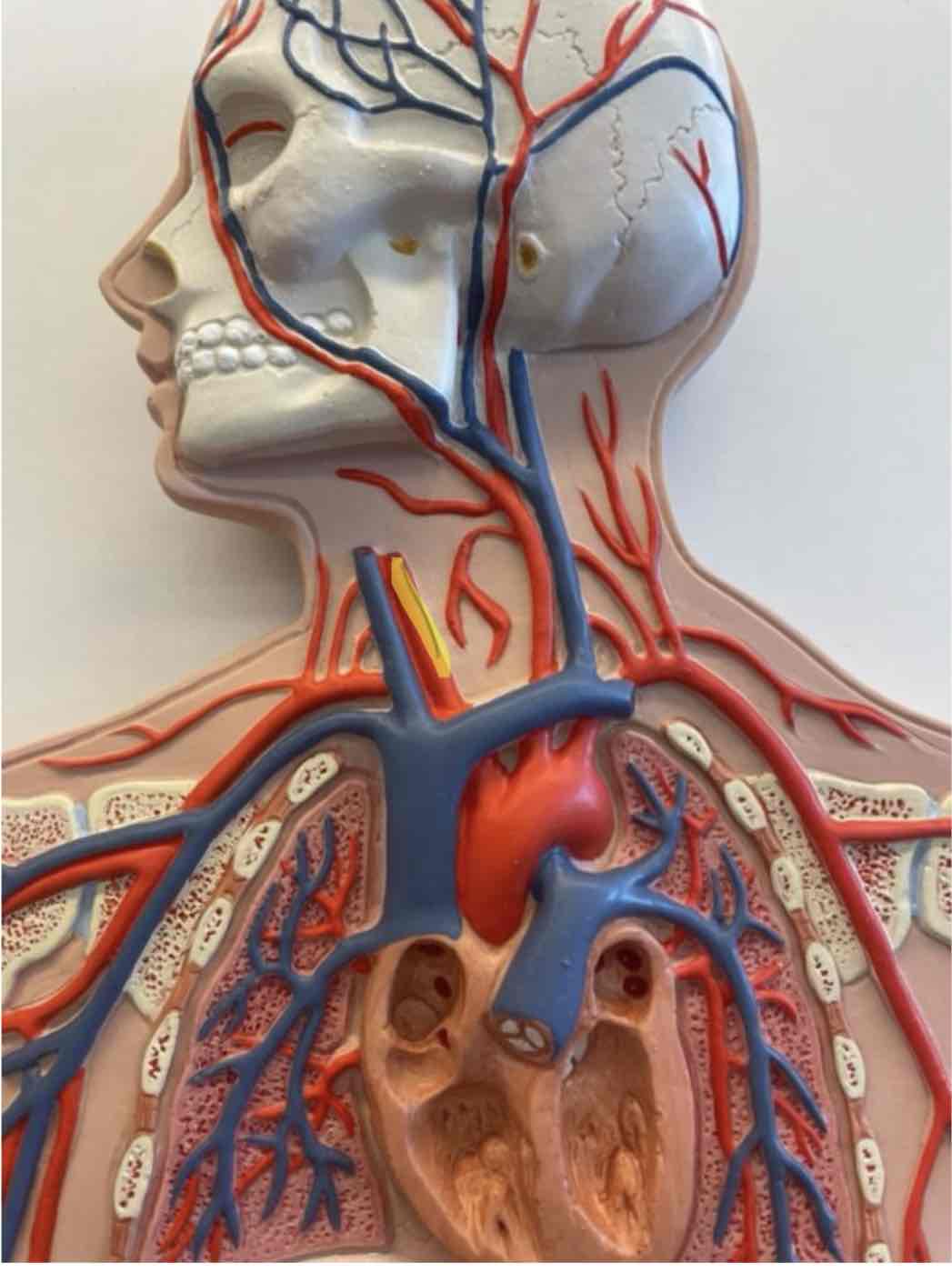
What is the highlighted area
Aortic arch
full body model
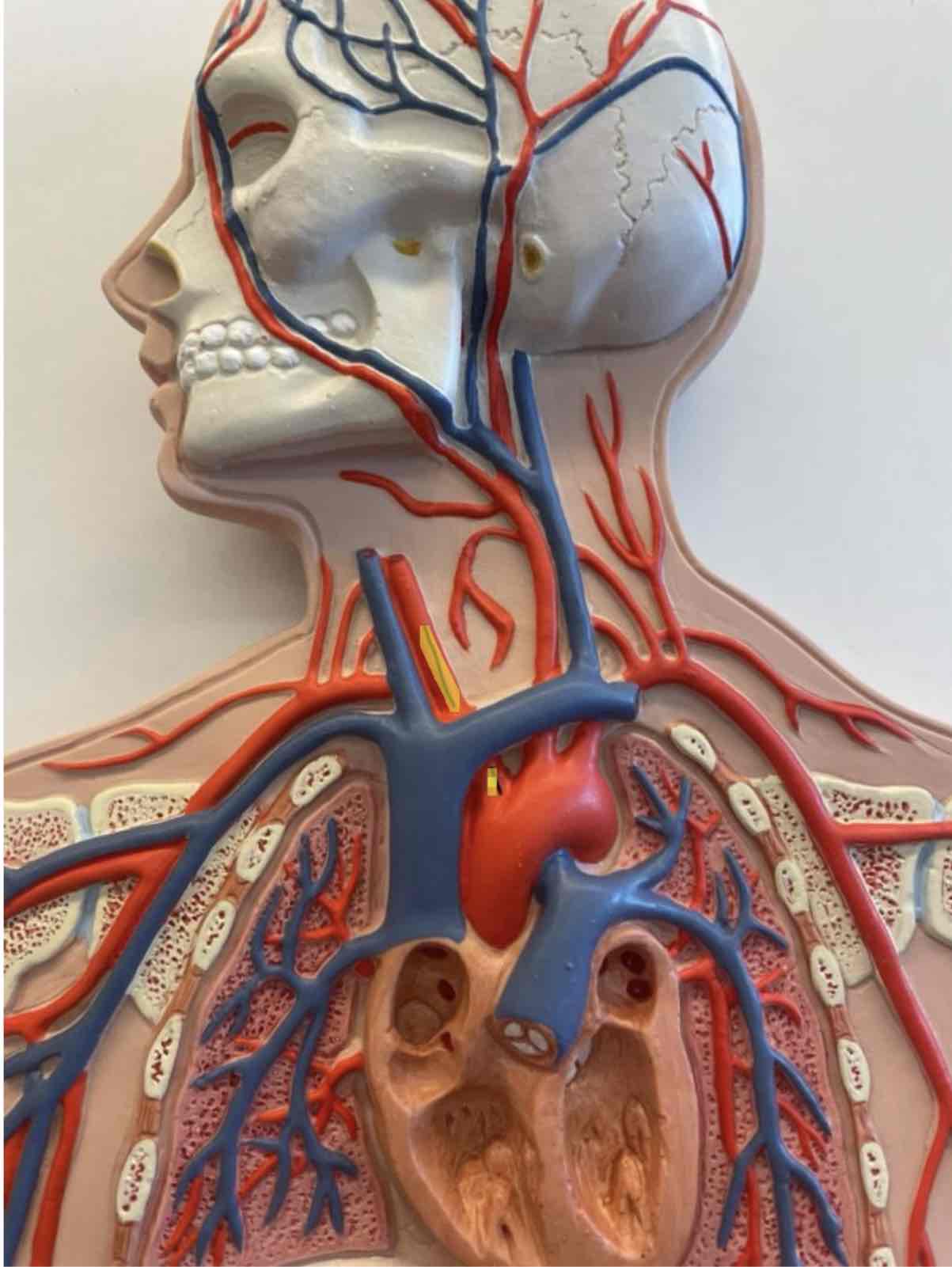
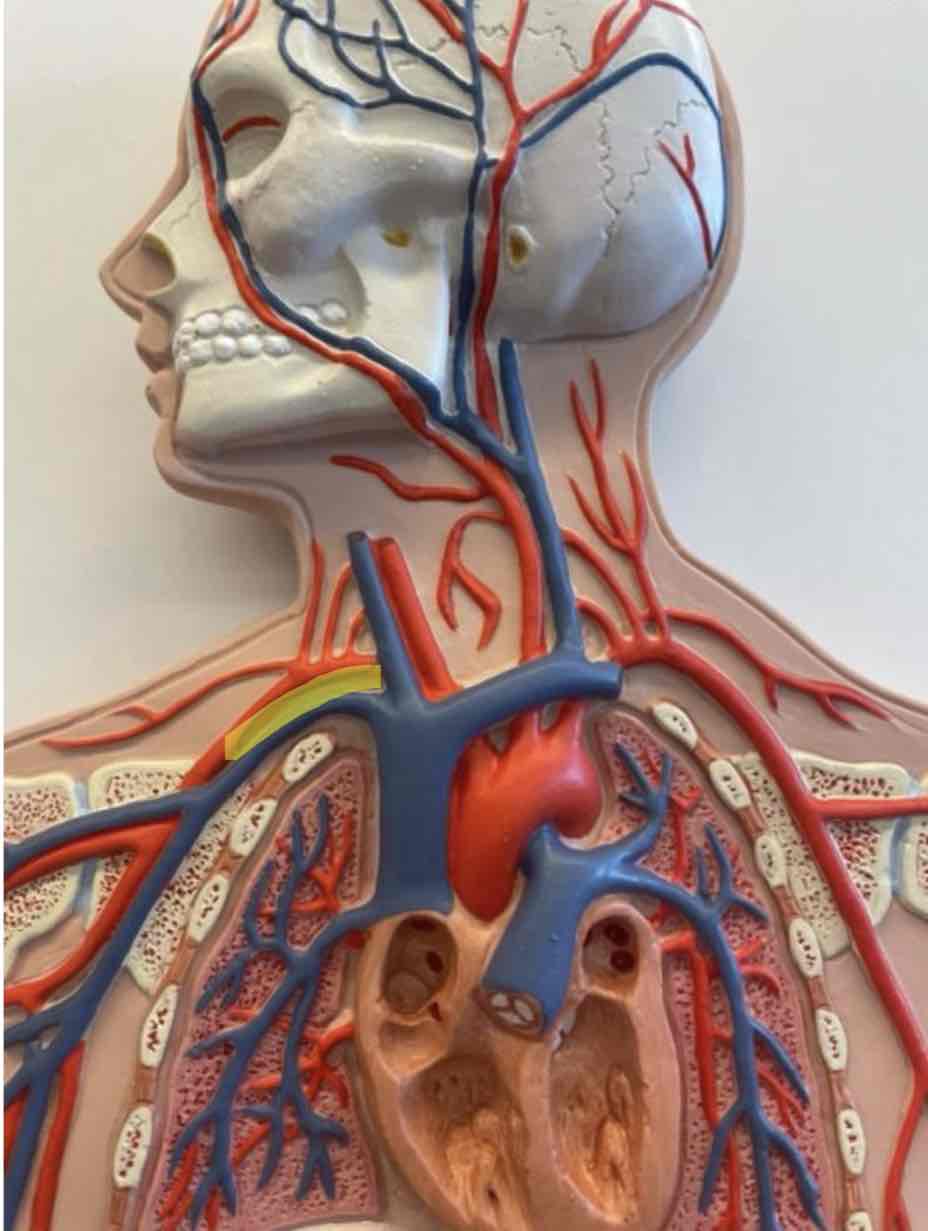
What is the highlighted area
Brachiocephalic trunk
full body model
What is the highlighted area
Left common carticoid artery
full body model
What is the highlighted area
left subclavian
full body model
What is the highlighted area
Right common carticoid artery
full body model
What is the highlighted area
Right subclavian
full body model
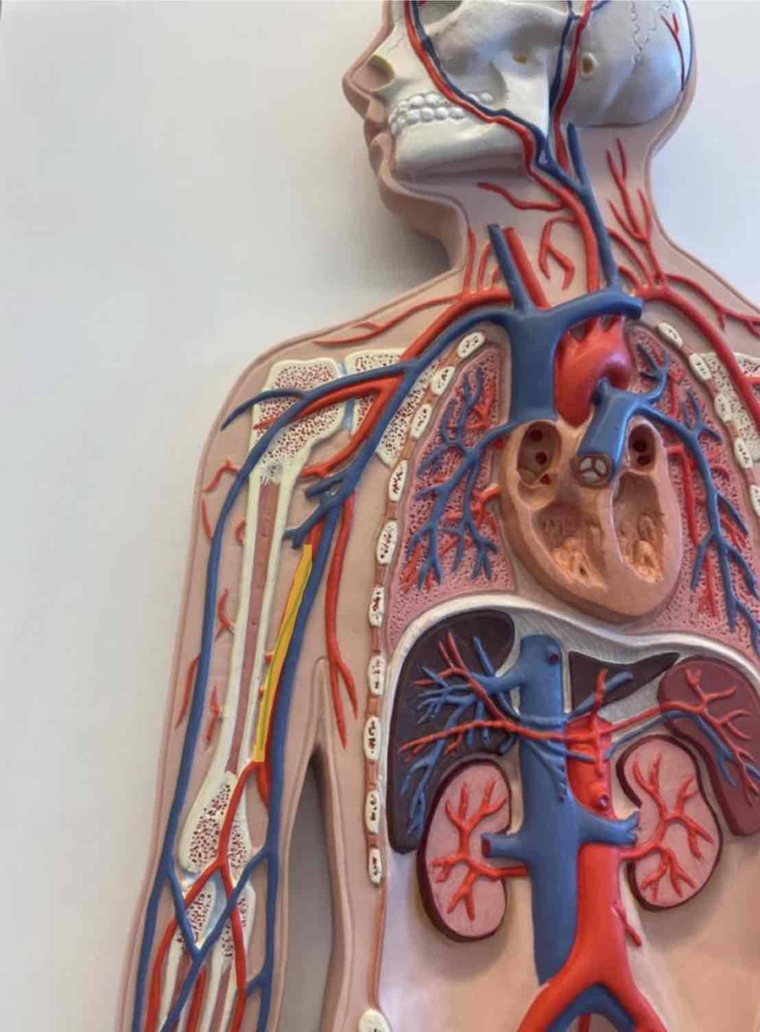
What is the highlighted area
Brachial artery
Full body model
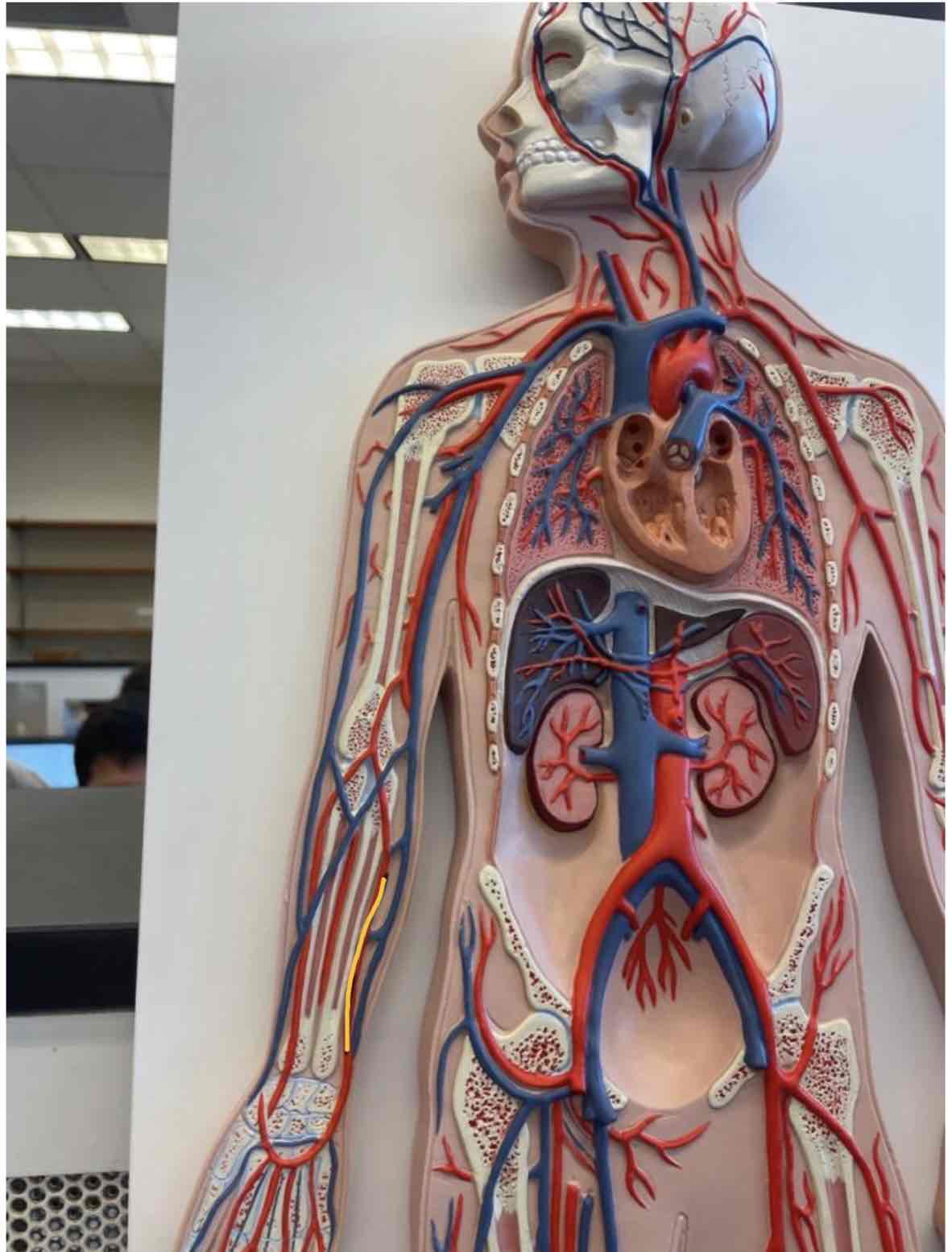
What is the highlighted area
Ulnar artery
Full body model
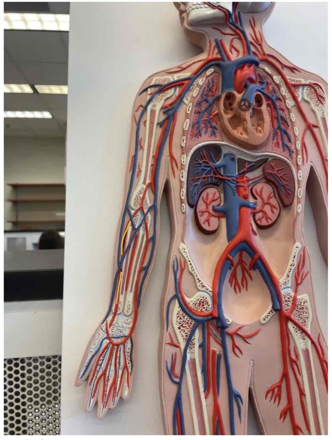
What is the highlighted area
Radial artery
Full body model
What is the highlighted area
Abdominal aorta
full body model
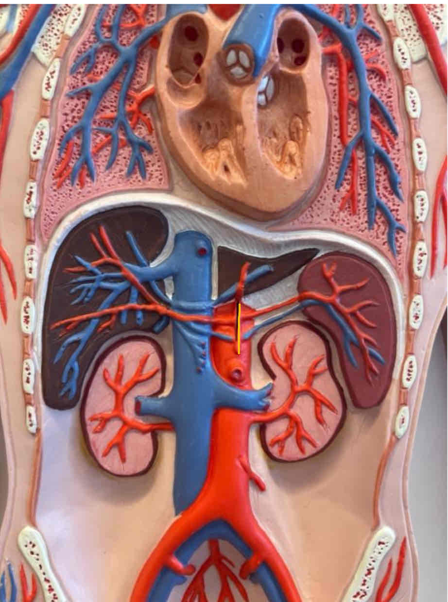
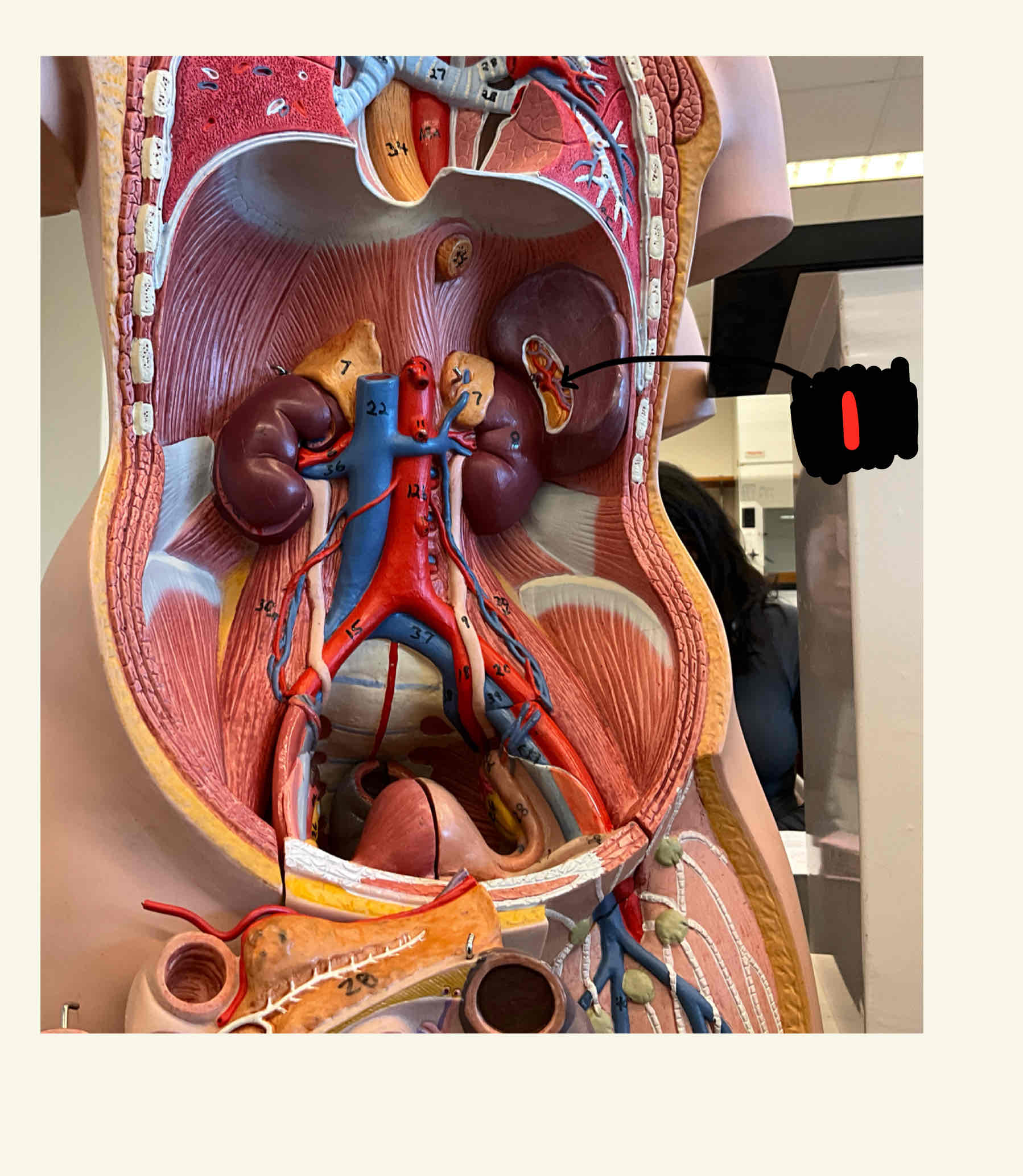
What is the highlighted area
Celiac trunk
full body model

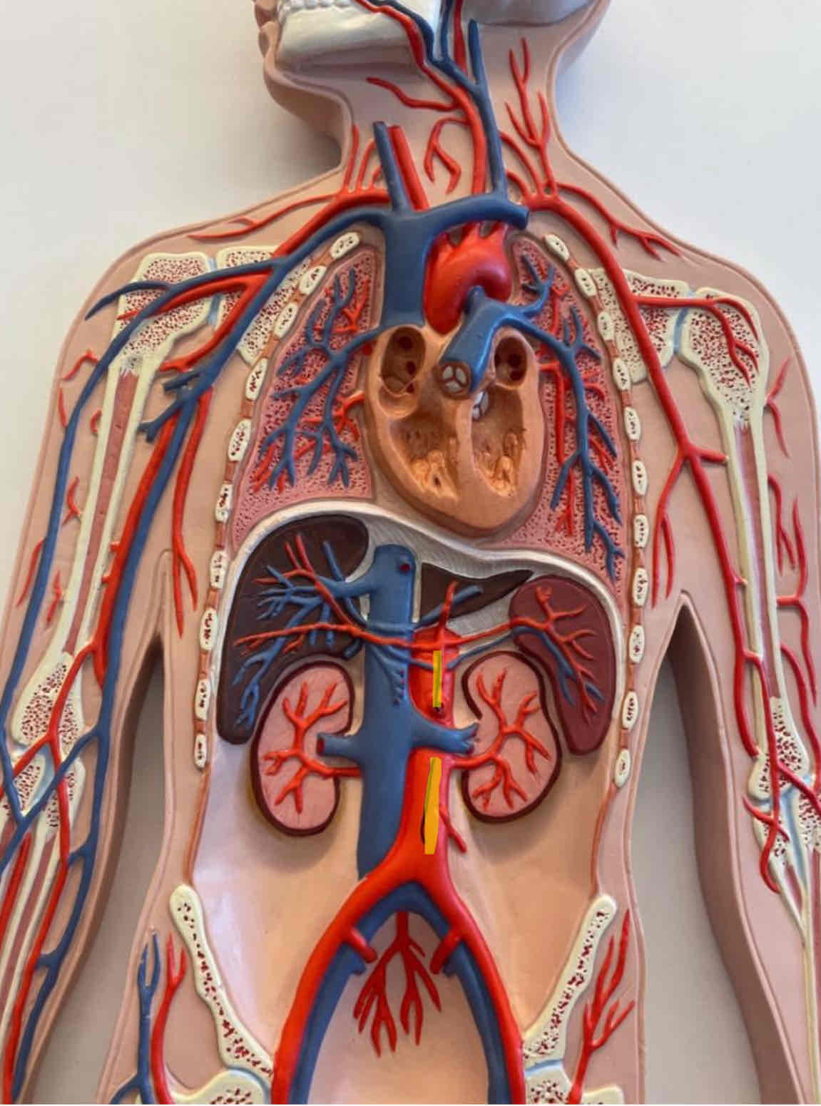
What is the highlighted area
Superior mesenteric artery
full body model
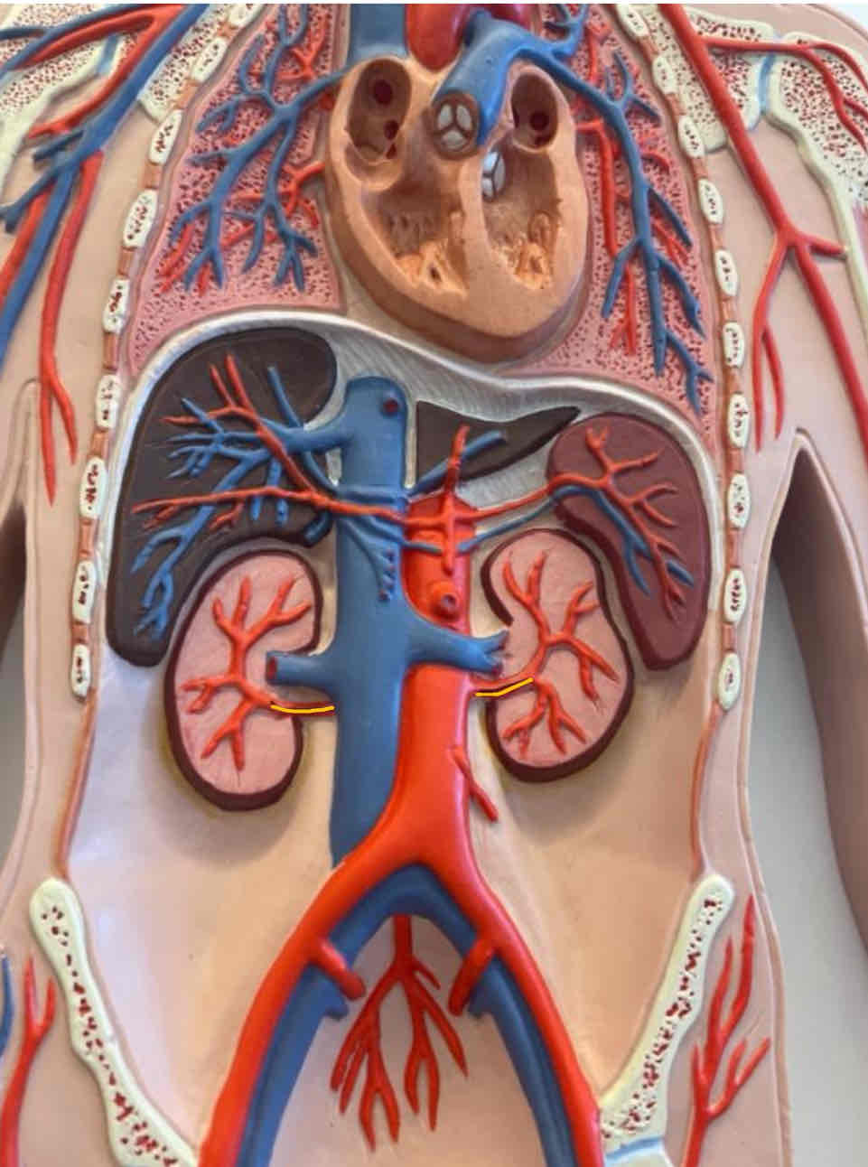
What is the highlighted area
Right and left renal arteries
full body model
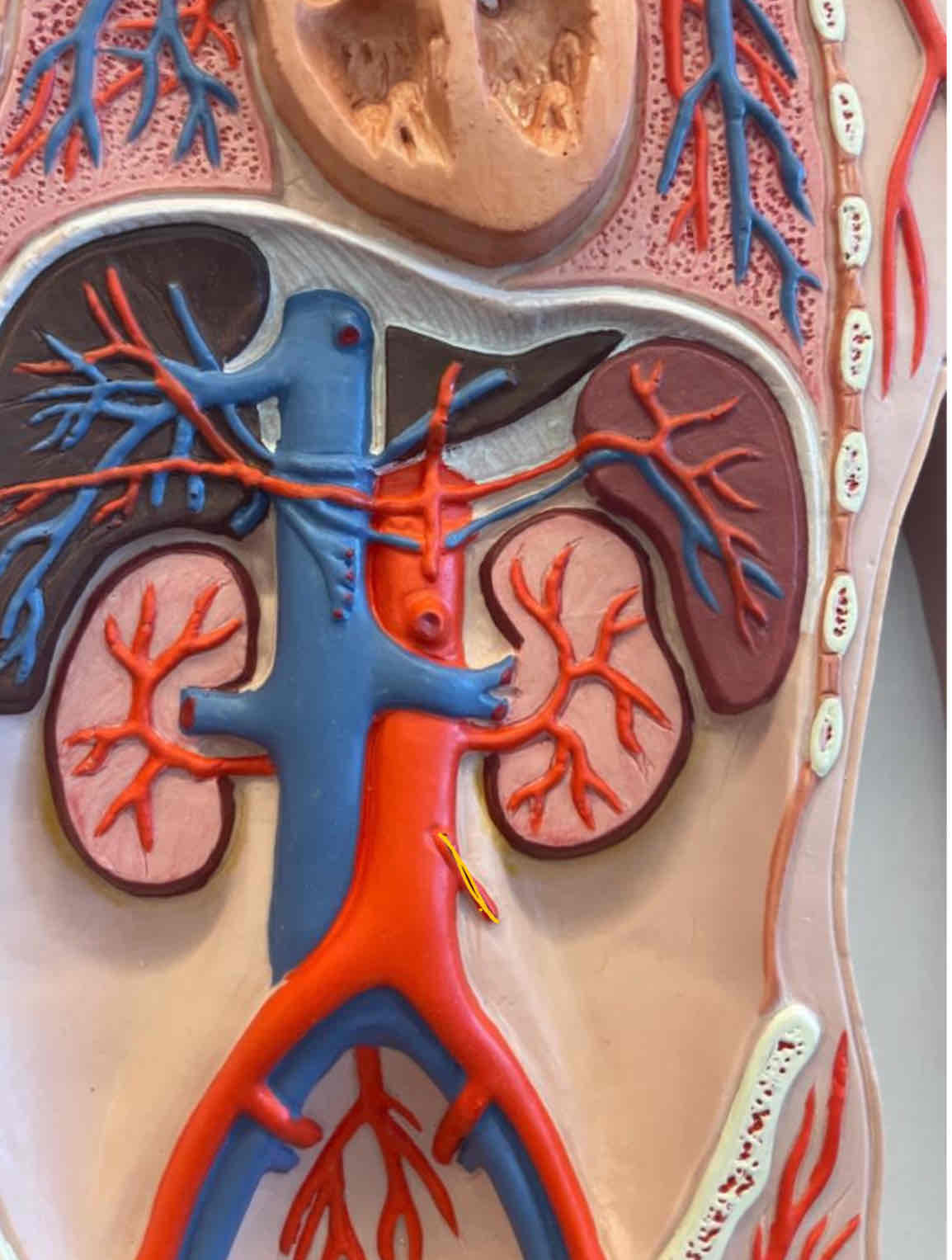
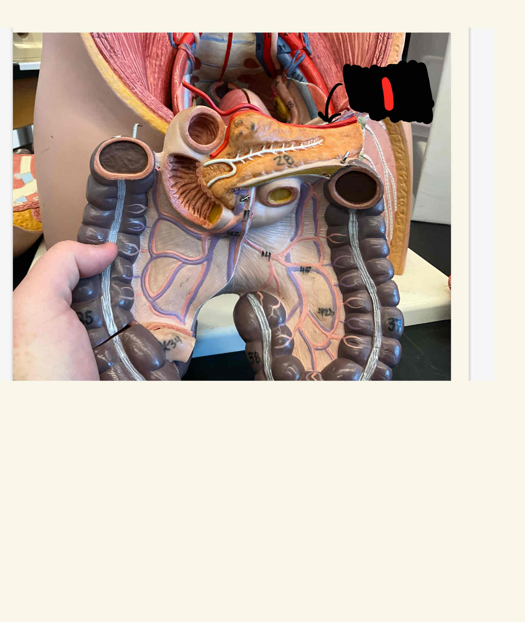
What is the highlighted area
Inferior mesenteric artery
full body model
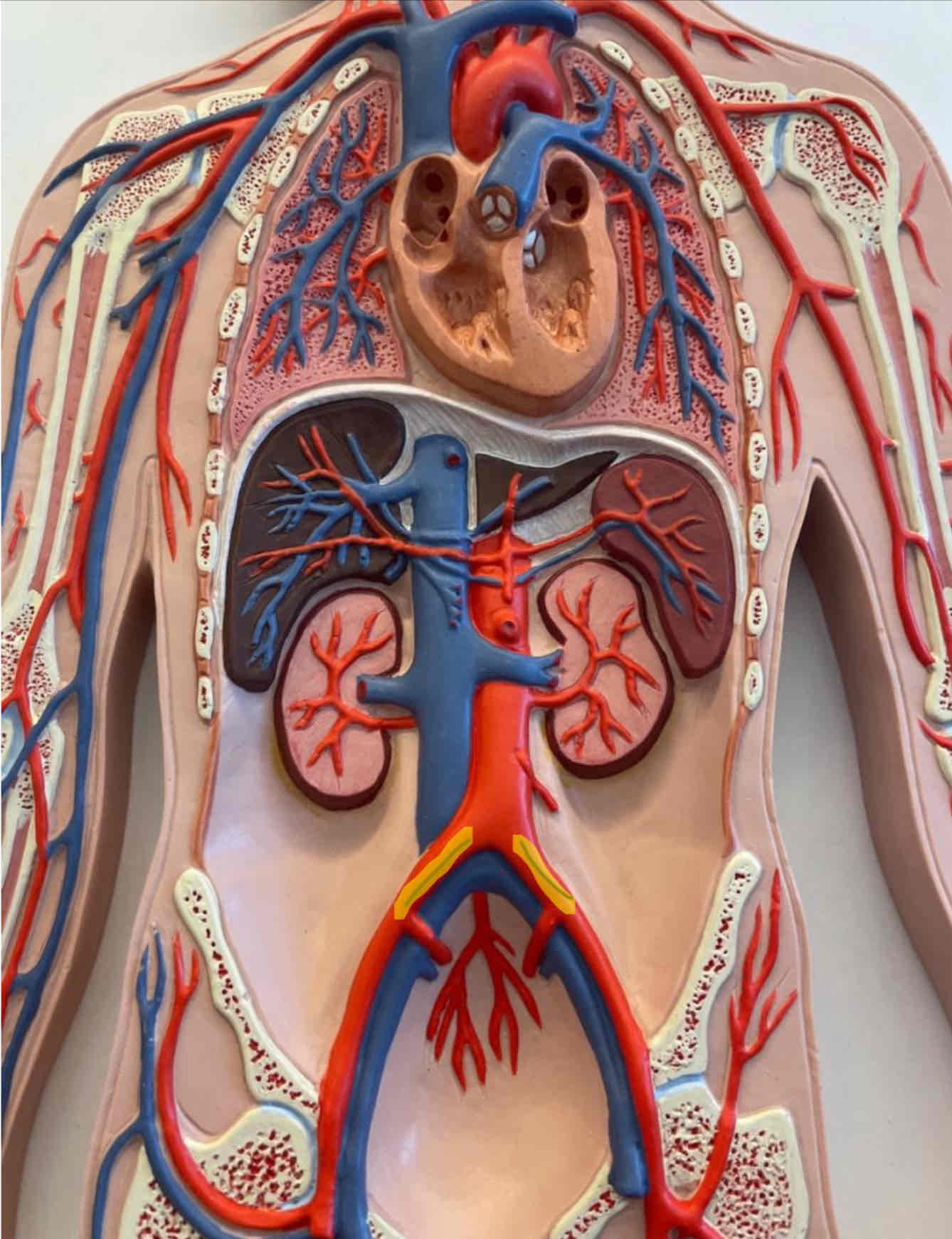
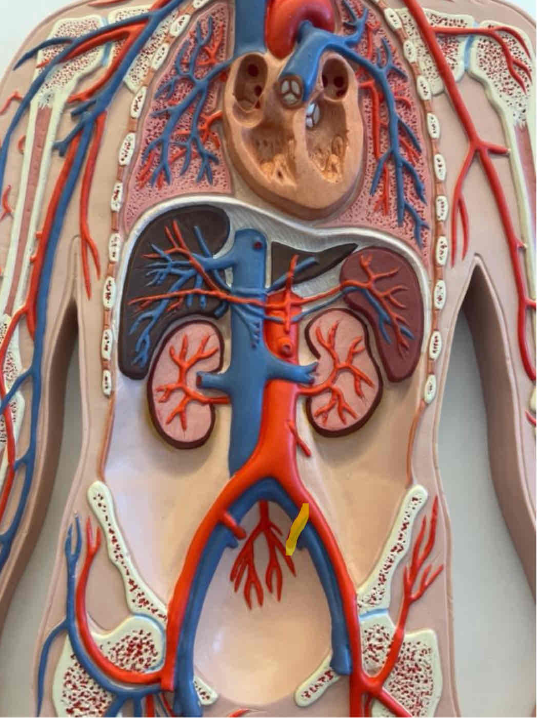
What is the highlighted structure
Right and left common iliac artery
full body model
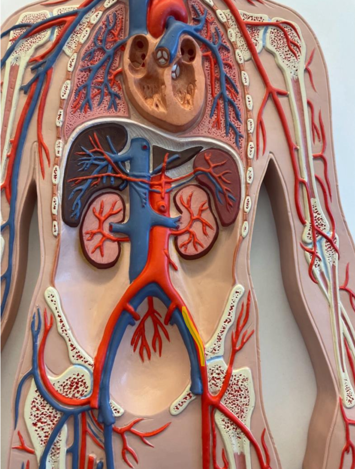
What is the highlighted structure
Left internal iliac artery
Full body model
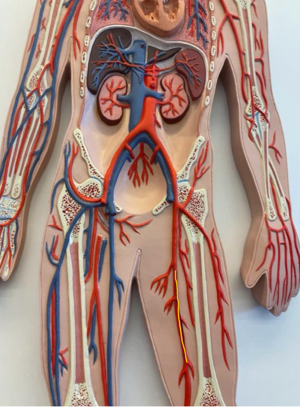
What is the highlighted structure
Left external iliac artery
Full body model
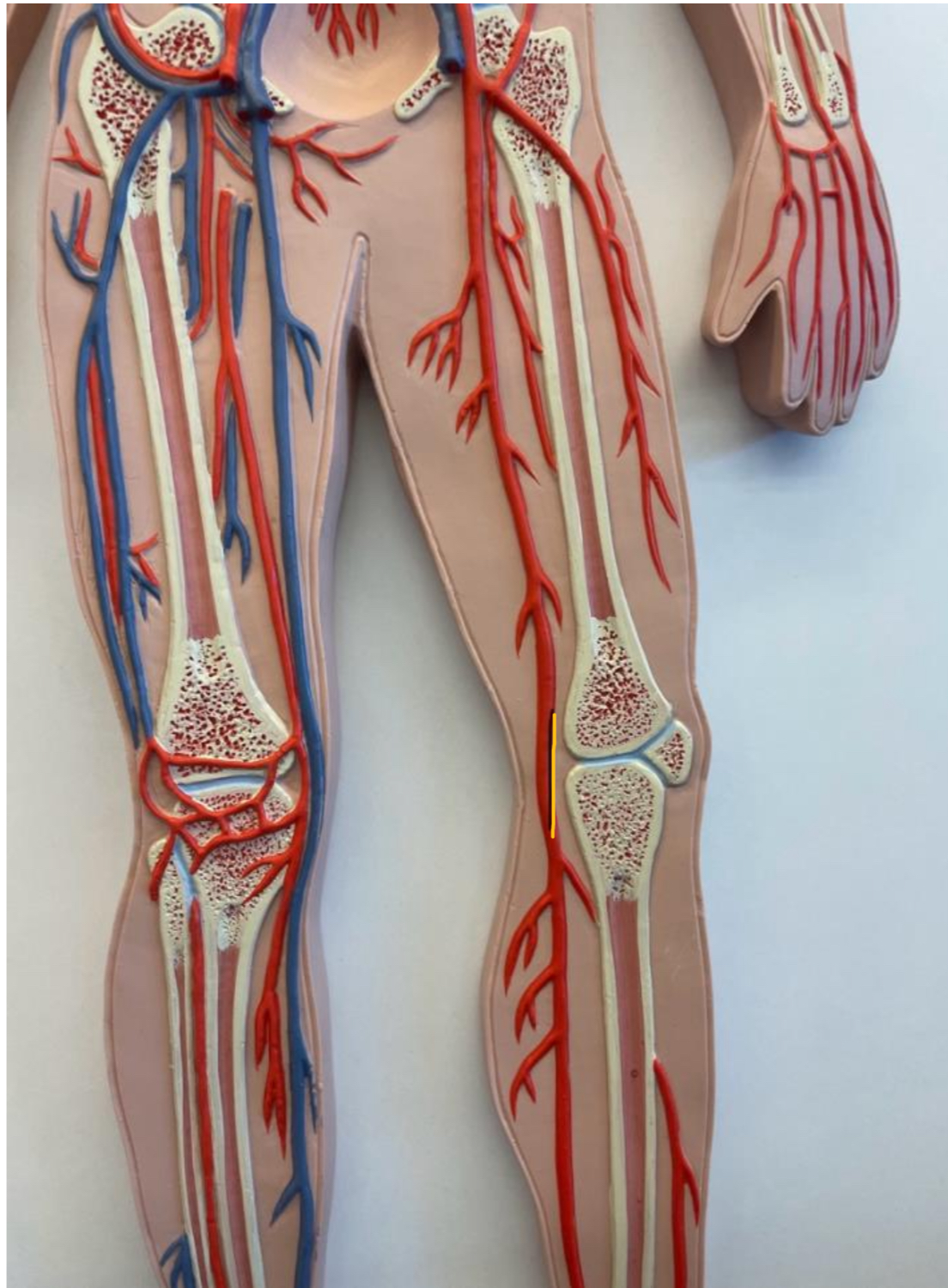
What is the highlighted structure
Femoral artery
Full body model
What is the highlighted structure
Popiterial artery
Full body model
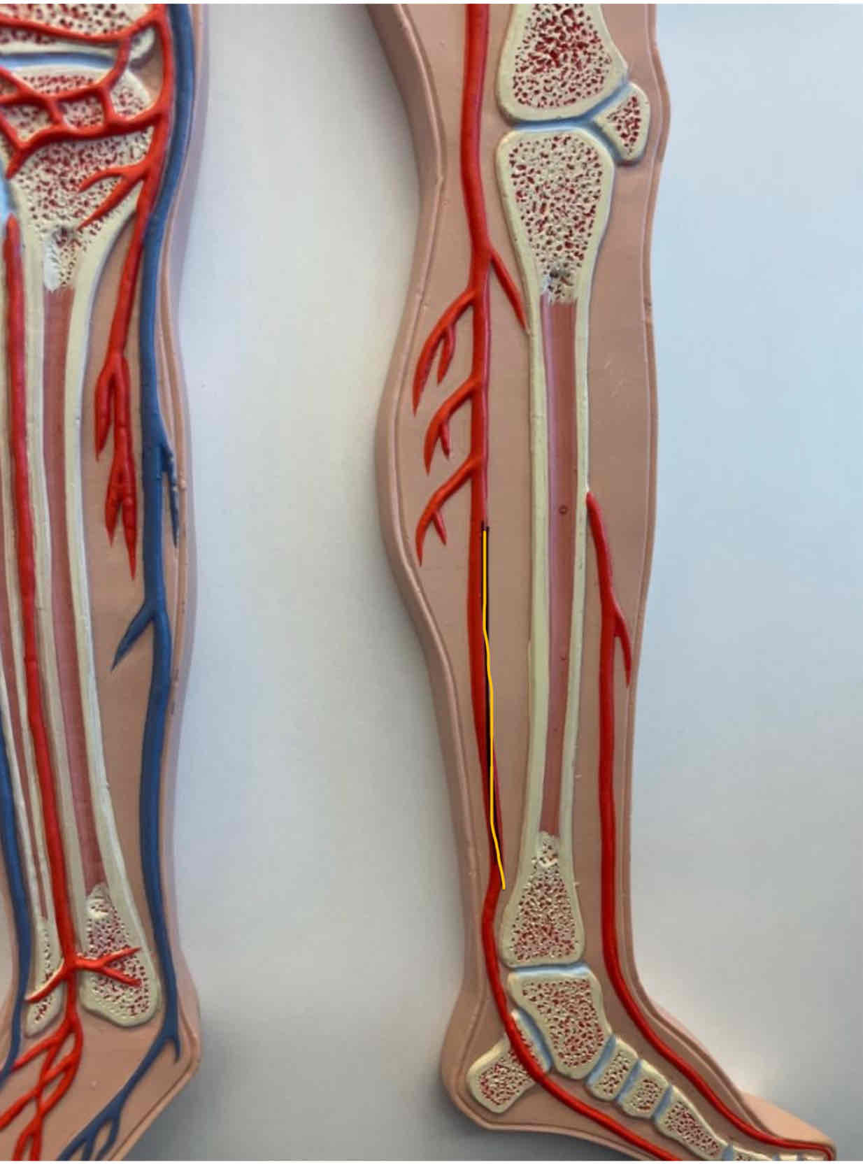
What is the highlighted structure
Posterior tibial artery
Full body model
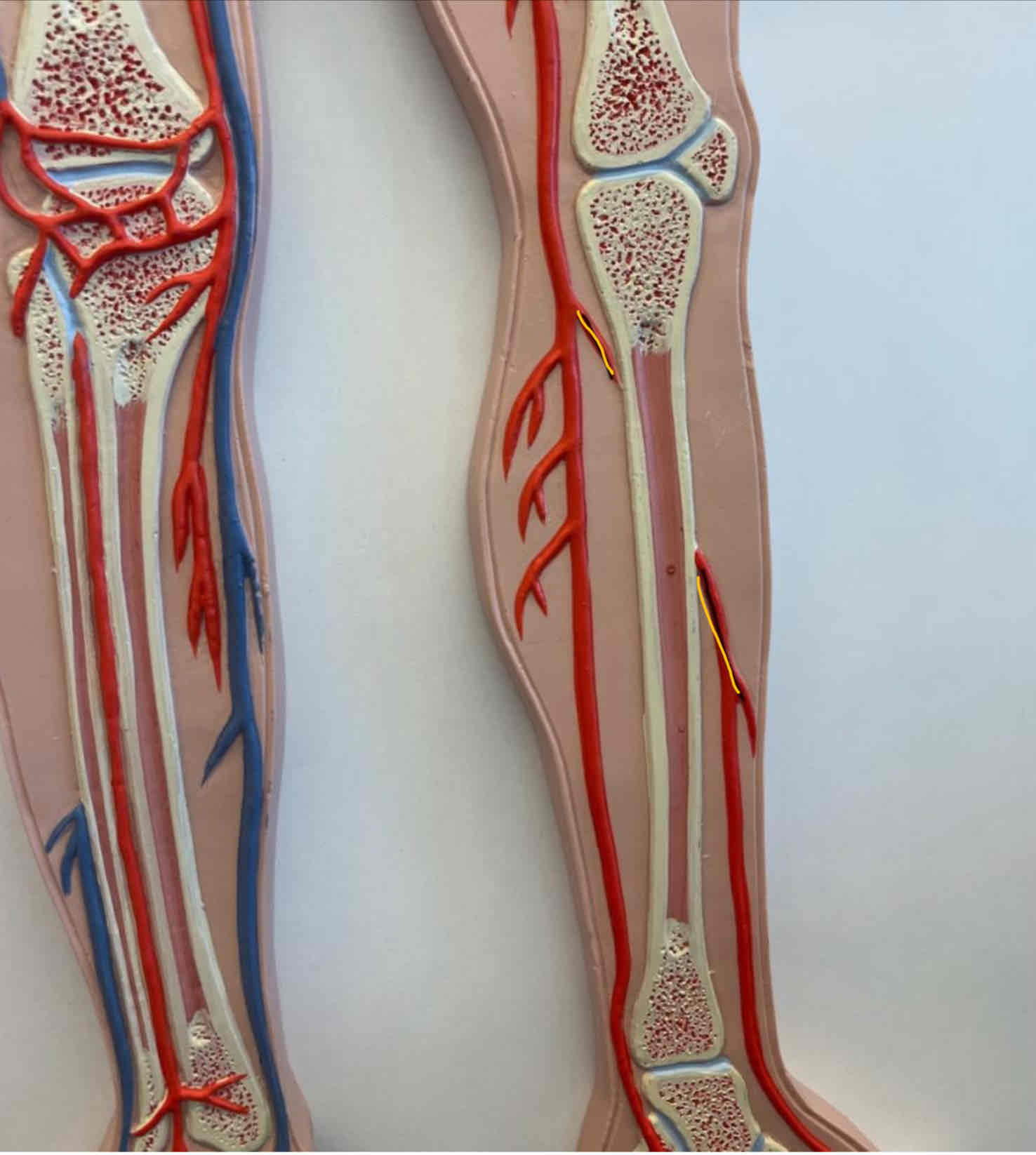
What is the highlighted structure
Anterior tibial artery
Full body model
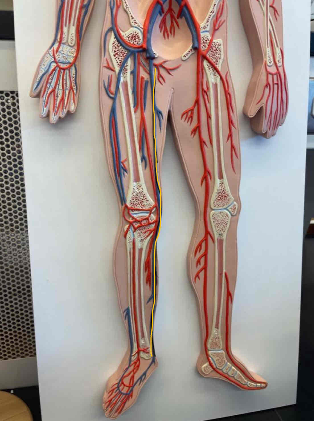
What is the highlighted structure
Great saphenous vein
Full body model