Inner Ear
1/19
There's no tags or description
Looks like no tags are added yet.
Name | Mastery | Learn | Test | Matching | Spaced | Call with Kai |
|---|
No analytics yet
Send a link to your students to track their progress
20 Terms
Inner Ear Anatomy
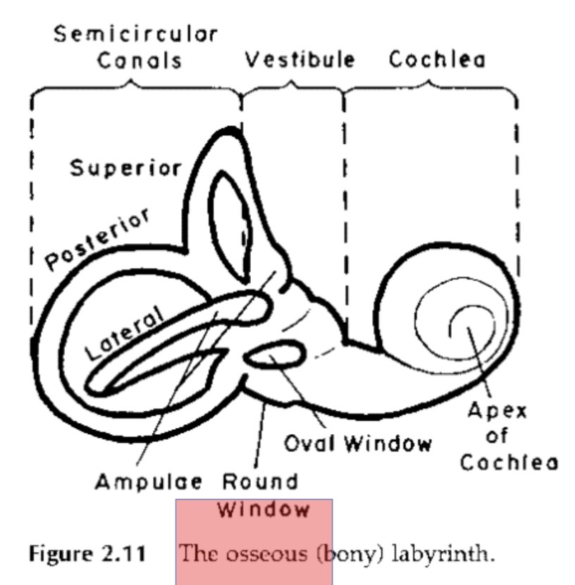
•Bony labyrinth: series of tunnels within which membranous labyrinth is housed
•Filled with perilymph
•Bony so it protects the organs within
•Membranous labyrinth: fluid filled sac within the bony labyrinth, separated by membranes
•Filled with endolymph
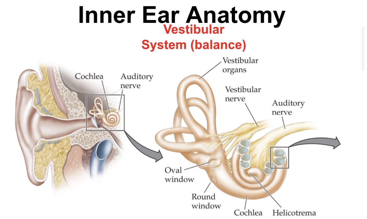
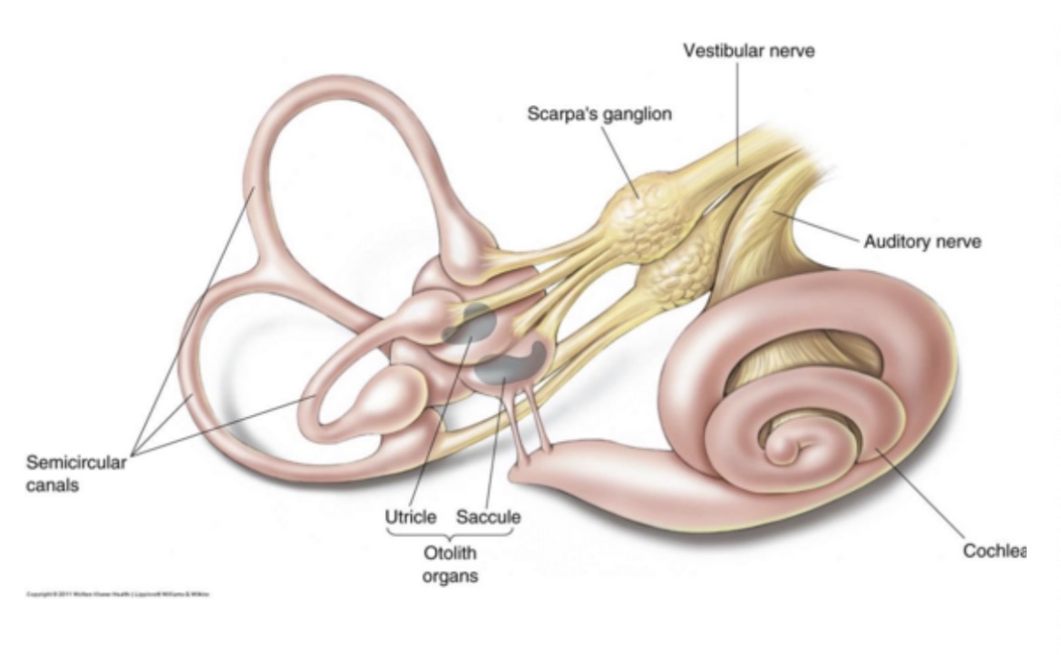
Inner Ear: Vestibular System Anatomy
•The vestibular system is responsible for maintaining balance (in coordination with vision and touch)
•Comprised of two types of end organs:
1. Semicircular canals
•Sense organs for movement of body in space
•Each ear has a superior, posterior, and lateral semicircular canal, at right angles to each other
2. Otolith organs
•Sense organs for acceleration of body through space
•Each ear has a utricle and saccule
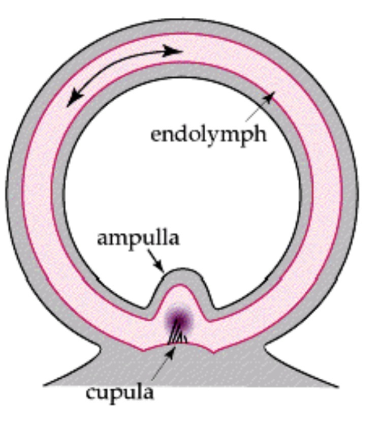
Semicircular canals
Detect angular acceleration (rotation)
•Each SC canal detects motion in a different plane
•Ampulla: contains sensory hair cells that project into a gelatinous mass called the cupula
Otolith organs
Utricle and saccule detect linear acceleration, position relative to gravity
Sensory hair cells project into gelatinous mass covered with calcium carbonate crystals (otoliths)
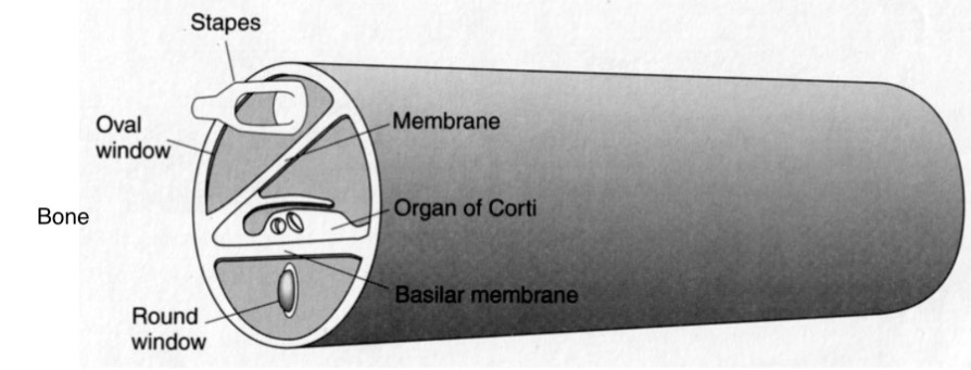
Oval Window
membrane at the cochlea base, connects the middle ear (via the stapes) to the scala vestibuli
Round Window
membrane at the cochlea base, end of the scala tympani, allows for release of fluid pressure
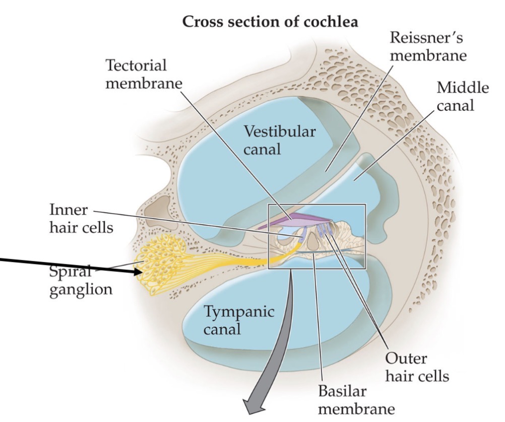
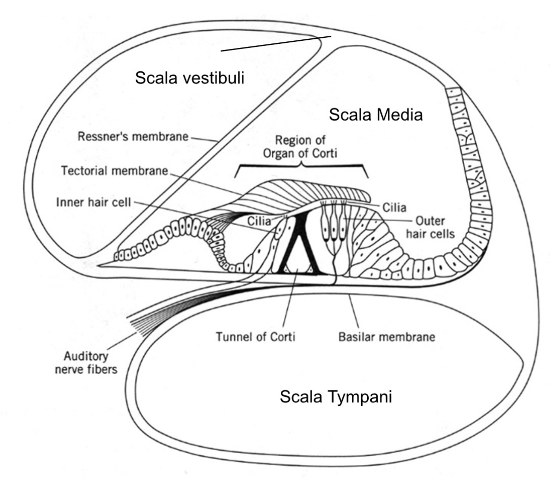
A closer look at the scala media, or cochlear duct
•Reissner’s membrane (roof of SM): separates perilymph of scala vestibuli and endolymph of scala media
•Basilar membrane
(floor of SM): separates perilymph of scala tympani and endolymph of scala media; is the base of the Organ of Corti (box in figure)
Sound transduction to the inner ear
•Sound travels from outer ear to middle ear
•Footplate of the stapes pushes the oval window
•Pressure differential created along the cochlear partition
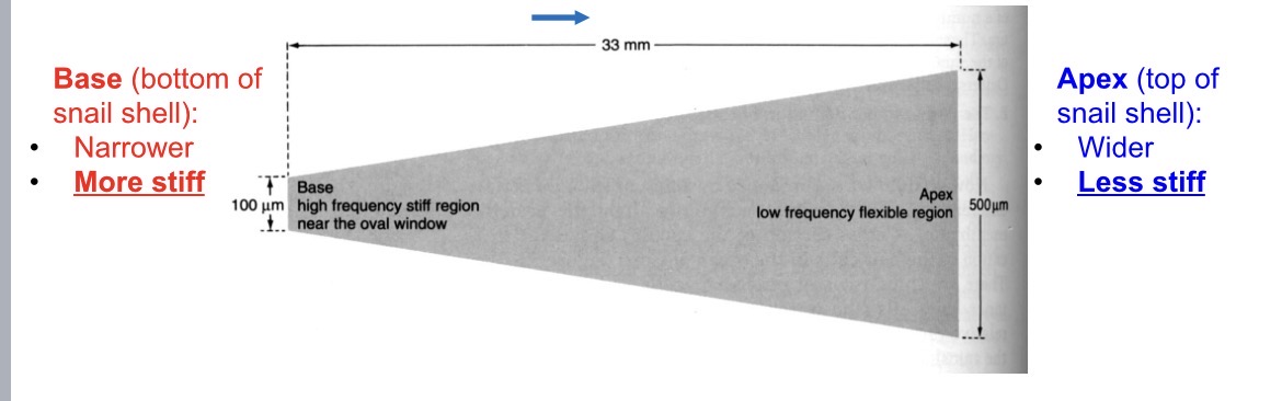
Cochlear Macromechanics
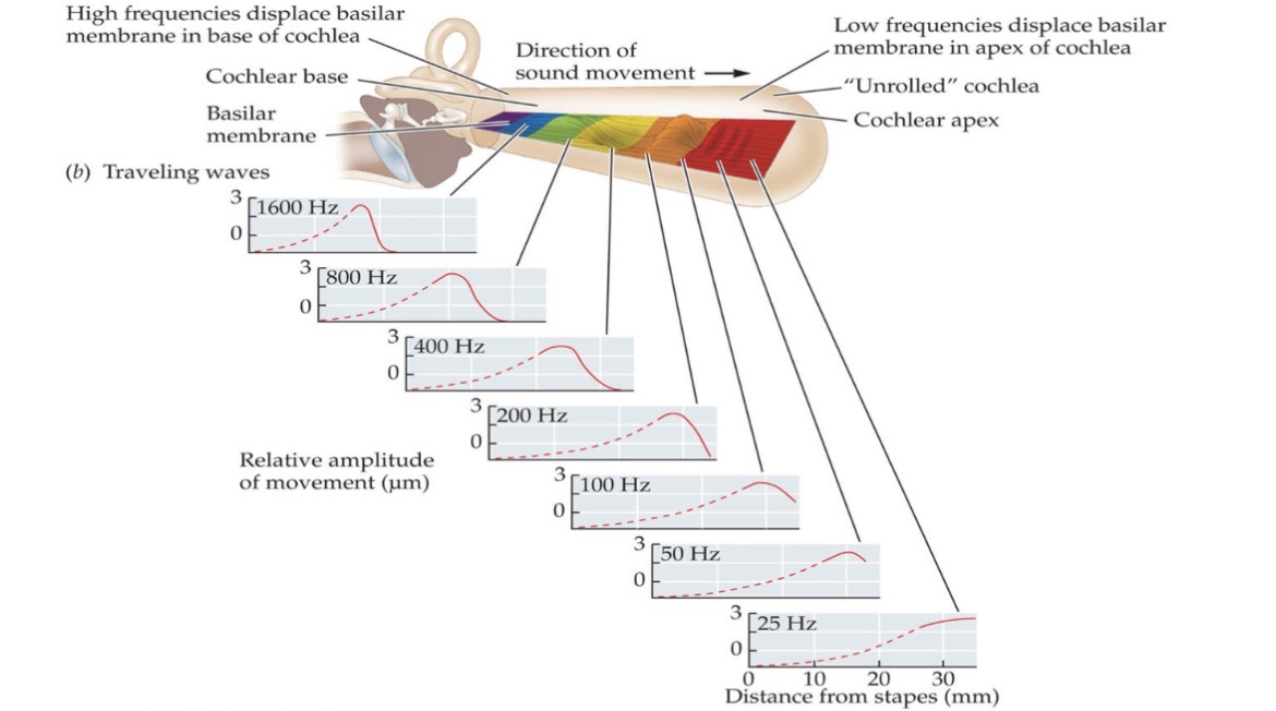
•The basilar membrane varies in thickness and stiffness along the cochlear spiral
•Different frequencies of sound will have maximum displacement (vibration) at different locations (based on resonance)
The cochlea unrolled
•Basic idea: vibrations at oval window cause vibrations in the cochlea. These vibrations cause voltage changes in hair cells in Organ of Corti. The electrical signals are carried up the auditory nerve to the brain.
•The details of the cochlea have been difficult to study because it so small and embedded in hard bone
Cochlear partitions and
the Organ of Corti
•Basilar membrane (floor of SM): separates perilymph of scala tympani and endolymph of scala media and is the base of the Organ of Corti (OoC)
•The OoC contains the sensory cells of the cochlea (i.e. hair cells)
•The auditory nerve (i.e. the 8thcranial nerve, CN VIII) connects to the base of hair cells through the basilar membrane and then exits the cochlea through the modiolus (center of the cochlear spiral)


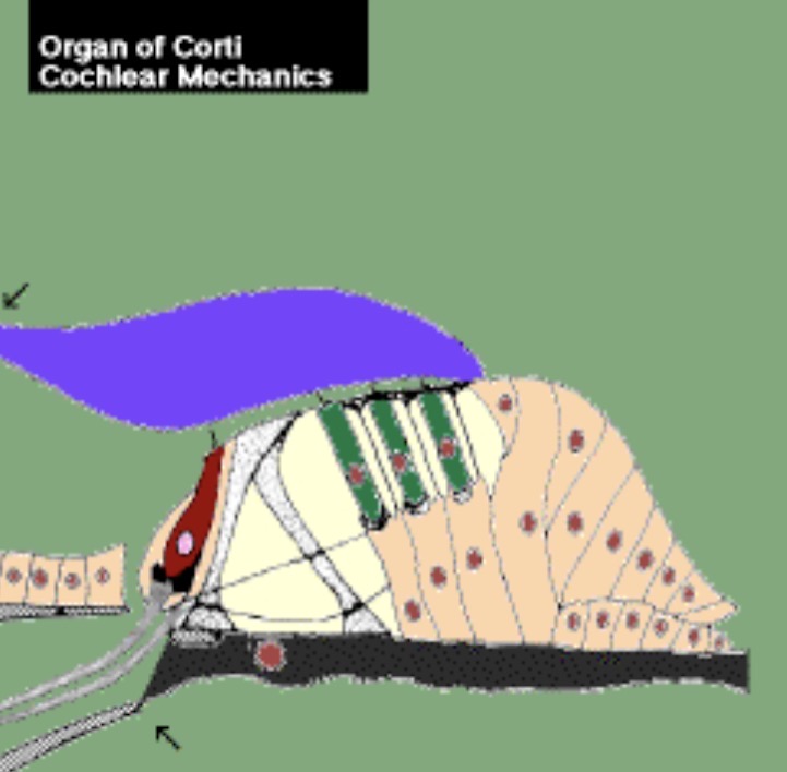
Organ of Corti
•Sits on the basilar membrane (BM)
•The organ of Corti is made up of sensory cells
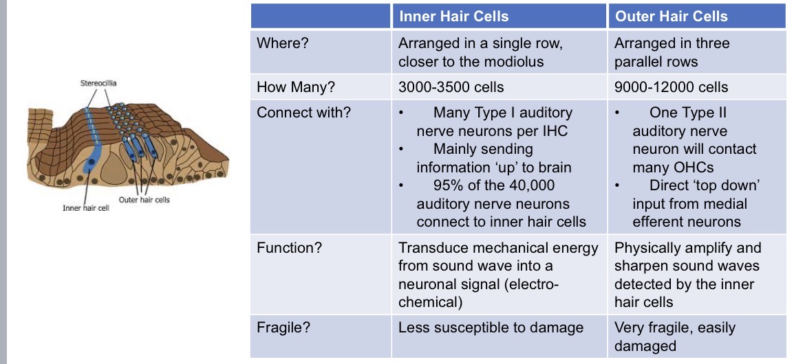
•Inner hair cells
•Outer hair cells
•Supporting cells
•Tectorial membraneoverlays hair cells

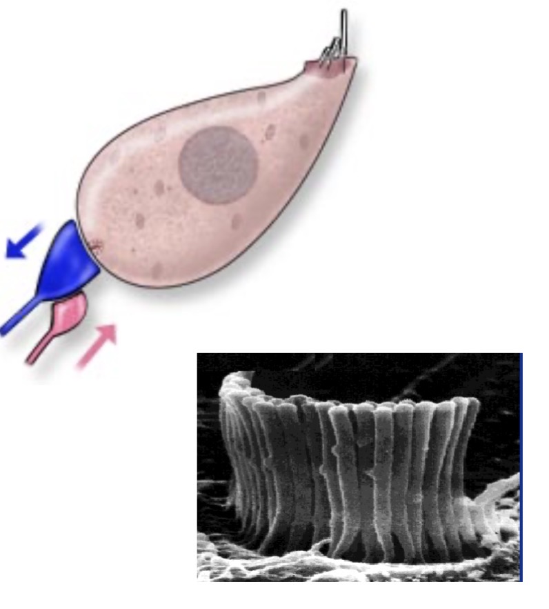
Organ of Corti/Hair Cells
Hair cells sit on top of the basilar membrane (black)
Hair cells are NOT hair!
•They are named after the hair-like projections sticking out of the top of the cells, called stereocilia
•The tectorial membrane (purple) lies above the surface of the hair cells
•There are two types of cochlear hair cells
•Inner Hair Cells (red, closer to modiolus)
•Outer Hair Cells (green)
Inner Hair Cells (IHCs)
•Single row, flask or bean shaped
•Stereocilia are arranged in a linear
or crescent shape
•Stereocilia are not embedded in the tectorial membrane
•Send information about sound to the auditory nerve (IHCs are the primary sensory cells of the cochlea)
•Required for sound detection
•~3500 in human cochlea
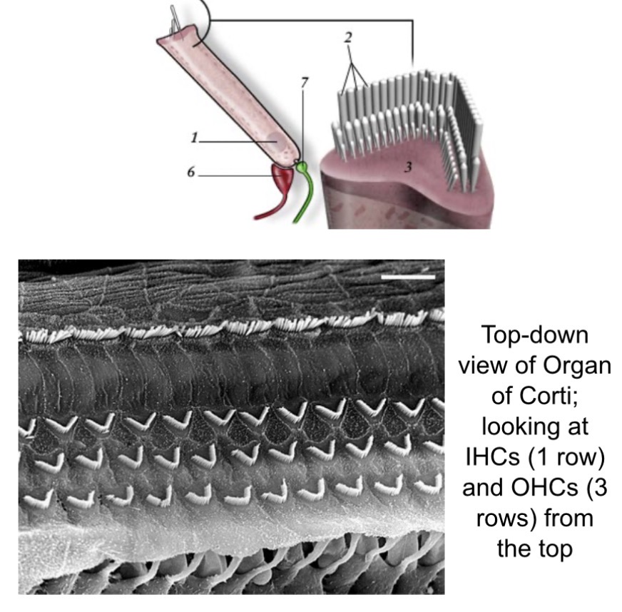
Outer Hair Cells (OHCs)
•3 rows, test-tube shaped
•Stereocilia are arranged in a U, V, or W pattern
•Stereocilia are embedded in the tectorial membrane
•Change stiffness and length with voltage – “cochlear amplifier”
•Improve sound detection and frequency selectivity
•~12,000 in human cochlea
Each place along the basilar membrane corresponds to a specific frequency
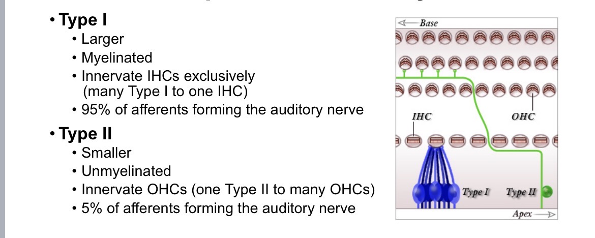
There are two types of afferent neurons that comprise the auditory nerve
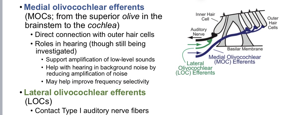
There are two types of cochlear efferent neurons
Prestin: The protein that makes outer hair cells move
•When an outer hair cell is depolarized (i.e., excited) by stereocilia deflections, prestin causes the outer hair cell to shorten
•When the outer hair cell is hyperpolarized (i.e., inhibited) by stereocilia deflections in the opposite direction, prestin causes the outer hair cell to lengthen
•Alternating patterns of contraction and extension = OHC movement amplification of basilar membrane movements
Summary of differences between hair cell types