IB SEHS Exam paper 1 and 2
1/196
There's no tags or description
Looks like no tags are added yet.
Name | Mastery | Learn | Test | Matching | Spaced | Call with Kai |
|---|
No analytics yet
Send a link to your students to track their progress
197 Terms
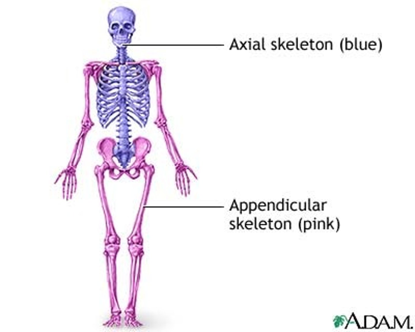
Distinguish anatomically between the axial and appendicular skeleton
AXIAL
•skull, spine, ribs, and sternum
•provides central support, attachment points, and protects nervous system
APPENDICULAR
•includes appendages of the body
-shoulders, arms, hips, and legs
•provides movement and appendages
state the 4 types of bone
1. Long (humerus)
2.Short (carpals)
3. Flat (parietal of skull)
4. Irregular (vertebrae)
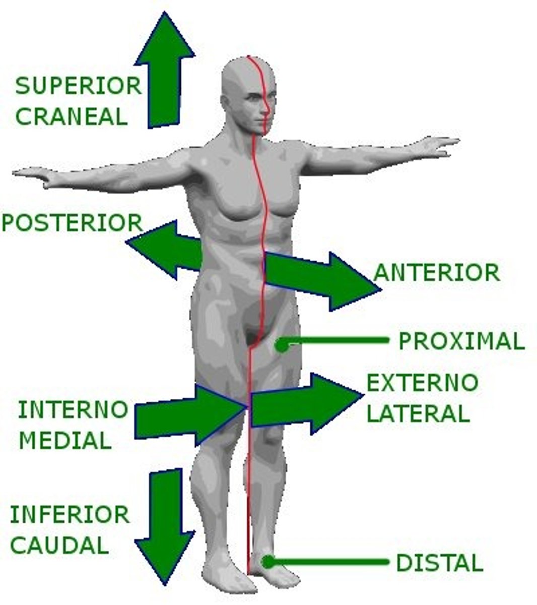
Apply anatomical terminology to the location of bones
•inferior
-below or further away from the head
•superior
-above or nearer to the head
•proximal
-nearer to where a limb attaches to the body
•distal
-further away from where a limb attaches to the body
•posterior
-behind or nearer to the back
•anterior
-in front or nearer to the front
•external
-on or near the surface of the body
•lateral
-further away from the midline of the body
•medial
-closer to the midline of the body
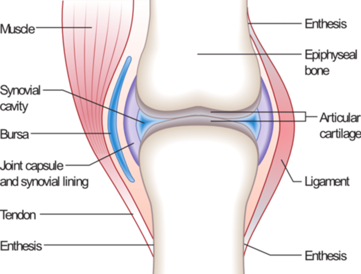
ID and Outline the functions of connective tissue
•Tendons
-connect muscles to bones
•ligaments
-tough, elastic fibers that link bones to bones
•cartilage
-prevents ends of bones from rubbing together
-lubricates joint
Define the term joint
•place where two or more bones meet
•provides movement
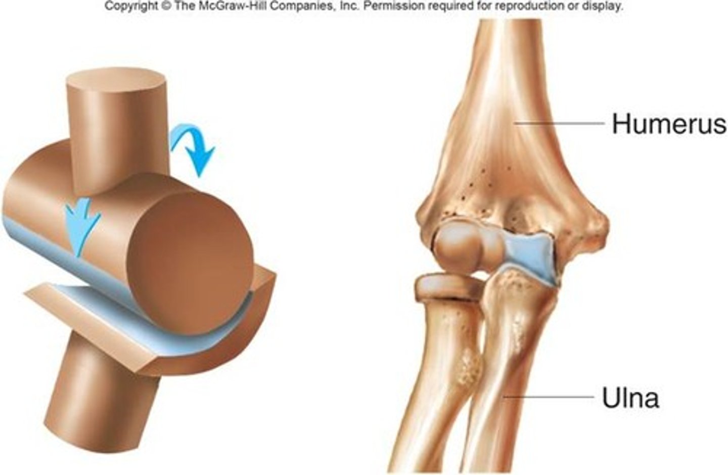
what are the types of joints
•immovable (fixed)
-skull
•slightly movable (cartilaginous)
-limbro-sacral vertebrae
•movable (synovial)
-knee
Outline the features of a synovial joint
•articular cartilage covers the ends of bones
•joint surfaces are enclosed by a fibrous articular capsule
•the joint cavity is filled with synovial fluid
•ligaments reinforce the joint
•90% of joints are synovial
•freely movable
•contain synovial fluid which is in the synovial membrane. Lubricates joints and absorbs shock
•all moving parts are held together by ligaments
•highly mobile
BURSAE
•flattened fibrous sacs
-lined with synovial membranes
-filled with synovial fluids
-not actually part of the joint
TENDON SHEATH
•elongated bursa that wraps around the tendon
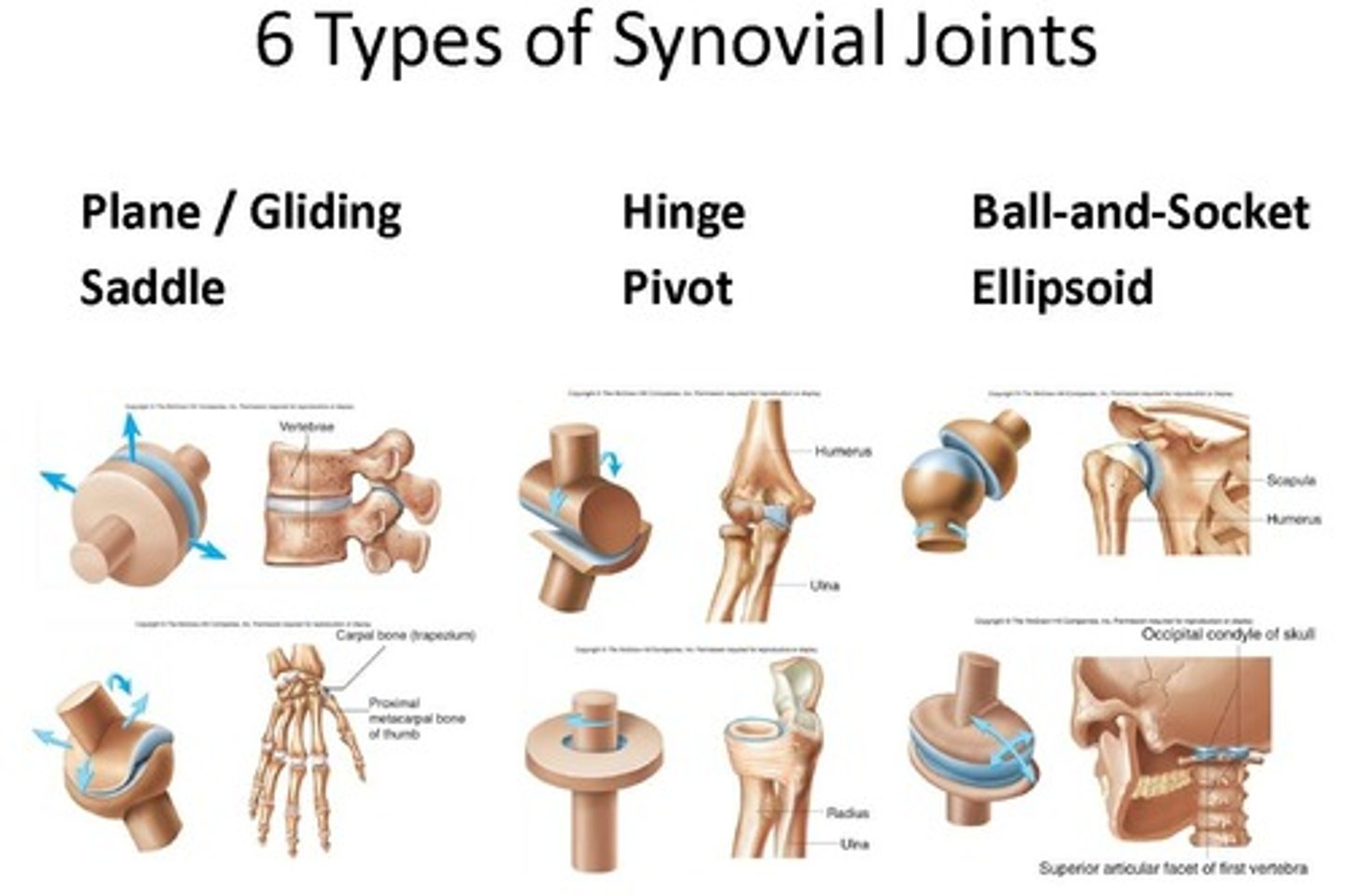
List the different types of synovial joints and Distinguish between the different types of joint in relation to movement permitted.
BALL AND SOCKET
•rounded end of the bone fits inside the cup-shaped ending
•allow movement in all directions and rotation
•most mobile joints in body
ex/ shoulders and hips
PIVOT
•have a ring of bone that fits over a bone protrusion, around which it can rotate
•only allows rotation
ex/ joint between atlas and axis in neck
CONDYLOID
•have an oval shaped bone end which fits into a correspondingly shaped bone end
•forward, backward, left, and right
•no rotation
ex/ between metacarpals and phalanges in hand
GLIDING
•have two flat faces of bone that slide over one another
ex/between tarsals in the ankle
SADDLE
•ends of two bones fit together in a special way
•forwards, backwards, and left to right rotation
ex/ thumb
HINGE
•only forwards and backwards movement
ex/ knee and elbow
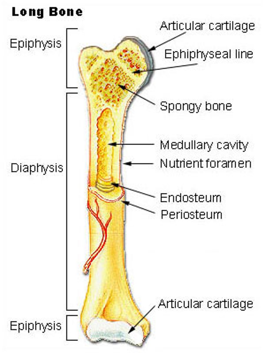
Draw and annotate the structure of a long bone.
*look at diagram quizlet
DIAPHYSIS
•shaft
•composed of compact bone
EPIPHYSIS
•proximal and distal
•ends of bone
•composed mostly of spongy bony
PERIOSTEUM
•outside covering of the diaphysis
•fibrous connective tissue membrane
ARTERIES
•supply bone cell with nutrients
-compact bone+bone marrow
ARTICULAR CARTILAGE
•covers the external surface of the epiphysis
•made of hyaline cartilage
•decreases friction at joint
MEDULLARY CAVITY
•cavity of the shaft
•yellow marrow in adults
-found in hollow interior of diaphysis
•contains red marrow (for cell formation) in infants
ENDOSTEUM
•covers medullary cavity
•delicate membros lining
YELLOW BONE MARROW
•store triglycerides that can serve as an energy source
Outline the general characteristics common to muscle tissue
CONTRACTIBILITY
•ability to receive and respond to stimuli via generation of an electrical pulse which causes contraction of the muscle cells
EXCITABILITY
•ability to shorten
EXTENSIBILITY
•ability to lengthen
ELASTICITY
•ability to return to normal size
ATROPHY
•wasting of muscle tissue
HYPERTROPHY
•increase in size of muscle tissue
•controlled by nerve stimuli
•fed by capillaries
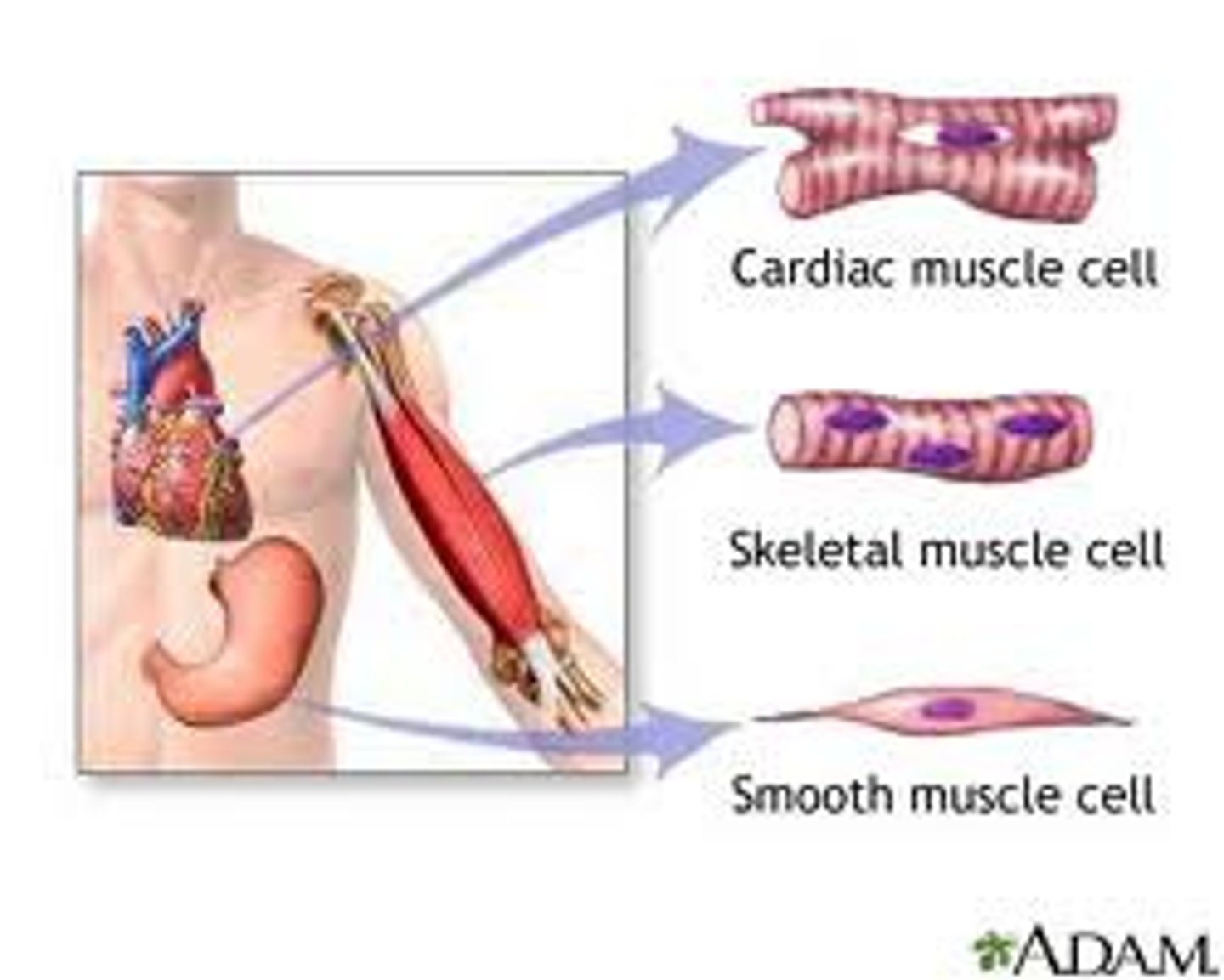
Distinguish between the different types of muscle
SKELETAL
•striated and voluntary
•attach to bones
•contract to facilitate movement
striated- appearance of light and dark stripes
SMOOTH
•unstriated
•involuntary due to our inability to control its movement
•found in walls of hollow organs such as stomach, esophagus, bronchi, and blood vessels
CARDIAC
•striated, tubular, branched, uninucleate fibers
•walls of heart
•involuntary
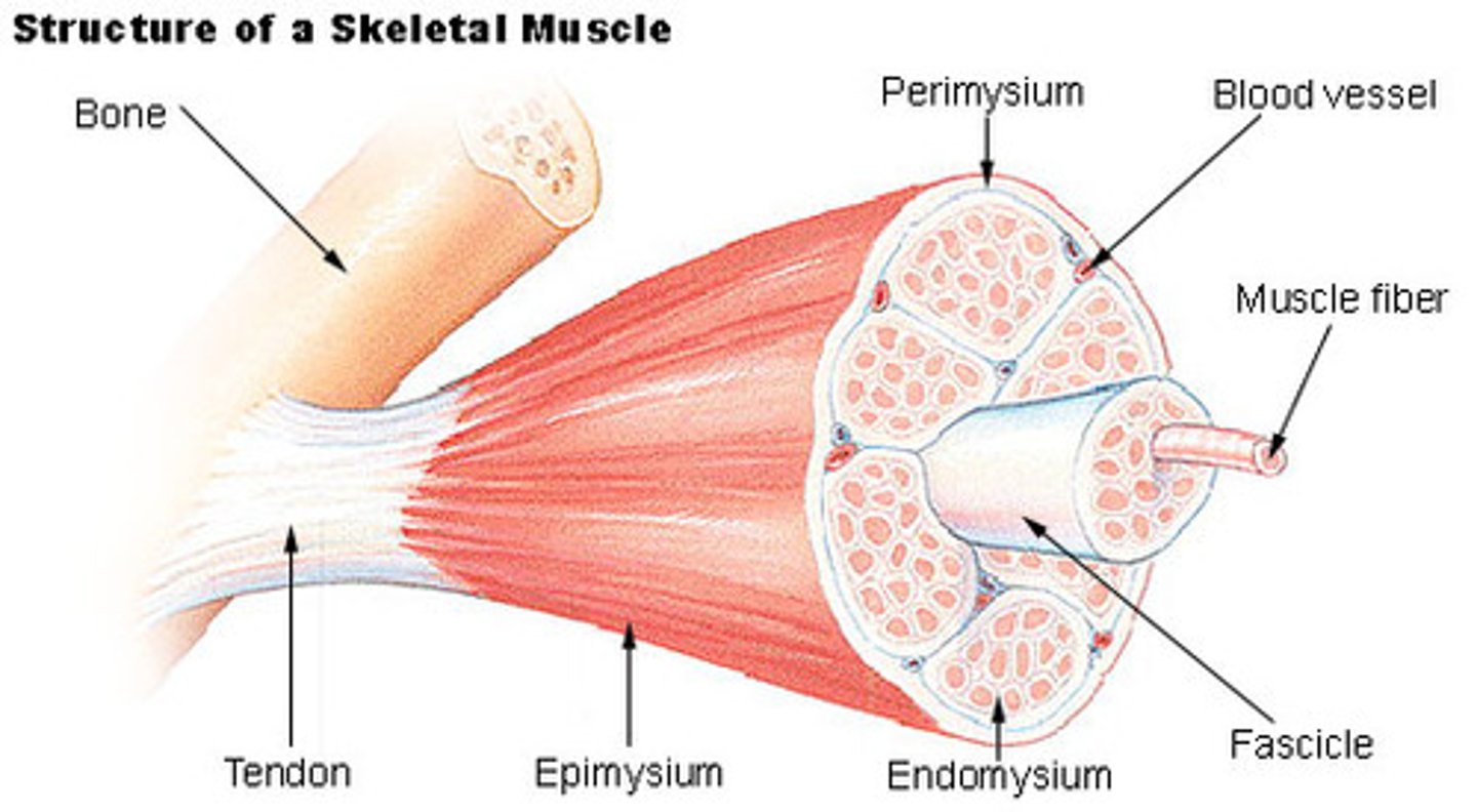
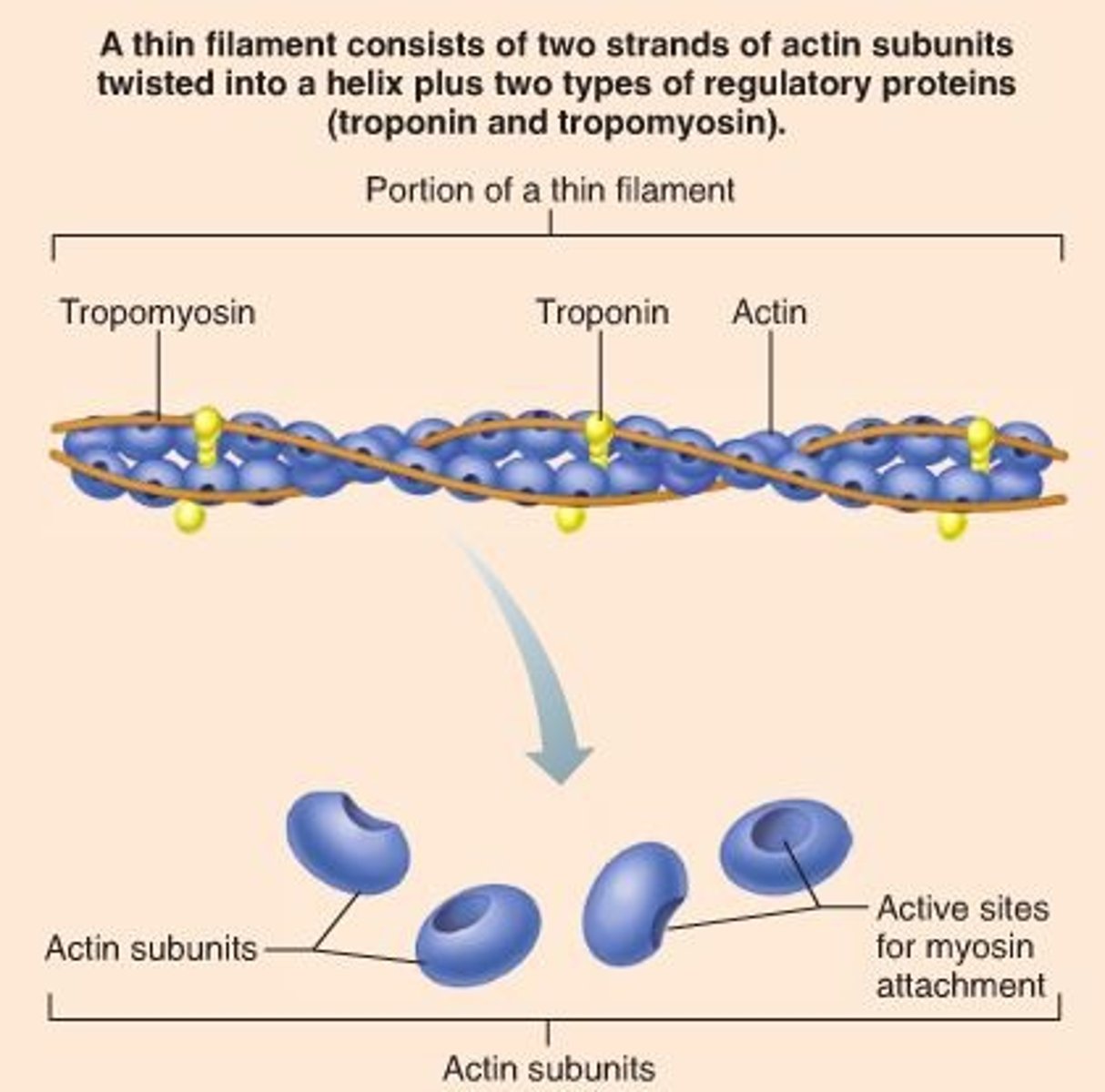
Annotate the structure of skeletal muscle
*look at quizlet diagram
ENDOMYSIUM
•connective tissue wrapped around each individual muscle cell (fiber)
PERIMYSIUM
•surrounds each fascicle (bundle of muscle fibers)
EPIMYSIUM
•connective tissue wrap just under deep fascia that surrounds the entire muscle
***Muscle Fibers
•made up of a group of myofibrils
•myofibrils contain myofilaments whose action is responsible for the contraction of myofibrils and therefore the whole muscle
Define the terms origin and insertion of muscles
ORIGIN
•attachment of a muscle tendon to a stationary bone
INSERTION
•attachment of a muscle tendon to a movable bone
Identify the location of skeletal muscles in various regions of the body
look at quizlet diagram
List the principal structures of the ventilatory system.
•nose
•mouth
•pharynx
•larynx
•trachea
•bronchi
•bronchioles
•lungs
•alveoli
•smooth muscle tissue is found on some of the walls of internal hollow organs. Produces smooth rhythmic actions
•involuntary; movement of blood and air in lungs
•the trachea runs down the posterior wall and is composed of smooth muscles
-thin walled tube a diameter wide
-composed of thin, tough connective tissue
-strengthened at intervals by complete rings of cartilage
purpose of conducting airways
•no gas exchange takes place here
•they filter chemicals and other harmful substances that are in the air
•warm and moisten air
1.2 Outline the functions of the conducting airways.
•nostrils are lined with hair (cilia) which protects the nasal cavity from invasion
-humidifies air and filters particles
•air passes through the pharynx
•then through larynx and trachea
-larynx is voice box which protects the trachea from invasion of foods and fluids
Define These Respiratory Terms
PULMONARY VENTILATION
•inflow and outflow of air between the atmosphere and lungs
TOTAL LUNG CAPACITY (TLC)
•volume of air in lungs after a maximum inhalation
•sum of vital capacity and residual volume
VITAL CAPACITY (VC)
•max volume of air that can be exhaled after maximum inhalation
TIDAL VOLUME (TV)
•volume of air breathed in and out in any one breath
EXPIRATORY RESERVE VOLUME (EVR)
•volume of air in excess of tidal volume that can be exhaled forcefully
INSPIRATORY RESERVE VOLUME (IRV)
•additional inspired air over and above tidal volume
RESIDUAL VOLUME
•volume of air still contained in lungs after max exhalation
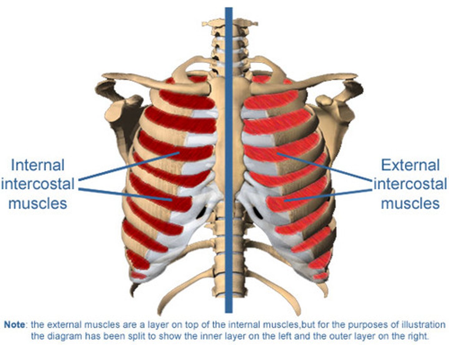
internal vs external intercostal muscles
INTERNAL
•skeletal muscles located between ribs
•innervated by intercostal nerve
•only used during forceful exhalation, coughing, or exercise
EXTERNAL
•muscles of inhalation
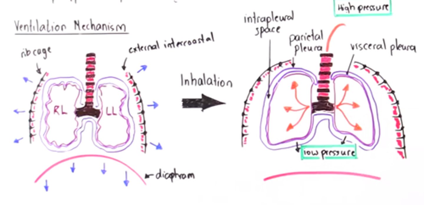
Explain the mechanics of ventilation in the human lungs
•airflow is due to pressure differences in the lungs and atmosphere
•intercostals and diaphragm contract to expand the chest cavity during inhalation
•diaphragm flattens and moves down
•intercostals muscles move rib cage up and out increasing space for lungs
•this increase in size decreases the internal air pressure
•now outside air is at a higher pressure and rushes into lungs to equalize
•exhaling diaphragm and intercostal muscles relax and return to resting position, reducing cavity size, increasing pressure
Describe nervous and chemical control of ventilation during exercise.
MEDULLA OBLONGADA
•in charge of involuntary functions in the hindbrain (oldest part of brain)
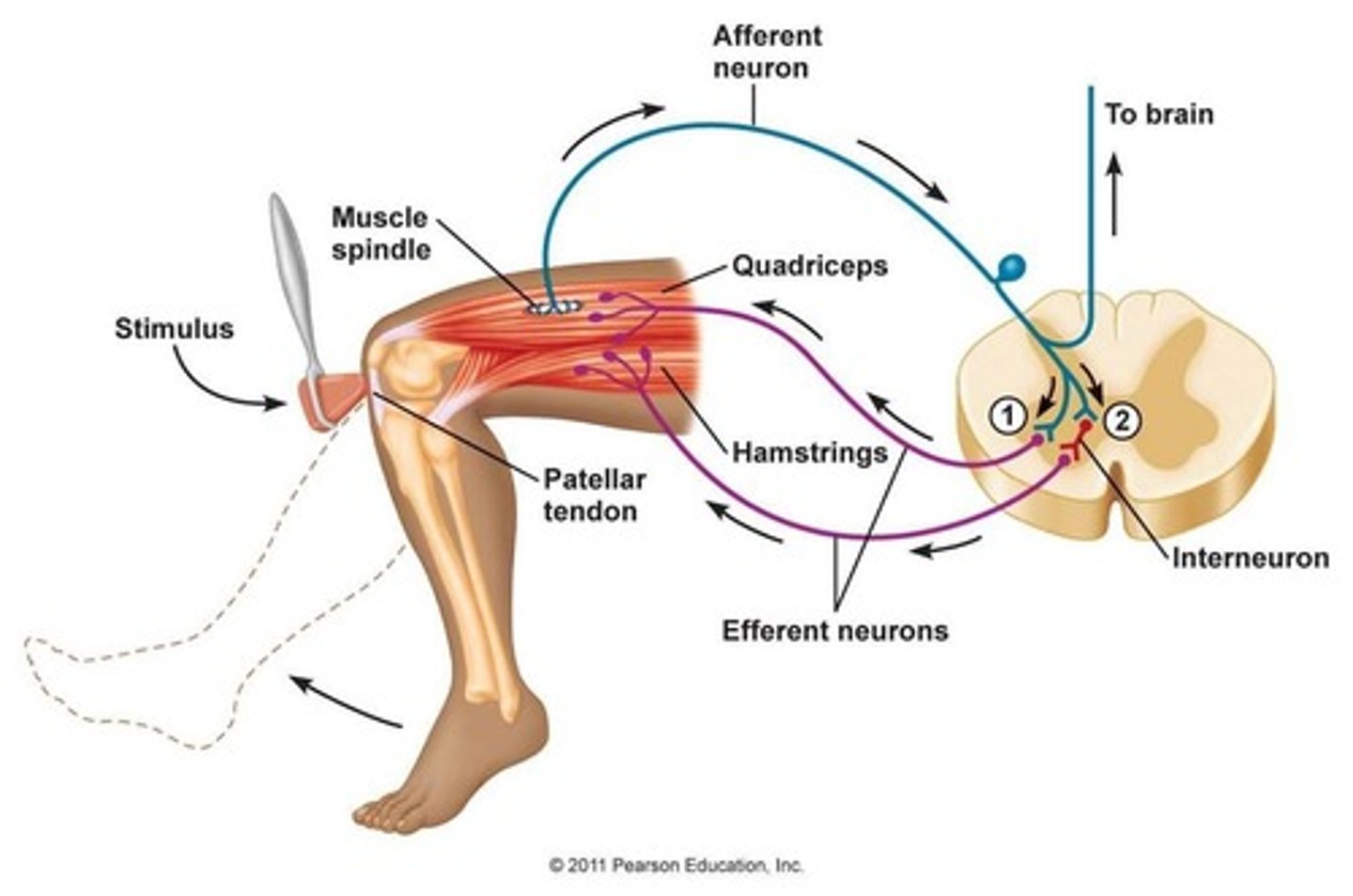
PROPRIOCEPTORS
•found in the muscles
•detect changes of length and contraction in the muscles
•monitors CO2 production and O2 levels
CHEMOCEPTORS
•found in the bloodstream and monitor CO2 and O2 levels as well
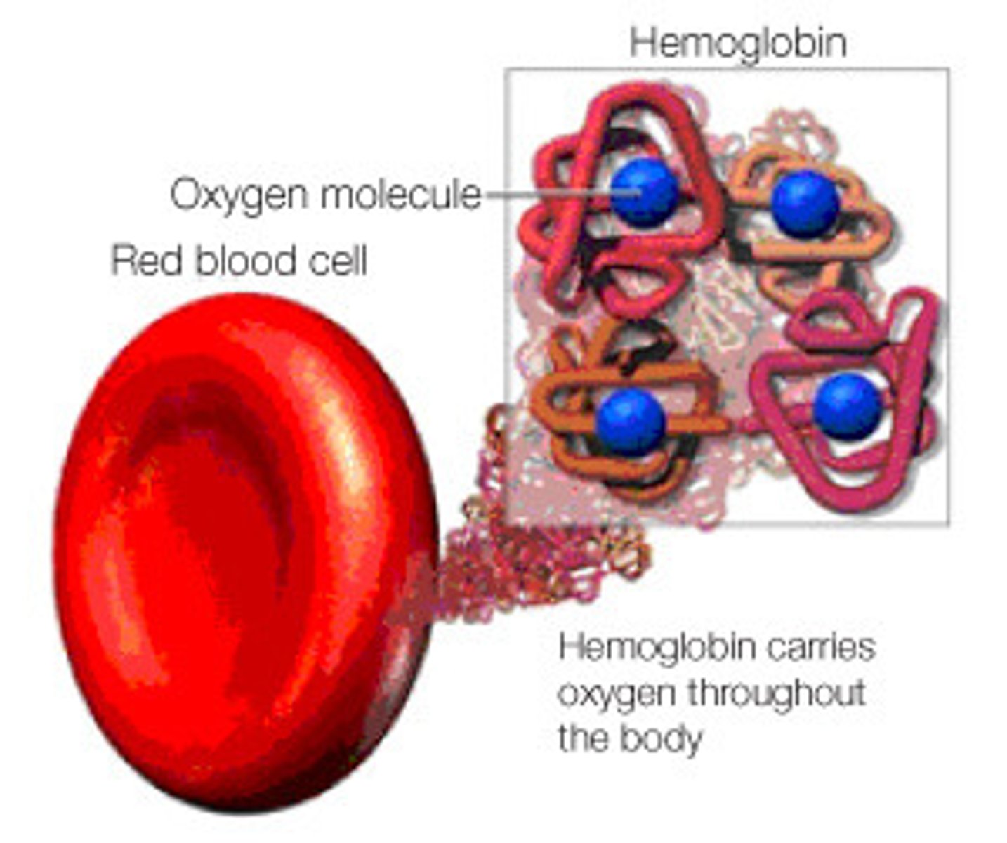
Outline the role of hemoglobin in oxygen transportation
•the protein that allows oxygen to bind to red blood cells
•the compound of protein and iron gives blood its red color
-98% carry oxygen in blood
-2% carry carbon dioxide in blood
•RBC effectiveness is due to the carrying capacity of the hemoglobin
•While oxygen atoms are diffused into tissues, they also pick up the CO2 to return to the lungs to exhale
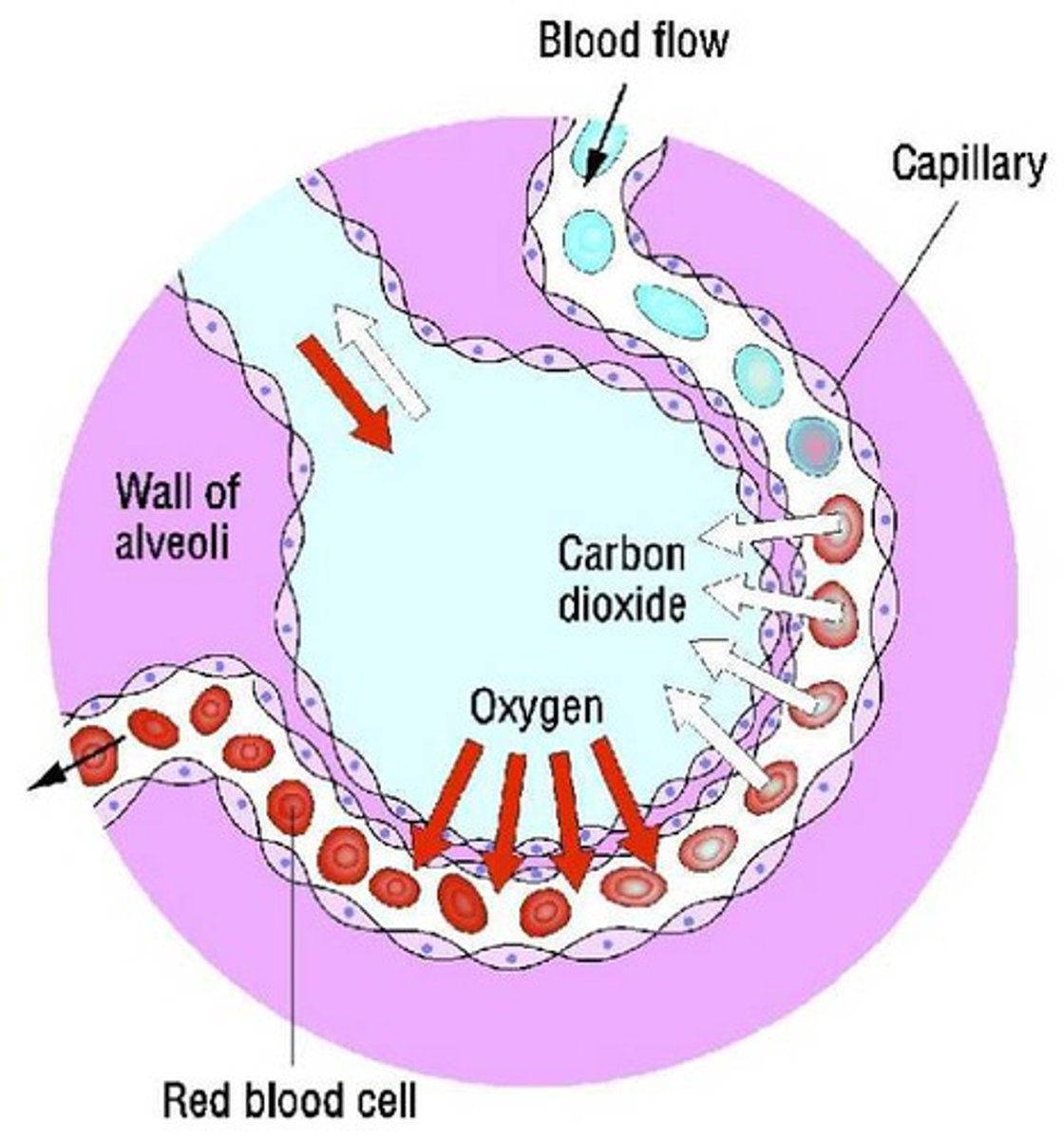
Explain the process of gaseous exchange at the alveoli.
•bronchioles control gas exchange
•alveoli are attached to the branches of the bronchial passages
-oxygen exchange takes place here
•the alveoli inflate and deflate with each breath and create a passage gradient
•alveoli fill up with air during inhalation and the oxygen diffuses from the air in the alveoli and into the blood
•the CO2 diffuses from the arriving venous blood and into the air which exits body during exhalation
State the Composition of Blood
ERYTHROCYTES
•red blood cells
•40-45%
LEUKOCYTES
•white blood cells
•<1%
PLATELETS
•thrombocytes
•<1%
PLASMA
•liquid portion of blood
•includes water, gases, dissolved nutrients)
•55%
Distinguish between the functions of erythrocytes, leucocytes and platelets.
ERYRTHROCYTES
•contain an oxygen carrying pigment called hemoglobin, which gives blood its red color
•RBC effectiveness is due to carrying capacity of hemoglobin
•blood transports oxygen, carbon dioxide, hormones, and waste
•platelets protect us from bleeding to death (clotting) and destroying pathogens
•acts as a regulator for temperature, water content in cells, and body pH
LEUKOCYTES
•exist in our bodies to combat infection and inflammation
•involved in immune function
•protects from infection
•retain antibodies from previous infections
-capable of preventing some virus
•destroy bacteria, fungi, and parasites
PLATELETS
•involved in the process of clotting and help repair slightly damaged blood vessels after injury
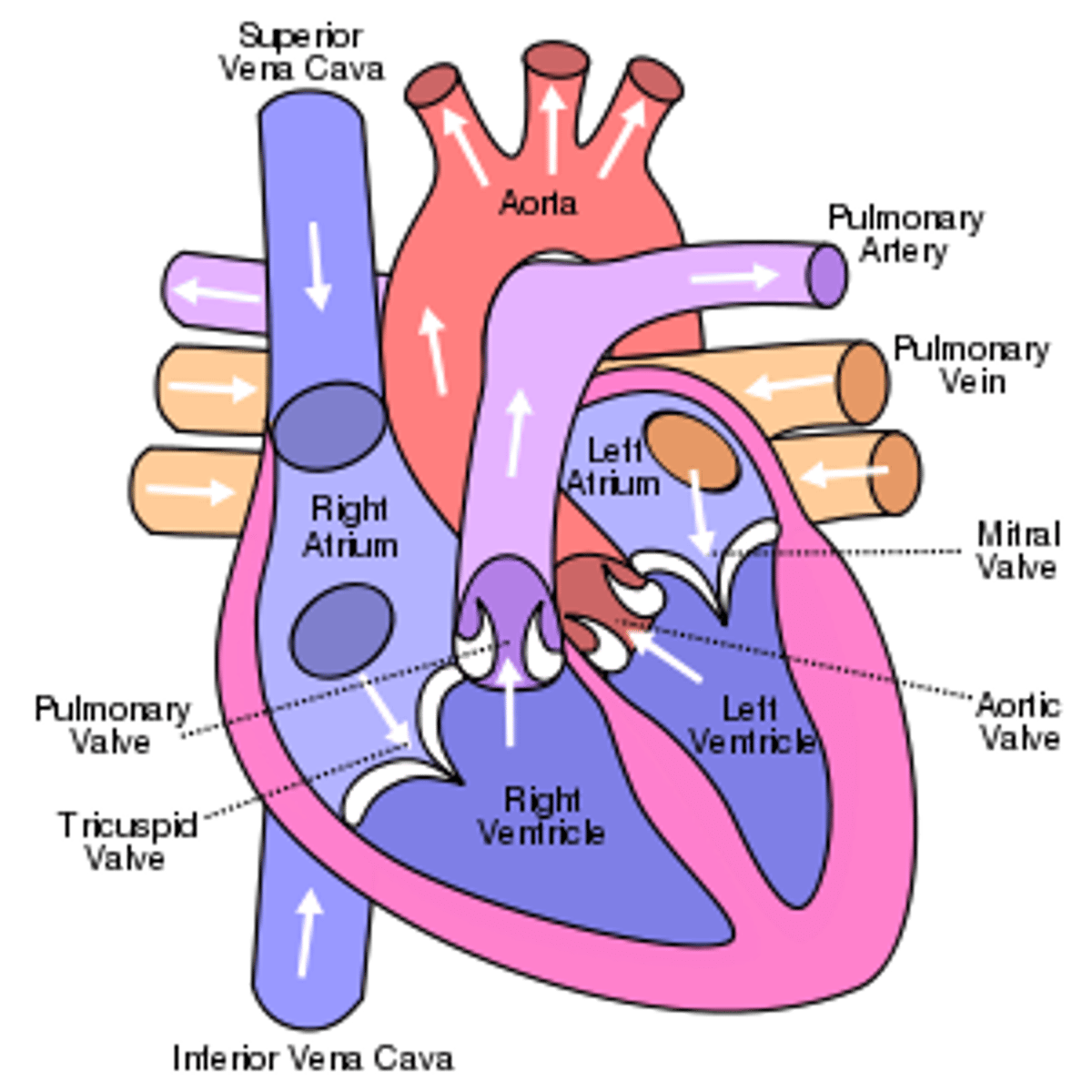
Describe the anatomy of the heart with reference to the heart chambers, valves and major blood vessels
•involuntary muscle with striated muscle fibers
•composed of 4 chambers separated by a septum and valves
PERICARDIUM
•triple layered bag surrounds, anchors, and protects the heart
CHAMBERS
•Atraia
-receiving chamber for blood returning to the heart
-small and thin walled, because they only have to pump blood a small distance into the ventricles
•ventricles
-large because they propel blood from the heart into circulation around the body
VALVES
•dense connective structures prevent back flow of blood by opening and closing when heart contracts and relaxes
•two lie between each atria and ventricle (atrioventricular valves)
-tricuspid on the right
-mitral (bicuspid) on the left
BLOOD VESSELS
•both arteries coming from the heart have a valve on them to prevent back flow (pulmonary and aortic valve)
•heart has its own blood supply via coronary arteries
*** arteries carry oxygenated blood to tissues out of the heart; veins carry deoxygenated blood back to the heart
Describe the intrinsic and extrinsic regulation of heart rate and the sequence of excitation of the heart muscle
INTRINSIC
•sinoatrial node (SA) is a small mass of specialized muscles in the posterior wall of the right atrium
•because automatic self-excitation of the SA node initiates each heartbeat, its known as the pacemaker
•the end of the fibers of the SA node fuse with surrounding atrial muscle fibers so that the contraction spreads, producing atrial contraction
•several groups of atrial muscle fibers conduct contraction to the atrioventricular (AV) node, which spreads action potential thoughout the rest of the heart via specialized muscle fibers
•next potential goes to the Bundle of His and then purkinje fibers
EXTRINSIC
•info collected by lung strength receptors, muscle proprioceptors, and chemoceptors provide info about low pH and high CO2.
•if CO2 is high, increased ventilation and increased heart rate occur to increase O2 levels.
•medulla also helps detect H+ to increase breathing frequency
•peripheral chemoreceptors in the aortic bodies which regulateO2, CO2, and pH balance
•sympathetic nerves
-increase HR
-increase arousal
-decrease digestion
-encourage adrenaline release
•parasympathetic nerves
-decrease HR
-Adrenaline and noradrenaline released by adrenal glands
-increase HR
-increase O2 spread to muscles
stretch reflexes
•if a muscle is being stretched, the stretch is caused by the contraction of its antagonist
•this sensory proprioception info contributes to maintaining proper muscle tone
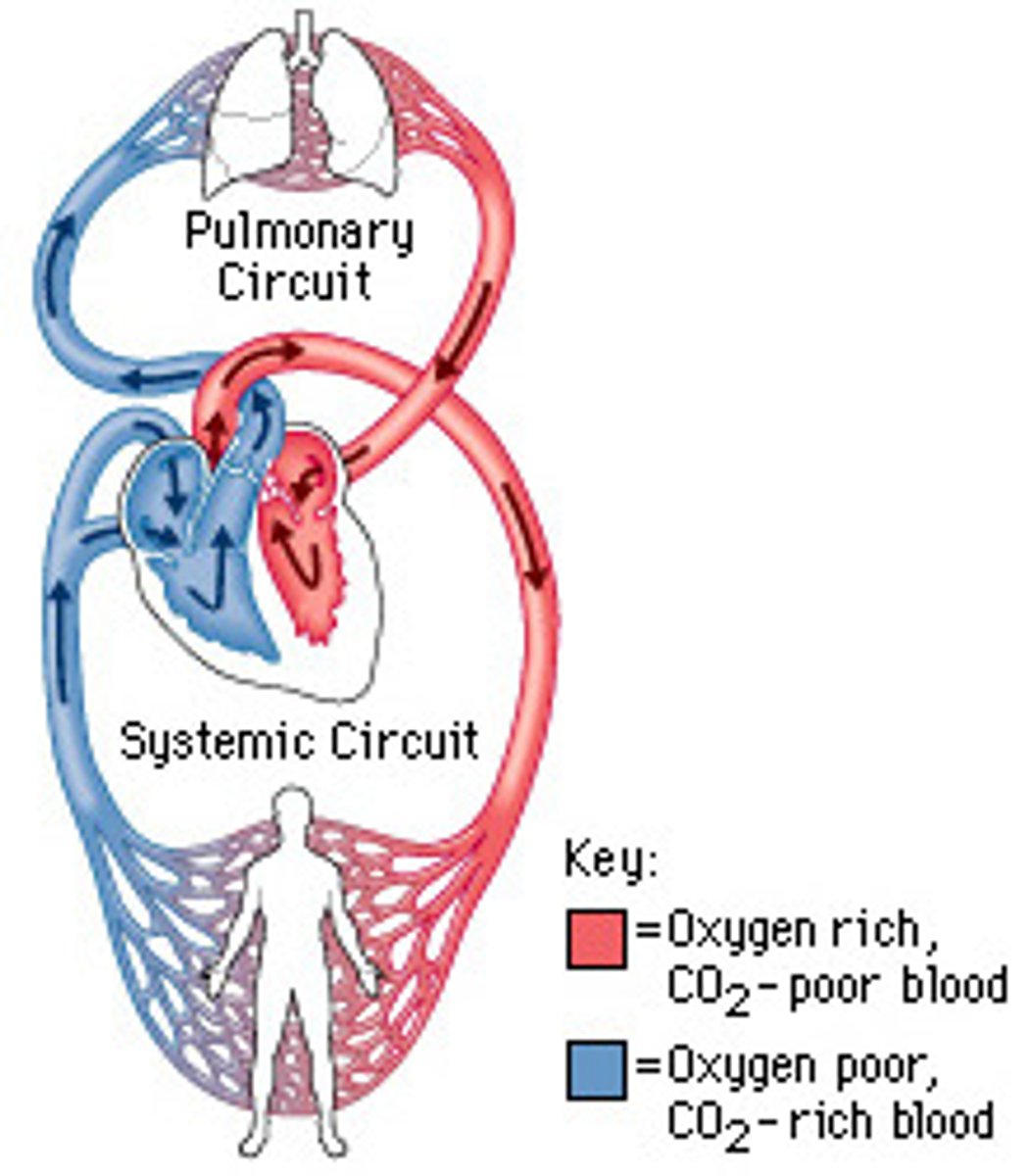
Outline the relationship between the pulmonary and systemic circulation
PULMONARY
•portion of the cardiovascular system which carries oxygen depleted blood back to the heart, to the lungs
•returns with oxygenated blood
SYSTEMIC
•portion of cardiovascular system which carries oxygenated blood away from the heart, to the body
Describe the relationship between heart rate, cardiac output and stroke volume at rest and during exercise.
HEART RATE
•number of beats per minute
CARDIAC OUTPUT (Q)
•amount of blood pumped from the heart in 1 minute (liters/min)
STROKE VOLUME
•amount of blood pumped by the left ventricle in each contraction
•average volume is 0.07 liters/beat
•increases depending on type of exercise and intensity
•during upright exercise like running, SV increases from 50mL at rest to 120 at max
*** Q=SV•HR
•since SV increases, Q will increase with increased HR
How does the heart control the rate at which it beats
•systole= contraction
•diastole= relaxation
•SAN creates an impulse and both atria contract (systole).
•Inpulse reaches AVN
•impulse passes down bundle of HIS into purkinje fibers and achieves ventricular systole
What effect does training have on resting cardiac output and stroke volume
•typical person has a Q of 51/min
•at rest, cardiac output remains the same regardless of fitness level
•SV will have increased-- cardiac hypertrophy
•low resting HR indicates high fitness level
what happens to SV, Q, and HR during exercise
•HR increases
•SV increases
-rest 70ml/beat --> exercise 100ml/beat
•Q increases
•venous return
-return of blood to the heart
STARLINGS LAW (increased SV during exercise)
•venous return increases
•heart will fill with blood quicker, which will stretch it more
•chambers will hold more blood
•heart will produce a more powerful contraction
Analyse cardiac output,
stroke volume and heart
rate data for different
populations at rest and
during exercise.
•HR can reach 200 beats/min in some individuals
•max cardiac output differs due to body size and training
•males have a higher Q and SV
•trained people have a lower HR, higher SV, and higher ventricular mass
•HR decreases with age
Explain cardiovascular drift
•core temperature rises, distributing blood to skin to release excess heat
•as exercise increases, this leads to lower venous return, lower SV, decrease in mean arterial pressure, water loss due to sweat, and lower blood plasma
•A portion of lost fluid (from sweat) comes from the plasma
•decrease in plasma volume will diminish venous return and SV
•HR increases to compensate and maintain cardiac output
•core temperature rises, distributing blood to skin to release excess heat
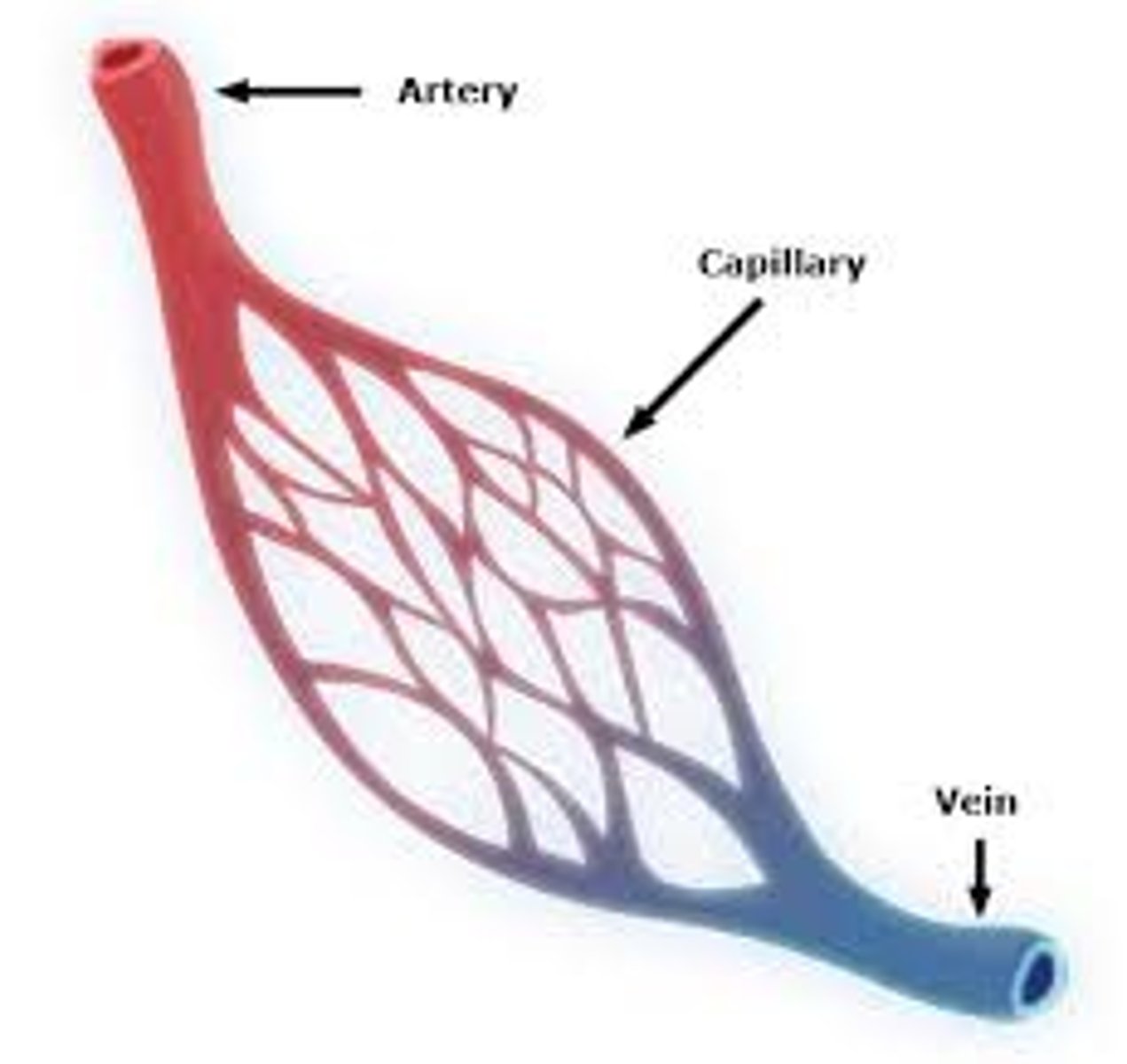
Arteries vs Veins
ARTERIES
•take blood away from heart
•oxygenated blood going to the tissue
•high pressure because heart is pumping blood out
•low volume
•no valves
•break in arteries look like fountains
VEINS
•bring blood back to the heart
•carry deoxygenated blood from tissues
•high volume
•low pressure
•valves to prevent back flow
•break in vein creates pool (bruise)
Define the terms systolic and diastolic blood pressure
SYSTOLIC
•the force exerted by blood on arterial walls during ventricular contraction
DIASTOLIC
•the force exerted by blood on arterial walls during ventricular relaxation
Discuss how systolic and diastolic blood pressure response to dynamic and static exercise. (ALSO includes changes from rest to exercise)
DYNAMIC
•requires muscular movement and elevated HR
•systolic bp increases at a lower rate (140-160)
•breathing frequency is higher in dynamic, so CO2 is expelled quickly
•Diastolic bp remains the same
-muscles are moving constantly, so there is no added pressure
-arteries are dilated as vasodilation is occurring
STATIC
•constant contraction
•systolic bp increases
-volume of blood and concentration rate of larger amounts of blood is pumped though the arteries of working muscles (200mm/mg)
•diastolic bp increases
-pressure on arterial walls is increased so vasoconstriction
-muscles squeeze veins to create venous return
-breathing is more constricted so there is less oxygen and more CO2, so the heart must work harder to pump
-it has to supply muscles with sufficient oxygen
Compare the distribution of blood at rest and the redistribution of blood during exercise.
•because arteries are large, walls offer little resistance to blood flow, even during exercise
•arterioles have a much smaller diameter and offer lots of resistance to blood flow
•as blood flows through capillaries, most of the pressure caused by the action of the heart is spent
•during exercise, increased muscle contraction results in increased flow of blood through veins and into heart
AT REST:
•20% of blood flow is directed to all organs, brain, stomach, kidney, muscles, etc
•blood flow is reduced and capillaries close
•vasoconstriction: skin receives minimum blood flow
DURING EXERCISE
•90% of blood is distributed to active muscles
•stomach and kidneys need less blood, but essential organs still get sufficient O2
•arterioles dilate to supply muscles and open capillaries
•vasodilation: skin receives more blood to cool body
Describe the cardiovascular adaptations resulting from endurance exercise training.
BLOOD:
•resting bp decreases as a result of improved cardio-vascular functions
-increased blood plasma
-RBC volume and hemoglobin
MUSCULAR and HR ADAPTATIONS
•increased capilarization in muscles
•Q increases during max exercise
•As the stroke volume increases the cardiac output can remain constant, therefore enabling the resting heart rate to be lower.
•more efficient CO2 expulsion
•resting HR decreases
•The myocardium (muscular tissue of the heart) increases in thickness
•The increase in size of the heart enables the left ventricle to stretch more and thus fill with more blood.
•The increase in muscle wall thickness also increases the contractility resulting in increased stroke volume at rest and during exercise, increasing blood supply to the body
Explain maximal oxygen consumption.
•VO2 max is the max amount of oxygen (ml) one can use in a minute
•more fit= higher VO2 max
Discuss the variability of maximal oxygen consumption in selected groups
•males have higher VO2 max than females
-due to higher hemoglobin concentrations
-and size difference
•VO2 max increases with age
-size
•athletes have higher VO2 max
-due to chronic adaptations
thermoregulation in the circulatory system
•when exercising:
-body produces heat, muscles are active and release heat
-body must maintain core temp of 98.6
•capillary beds have fine vessels with high surface area; allows exchange of 02 between blood and tissue
WHEN BODY IS HOT
•capillaries dilate (vasodilation)
-more blood passes
-greater loss of heat
WHEN BODY IS COLD
•capillaries constrict (vasoconstriction)
-traps heat underneath skin
** nerves tell muscles within arteries to contract or relax
Breathing in at rest vs exercise
REST
•diaphragm contracts (moves down)
•External intercostals contract
-outside of ribs
-pulls ribs up and out
•thoracic cavity (chest) increases
-decreases pressure
EXERCISE
•diaphragm and external intercostals contract
-just like breathing at rest
•more muscles work to make thoracic cavity as large as possible
-pectorals pull ribs out further
-sternocleomastoids (neck) contract to increase airflow
breathing out at rest vs exercise
REST
•diaphragm and eternal intercostals relax
-pushes diaphragm up
-pulls ribs in
-thoracic decreases
-pressure increases and air flows out
EXERCISE
•internal intercostals (inside of ribs) contract and pulls ribs down and in
-thoracic gets smaller
•diaphragm relaxes and abdominals contract
-thoracic decreases, forcing diaphragm up
artereo-venous difference
•difference between oxygen content of arterial blood and mixed venous blood
•represents extent to which oxygen is removed from the blood as it passes through the body
what factors affect bp in arteries
DIET
•fat deposits build up and increase resistance, increasing bp
EXERCISE
•training burns fat
VASODIALATION/CONSTRICTION
•changes blood flow
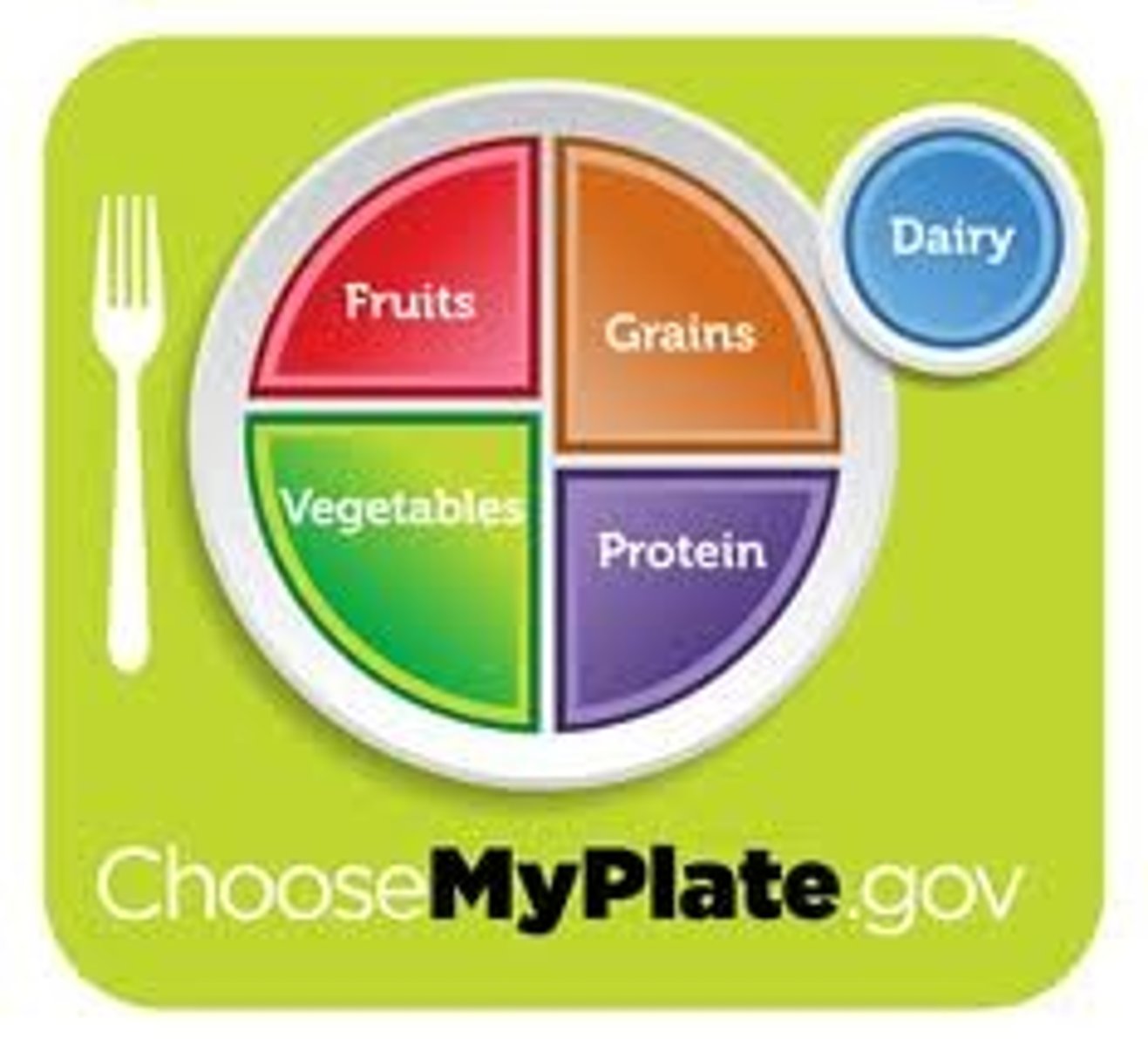
List the macronutrients and micronutrients.
MACRONUTRIENTS
•a substance required in relatively large amounts by living organisms
•breakdown:
-Carbohydrates: 55-65%
-Fat (lipid): 25-30%
-Protein: 10-15%
MICRONUTRIENTS
•a substance required in trace amounts
•fibers, minerals, and vitamins
outline the role and purpose of the macronutrients
CARB
•energy storage, cell membrane, DNA, RNA
•glycogen stored in muscle provides energy for performance
PROTEIN
•made of various combinations of more than 20 amino acids
-8 of these are essential because they can not be manufactured
-makes and repairs cells
•provides energy in extreme conditions
•structure (bones, muscles, etc)
LIPIDS
•concentrated source of energy
•assist in transport of fat-soluble vitamins (ADEK) to the small intestine for digestion
outline the role and purpose of micronutrients
VITAMINS
•growth, development, and metabolism
•assist enzymes that catalyze the breakdown of carbs, proteins, fats, and minerals
•Fat Soluble
-A,D,E, and K
-usually stored in the body
-high levels can be toxic
•Water Soluble
-B,C
-must be supplied regularly and frequently to the body through diet or supplementation
-because they dissolve in water they are easily expelled from the body
MINERALS
•calcium, potassium, iron, sodium, phosphorus, and chlorine
•important in cellular function such as muscle contraction, fluid balance, and energy systems
•deficiencies can impede athletic performance
FIBER
•part of a plant that can't be digested by the body
•slow rise in glucose level
•lower insulin requirement
•normal bowel function
•lower cholesterol levels
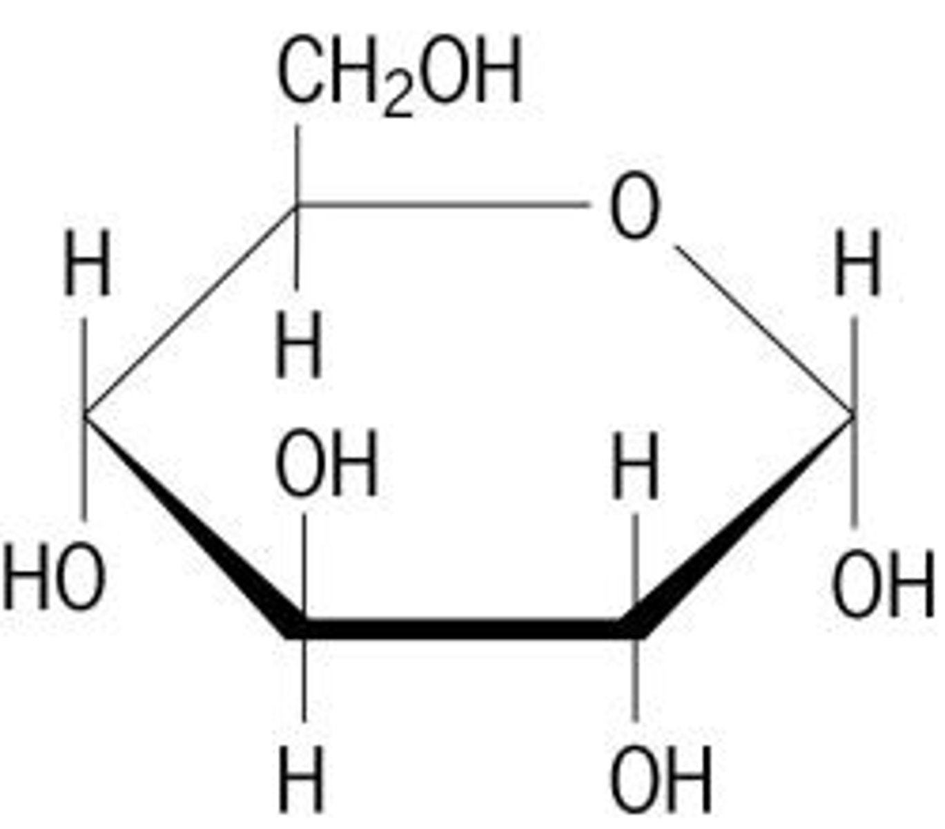
State the chemical composition of a glucose molecule.
•carbohydrate
•C6H12O6
•ratio is 1:2:1
Identify a diagram representing the basic structure of a glucose molecule.
Explain how glucose molecules can combine to form disaccharides and polysaccharides.
•two monosaccharides combine through combination to make a disaccharide
-condensation reaction- linking of a monosaccharide to another mono, di, or poly by the removal of water
•polysaccharides- chains with 3+ molecules
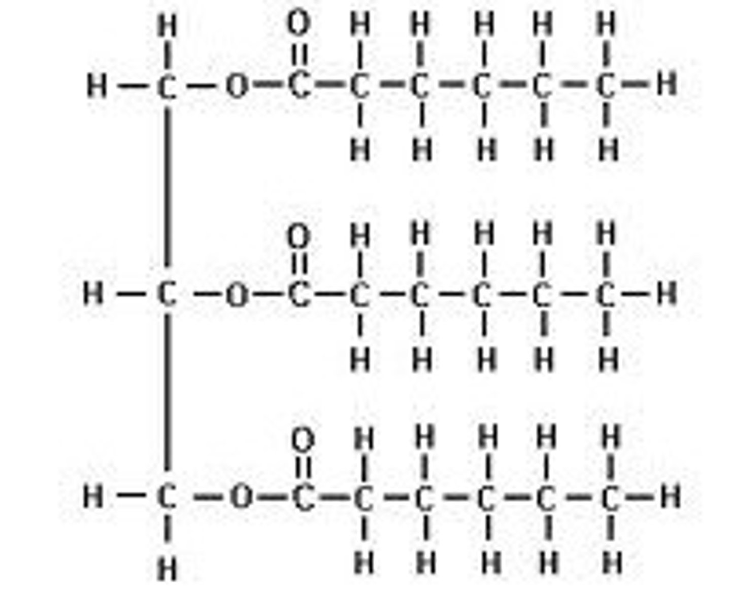
State the composition of a molecule of triacylglycerol.
•stored in adipose cells and tissues, which are highly concentrated stores of metabolic energy
•composed of 3 fatty acids and one glycerol
Distinguish between saturated and unsaturated fatty acids.
SATURATED
•mainly come from animals and full fat dairy
•no double bonds
•max # of hydrogen atoms
UNSATURATED
•plant based (olive oil, nuts)
•double bond between 2 carbon atoms and hydrogen radiating from the carbon
•liquid at room temp
State the chemical composition of a protein molecule.
• C H O N
Distinguish between an essential and a non-essential amino acid.
•essential (9) cannot be synthesized and must be provided by food
•non essential can be synthesized
•20 amino acids in total
Describe current recommendations for a healthy balanced diet.
CABRS
•250g
PROTEIN
•50g
FIBER
•50g
LIPIDS
•30g (unsaturated)
WATER
•2 liters
SALT
•<5g
State the approximate energy content per 100 g of carbohydrate, lipid and protein.
CARB
•1600kJ
LIPID
•3700kJ
PROTEIN
•1700kJ
Discuss how the recommended energy distribution of the dietary macronutrients differs between endurance athletes and non-athletes.
•sedentary people should consume a diet that has about 55-60% carbs, no more than 30% fat, and 10-15% protein
•endurance athletes require more carbs (60-70%)
•lipids slightly higher
•water intake must compensate for sweat
Outline metabolism, anabolism, aerobic catabolism and anaerobic catabolism.
METABOLISM
•all biochemical reactions that occur within an organism, including anabolic and catabolic
ANABOLISM
•energy requiring reaction
•building larger molecules
ex/ glucose to glycogen
CATABOLISM
•break down complex organic compounds into smaller ones
•net release of energy
ex/ triglyceride to glycerol and fatty acid
•aerobic catabolism requires oxygen but anaerobic does not
State what glycogen is and its major storage sites.
•carbohydrate
•polysaccharide
•stored as glycogen in the muscle and liver
Outline how a polysaccharide is formed
•glycogen is formed by linking together large numbers of glucose molecules
•condensation reaction with glucose molecules
State the major sites of triglyceride storage
• adipose tissue
•skeletal muscle
Explain the role of insulin in the formation of glycogen and the accumulation of body fat
•after eating, blood glucose level rises
•pancreas detects rise and secretes insulin
INSULIN
• hormone released from the pancreas
•increases transport of glucose into cells (especially skeletal muscle)
•stops most catabolic reactions
ex/ stops breaking down glycogen into glucose
ex/ stops lipids from breaking down to fatty acids and glycerol
ex/ stops proteins--> amino acids
ex/ stops lipolysis
•stimulates anabolic reactions
ex/ the reverse of all above examples
•stimulates glycogenesis (glucose--> glycogen) which lowers blood glucose level
Outline glycogenolysis and lipolysis
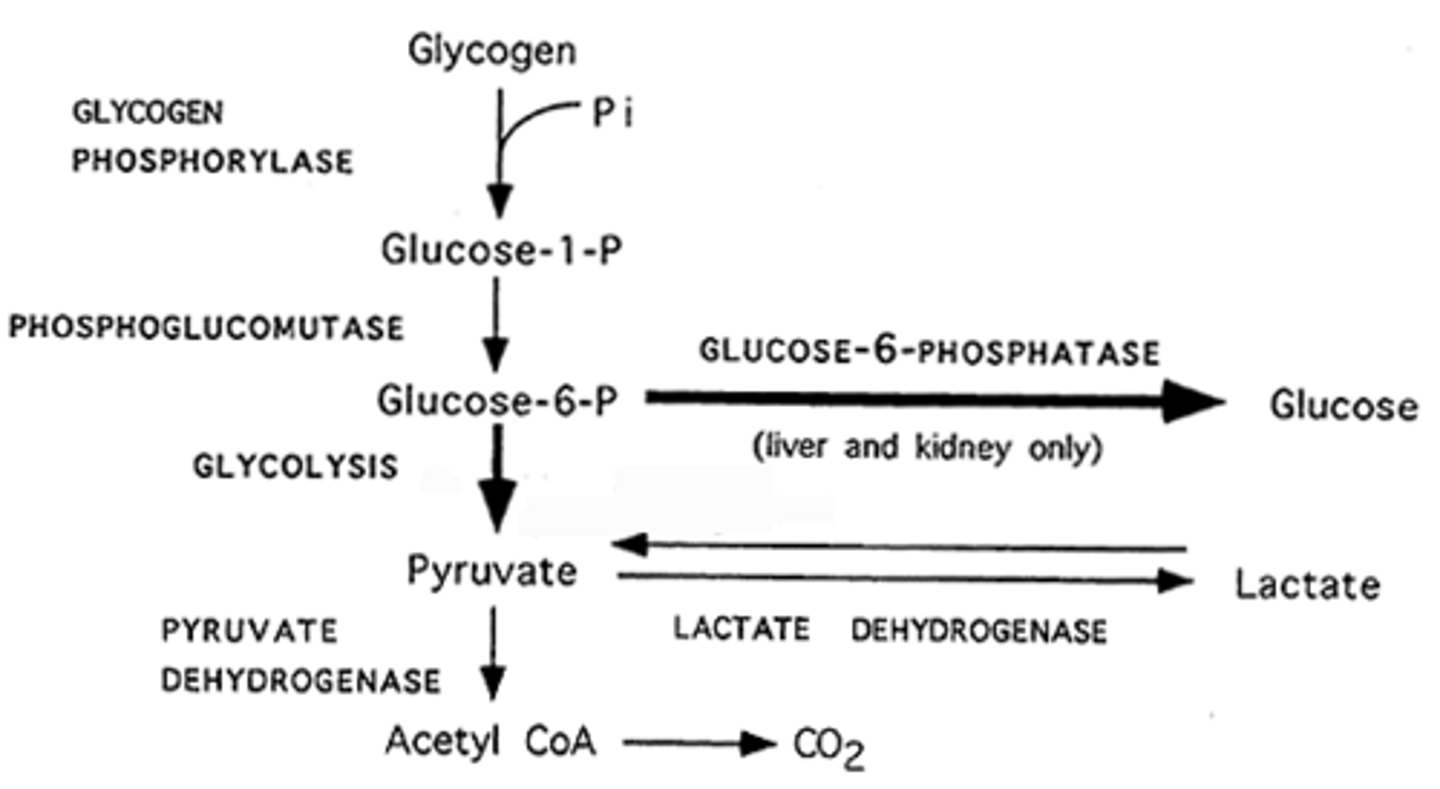
GLYCOGENOLYSIS
•process by which glycogen, stored in the liver and muscle cells, is broken down into glucose to provide energy.
•glycogen stored in liver and muscles is converted into glucose-1-phosphate
•then into glucose-G-phosphate
•hormone controlling it is glucagon from the pancreas and epinephrine from the adrenal gland; act upon enzymes to to stimulate glycogenolysis and inhibit glycogen synthesis (to stop glycogenesis)
•glucagon- released from pancreas in response to low blood glucose
•epinephrine- release in response to threat or stress
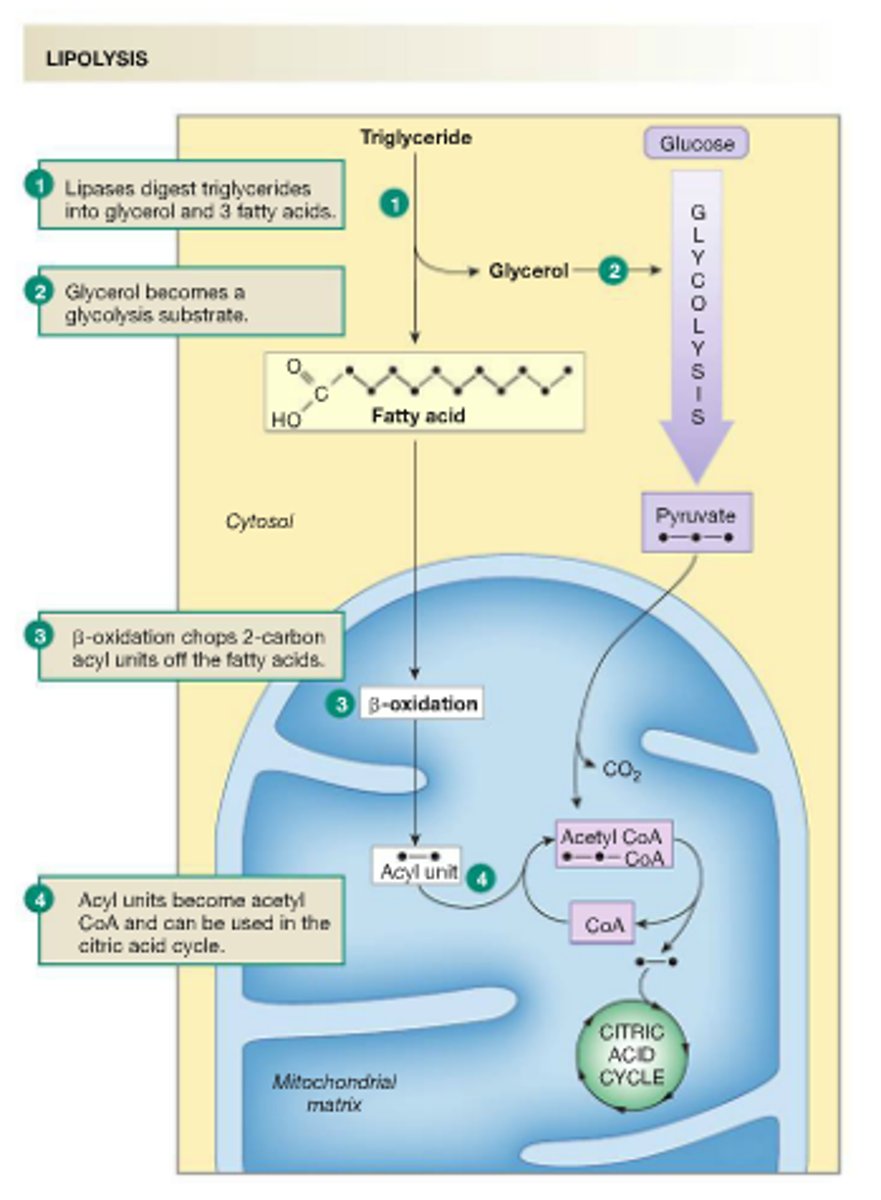
LIPOLYSIS
•the breakdown of lipids. Triglycerides are transported through the blood to appropriate tissues.
•breakdown of triglycerides stored in fat cells
•go through hydrolysis where they are broken down into glycerol and fatty acids
•fatty acids are released into blood stream and circulate through the body
•used in mitochondria (acetyl coA) to enter krebs cycle and ultimately produce ATP
•before fatty free acids can enter Krebs, they enter beta-oxidation where chemical reactions break them down to hydrogen ions and acetyl coA
•from there, the acetyl coA enters krebs and is metabolized like carbs
•
Outline the functions of glucagon and adrenaline during fasting and exercise.
•blood glucose level drops
•glucagon (from pancreas) and adrenaline (epinephrine from adrenal gland) increase
•glucagon stimulates glycogenolysis (glycogen--> glucose
•adrenaline acts like glucagon and dilates the arteries to the muscle
•blood glucose level can rise again
Explain the role of insulin and muscle contraction on glucose uptake during exercise.
•exercise lowers concentration of insulin in the blood; increase in glucagon and adrenaline
•exercise is a stimulus for skeletal muscle glucose uptake
•blood glucose decreases with exercise as the muscle takes it in
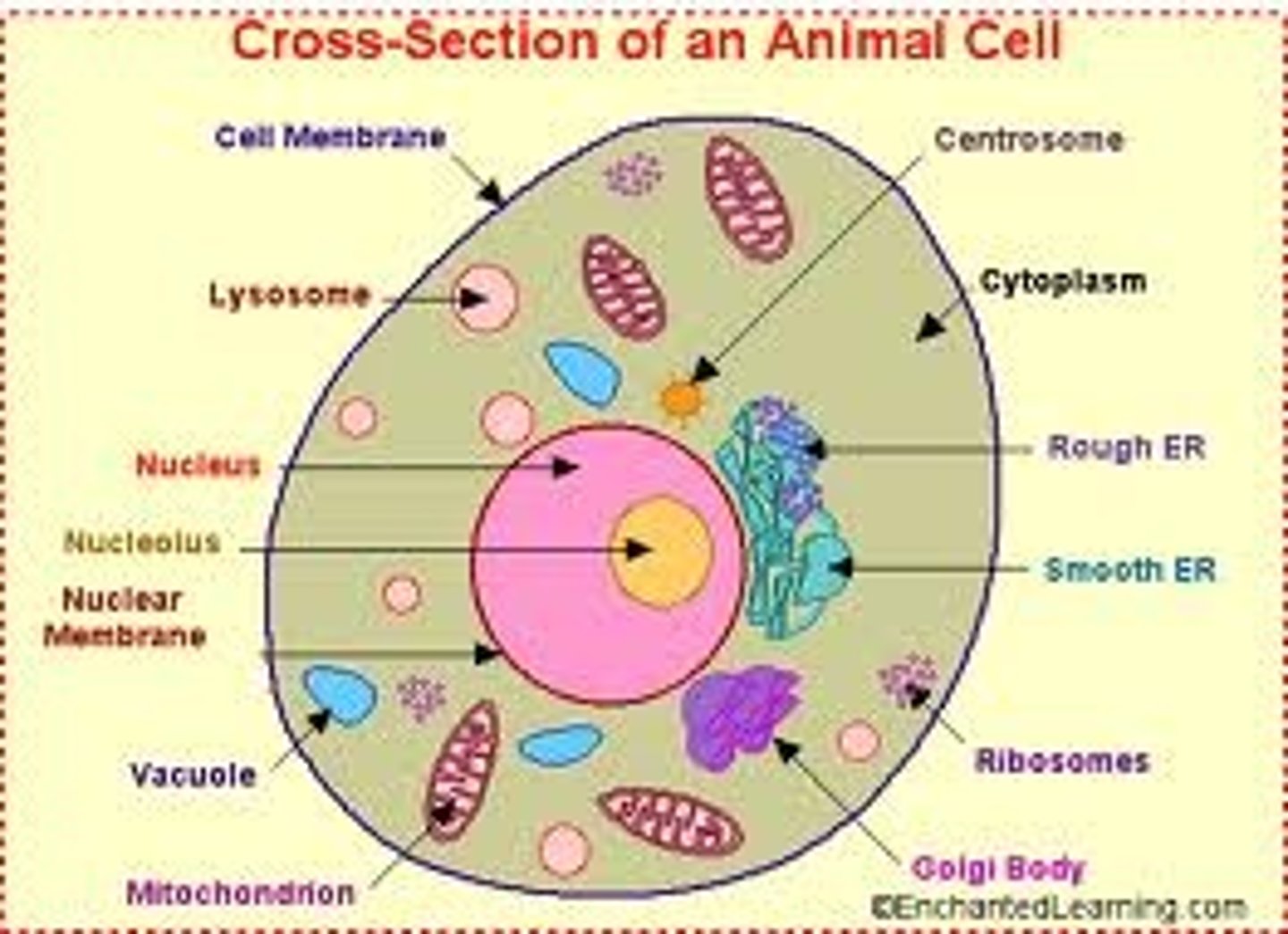
Annotate a diagram of the ultrastructure of a generalized animal cell.
***LOOK AT DIAGRAM
RIBOSOME
•cell organelle constructed in the nucleolus and functioning as the site of protein synthesis
ENDOPLASMIC RETICULUM
•an extensive membranous network
LYSOSOME
•membrane-enclosed sac of enzymes
GOLGI APPARATUS
•packing center, proteins mixed with chemicals
MITOCHONDRION
•organelle that serves as the side of cellular respiration
NUCLEUS
•an atoms central core, containing protons and neutrons
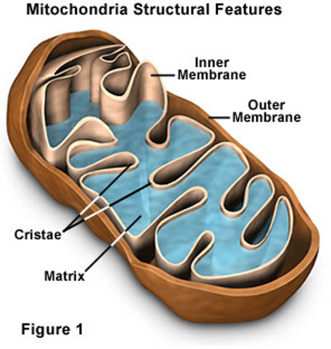
Annotate a diagram of the ultrastructure of a mitochondrion.
Define the term cell respiration.
•release of energy from the chemical bonds of food molecules (usually carbs) in the form of ATP

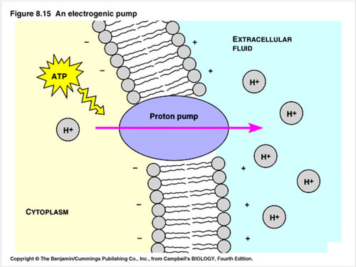
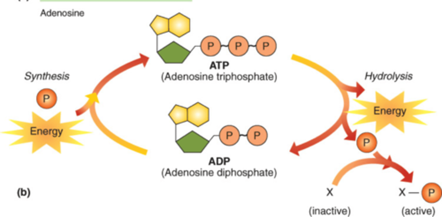
Explain how adenosine can gain and lose a phosphate molecule.
•adenosine triphosphate (ATP) is an energy rich chemical compound which serves as the immediate source of energy of most of the reactions in the body
•ATP is made up of a smaller compound (adenosine) and three phosphate groups
•ATP is broken down into adenosine diphosphate (adenosine + 2 phosphates and a separate phosphate)
***
•ATP combines with water
•ATP works by losing the endmost phosphate group when told by an enzyme
-big release of energy
-end product is ADP
•when body is resting, the reverse action takes place
-phosphate group is reattached to molecule using energy from food
Explain the role of ATP in muscle contraction
•ATP is converted to ADP when the phosphate molecule is released
•this liberates chemical energy for muscle contraction (told through nervous system)
Cellular respiration and Exercise
Carb--> inspiration--> expiration--> sweat/heat
.
.
glycolysis: 2 ATP
.
.
krebs: 2 ATP
.
.
E Transport Chain: 34 ATP
=38 ATP
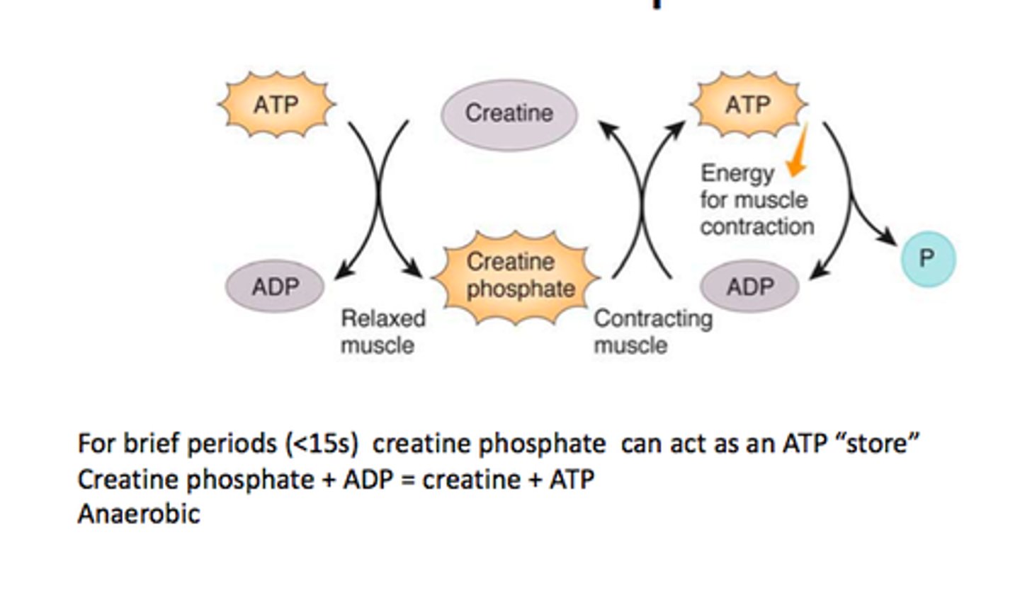
Describe the re-synthesis of ATP by the ATP-CP system.
CREATINE PHOSPHATE (high energy molecule)
•is broken down to provide a phosphate molecule for the resynthesis of ATP that has been used during the initial stages of exercise
•when should this system be used?
-interval sprints
-boxing
-benching 1 rep max
*rests are needed
•can be regenerated rapidly (30 secs= 50%; 3 mins= 100%)
•limited supply in muscles, short lived
•regeneration only takes place with O2
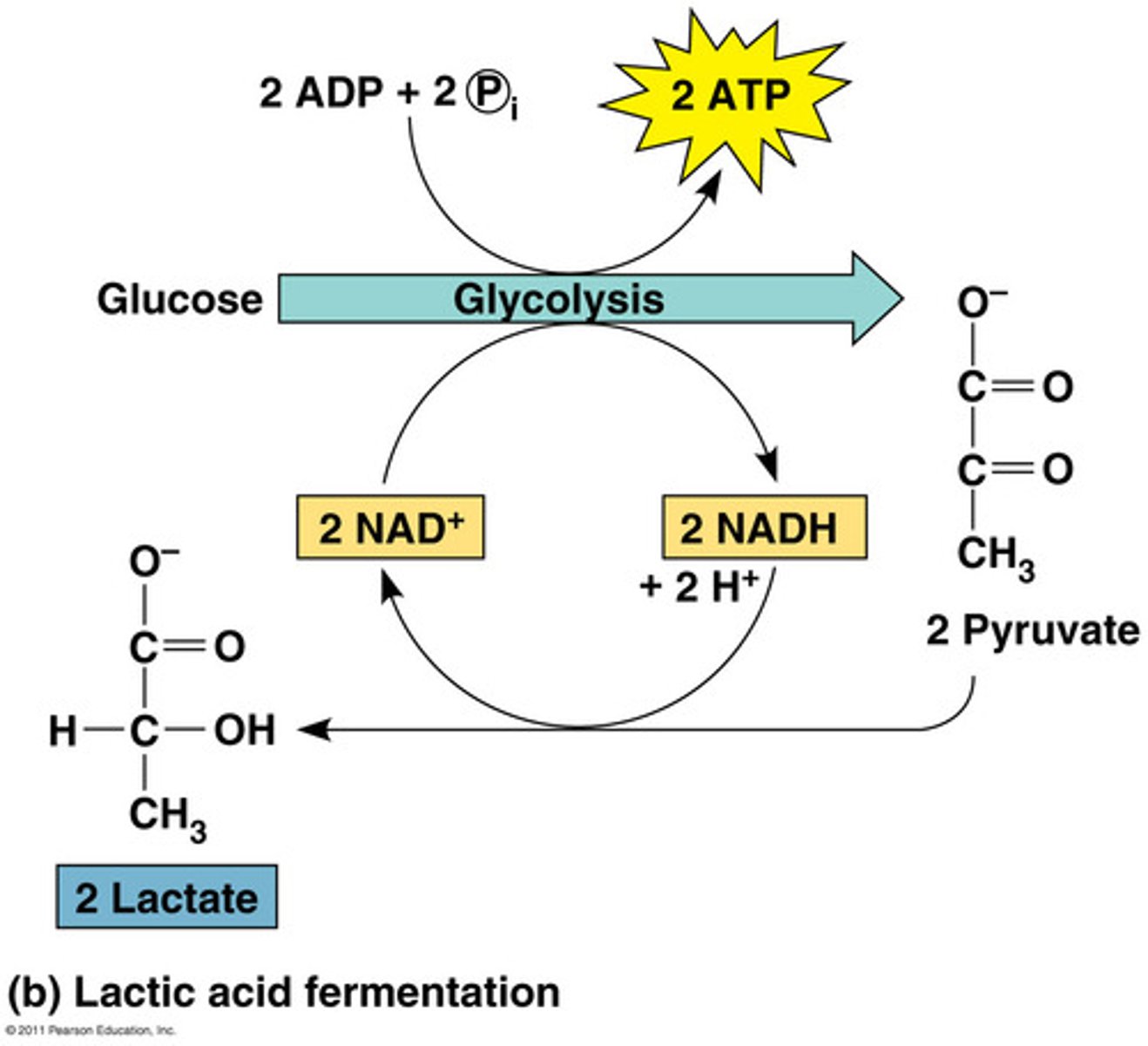
Describe the production of ATP by the lactic acid system.
•aka anaerobic glycolysis
•breakdown of glucose to pyruvate without the use of Oxygen
•pyruvate is then converted to lactic acid which limits the amount of ATP (2 molecules)
•lasts no longer than 2 minutes
•can be regenerated quickly
•lactic acid can be converted back into glycogen in the liver
•lactic acid is the by-product
•only a small amount of energy can be released without O2
Explain the phenomena of oxygen deficit and oxygen debt
Oxygen deficit- oxygen demand is greater than supply; start of exercise
•EPOC (excess post oxygen consumption) is in addition to the oxygen normally consumed at rest
•respiratory rate remains elevated to clear out the CO2 that has accumulated in the tissues as a byproduct of metabolism
•higher arousal has elevated adrenaline, increases respiration and thus need for oxygen
•EPOC helps rebuild ATP and Par and clear lactate from anaerobic metabolism (oxygen debt)
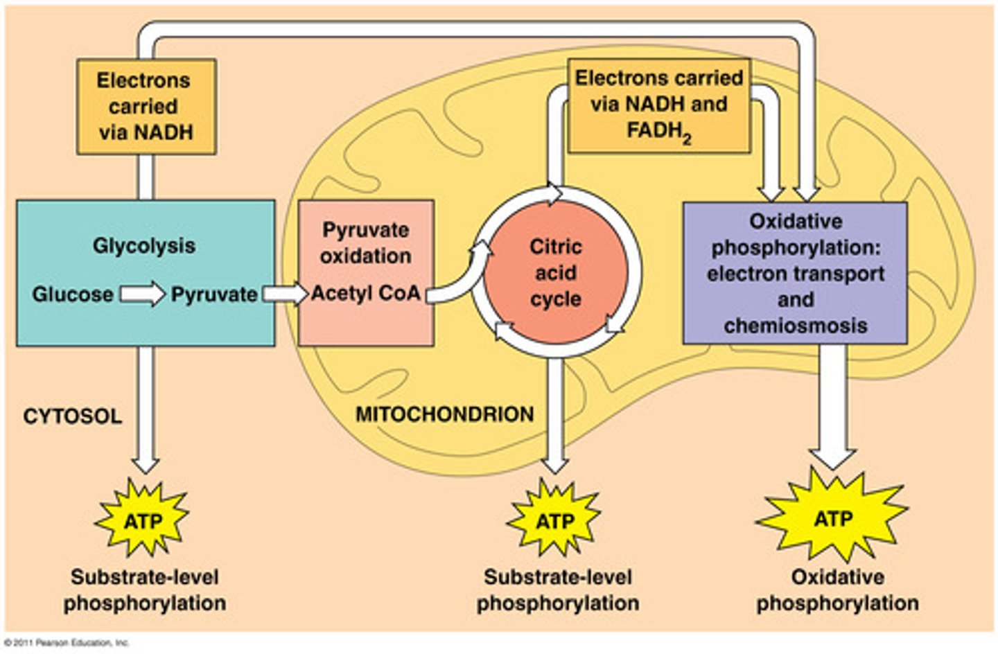
Describe the production of ATP from glucose and fatty acids by the aerobic system.
•glucose-->pyruvate (glycolysis)
•pyruvate--> H2O+CO2+H+(krebs cycle in the mitochondria)
-this happens in the presence of oxygen
•hydrogen ions carried to electron transport chain where energy is produced (ATP)
•38 molecules produced
•no fatiguing by products
•glycogen and triglyceride scores are large, so energy lasts long
•cannot be used right away
•O2 needed to breakdown glycogen or fatty acids (need more O2)
Discuss the characteristics of the three energy systems and their relative contributions during exercise.
ATP-PCr
•produces energy the quickest
•peaks around 5 secs and is exhausted by 12-15 secs
LACTIC ACID
•peaks at 15 secs and then declines
•there is an opportunity to oxidize metabolic by-products, therefore allowing lactic acid energy system to contribute additional energy
AEROBIC
•dominant at 55 secs
•if steady state is achieved, then energy demands are met
•continues to increase until demands are met or max O2 consumption occurs
Compare the fuel sources and by-products of anaerobic glycolysis (lactic acid) and the aerobic energy systems
LACTIC ACID
•fuel source
-glycogen/glucose
•by-product
-heat, energy, hydrogen ions, lactic acid
AEROBIC SYSTEM
•Fuel source
-glycogen, fats, proteins, glucose, lipids, amino acids
•by-product
-CO2, water, heat, energy
Discuss how the production of ATP by the various energy systems differs in an individual who is performing a 60 meter sprint, a 400 meter sprint, and a 26 mile marathon
60m SPRINT
•performance requires rapid resynthesis of ATP via ATP-PCr system
400m SPRINT
•rapid resynthesis of ATP via ATP-PCr system and lactic acid system
26 MARATHON
•prolonged resynthesis of ATP via aerobic system
Carbohydrate Metabolism
•glucose is broken down to provide energy (GLYCOLYSIS)
•when O2 is available, pyruvate enters the mitochondria where it is oxidized to CO2 and H2O
•without O2, pyruvate is converted to lactate, and then transported back to the liver where glucose is reformed or oxidized to pyruvate in the muscles
•glucose not immediately stored as glycogen
•glucose--> glycogen through glycogenesis
•glycogen--> glycogenolysis
Describe the process of glycogenolysis and glycolysis
GLYCOGENOLYSIS
•glygogen (sugar polysaccharide)-->glucose-1-phosphate--> glucose-G-phosphate
*catabolic reaction
*broken down by glucagon and epinephrine
•glucose goes through blood stream and into mitochondria, and with O2...
GLYCOLYSIS
•glucose--> pyruvate (2 ATP)--> actetol coA--> Krebs (2 ATP)--> e- transport chain (34 ATP) = 38 ATP
*results in CO2 and Heat
explain the process of lipolysis
triglycerides-->3 fatty acids + 1 glycerol--> acetyl coA (though beta oxidation)--> Krebs Cycle (2 ATP)--> e- transport chain (34 ATP)= 129 ATP
*more energy because of the breakdown of fats
explain the process of the Lactic Acid System (anaerobic glycolysis)
glucose-->(glycolysis)pyruvate--> lactate= 2 ATP
•1-2 mins- max intensity
•quick production bc theres no O2
•lactic acid is painful
explain the ATP-PC process
*ATP= adenosine triphosphate
•phosphocreatine-->creatine+free energy+ P1-->free energy-->ATP
•energy lasts for 8-10 secs (max/anaerobic)
•once you've worked hard for 10 secs, PC stores run out
-takes 2-3 mins to rebuild them
explain the aerobic energy system process
glucose(2ATP)(glycolysis)-->pyruvate-->Krebs(2ATP)(beta oxidations)-->Hydrogen ions--> e-transport chain(34ATP)
•can break down glucose/fats/protein
•takes a lot of time- sub max activities

:Define the term skill
•consistant production of goal-oriented movements
•learned
Describe the different types of
skill
MOTOR
•movement; no thinking
ex: sprint racing, weight lifting
COGNITIVE
•lots of thinking; little movement
•skills in team games such as rules, tactics, and game objectives
•decision making
ex: chess
PERCEPTUAL
•sense things, interpret them
•vision, vestibular (balance related to hearing, haptic (touch), and auditory
ex: rock climbing
PERCEPTUAL-MOTOR
•thought, interpretation of environmental stimuli, and motor response to the sensory information
•performer must adapt to environment
ex: dribbling a soccer ball to beat a defender
types of skill classification
DISCRETE
•clear start and finish
•brief and well defined
ex: golf swing
SERIAL
•linking together of skills to form a more complex movement
ex: flips and somersaults, lay up
CONTINUOUS
•end of one cycle is the beginning of the next
ex: swimming, running
stability of the environment related to skills
OPEN
•affected by environment
•variable and unpredictable
•athlete must adapt quickly
ex: rebounding in basketball
CLOSED
•more controlled environment
•stable and predictable
ex: archery
size of musculature involved in motor skills
GROSS
•large muscle group
ex: swimming, cycling
FINE
•small muscle groups
ex: piano
pace of skills
EXTERNALLY PACED
•environment may include opponents
•performer must pay attention to external events in order to control rate of movment
•usually open skills
ex:wind sailing
INTERNALLY PACED
•performer controls rate at which the skill is executed
•usually closed
ex: gymnast floor routine, climber
skills performed in relation to other actors
INDIVIDUAL
•performed in isolation
ex: high jump
COACTIVE
•performed at the same time as others but no confrontation
ex:swimming
INTERACTIVE
•performed where other performers are directly involved
ex: football
Compare skill profiles for contrasting sports.
Use the following terms when answering this question
•motor/cognitive/perceptual/perceptual-motor
•discrete/serial/continuous
•open/closed
•gross/fine
•externally paced/internally paced
•individual/coactive/interactive
Define ability
•general trait or capacity of the individual that is related to performance
•something we are born with
describe the types of abilities
PERCEPTUAL
•enable individual to process info about when to move
ex:football player notices someone coming at him so he gets rid of the ball
MOTOR
•related to actual movement
ex: strength and speed of limb movement of a sprinter
PERCEPTUAL-MOTOR
•combo of motor and perceptual
•capability to process, interpret, and then use sensory stimuli for performing a task
•make sense of info you receive from environment
Distinguish between Fleishman's physical proficiency abilities (physical factors)
and perceptual motor abilities
(psychomotor factors).
TAXOTOMY
•fleishman tested over 200 tasks
•used factor analysis to identify a number of abilities
•divided them by categories
-physical proficiency ability
-perceptual motor ability (combo of how we make sense of environment and how we act)
PHYSICAL PROFICIENCY
•Strength
•Flexibility
•Balance
•Coordination
•Endurance
PSYCHOMOTOR
•Control precision
•Rate control
•Aiming
•Response orientation
•Reaction Time
•Manual dexterity
•Finger Dexterity
•Arm- Hand steadiness
•Wrist and finger speed
Define the term technique.
•way of doing something
State the relationship between
ability, skill, and technique.
skill= ability + selection of correct technique