Neuroscience - Anterior Triangle of the Neck: Part 1
1/65
Earn XP
Description and Tags
Name | Mastery | Learn | Test | Matching | Spaced | Call with Kai |
|---|
No analytics yet
Send a link to your students to track their progress
66 Terms
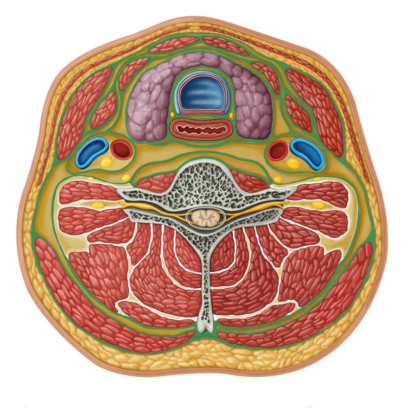
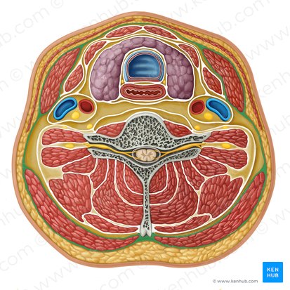
superficial cervical fascia
contains neurovascular supply to the skin, superficial veins, superficial lymph nodes, fat, platysma muscle
lies between the dermis and the deep cervical fascia
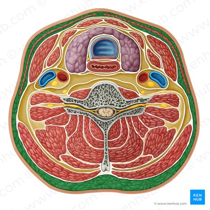
deep cervical fascia
deep to the superficial fascia and platysma muscle and organized into several layers
investing layer
pretracheal layer (muscular and visceral parts)
prevertebral layer
carotid sheath
Name the 4 layers of deep cervical fascia

investing layer of deep cervical fascia
most superficial layer
surrounds all the structures in the neck
completely surrounds the trapezius and sternocleidomastoid muscles
can be thought of as a tube with superior, anterior, inferior, and posterior attachments
external occipital protuberance
superior nuchal line of skull
inferior portion of mandible
superior attachment of investing layer of deep cervical fascia

hyoid bone
anterior attachment of investing layer of deep cervical fascia

spine and acromion process of scapula
clavicle
manubrium
inferior attachment of investing layer of deep cervical fascia

nuchal ligament
spines of cervical vertebrae
posterior attachment of investing layer of deep cervical fascia

pretracheal layer of deep cervical fascia
spans between the hyoid bone superiorly and the thorax inferiorly (where it fuses with the pericardium)
infrahyoid muscles
What does the muscular part of the pretracheal layer enclose?

thyroid gland
trachea
esophagus
Name the 3 structures that the visceral part of the pretracheal layer encloses

prevertebral layer of deep cervical fascia
surrounds the vertebral column and its associated muscles: scalene muscles, prevertebral muscles, and the deep muscles of the back

base of skull
superior attachment of prevertebral layer of deep cervical fascia

transverse processes and vertebral bodies of vertebral column
anterior attachment of prevertebral layer of deep cervical fascia

fused with endothoracic fascia of ribcage
inferior attachment of prevertebral layer of deep cervical fascia
nuchal ligament
posterior attachment of prevertebral layer of deep cervical fascia

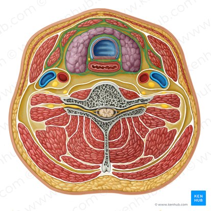
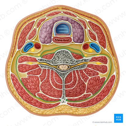
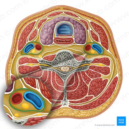
carotid sheath
bilateral structures which enclose an important neurovascular bundle of the neck
formed by fascial contributions from the pretracheal, prevertebral, and investing fascia layers
contain common carotid artery, internal jugular vein, vagus nerve, cervical lymph nodes, AND MORE
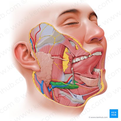
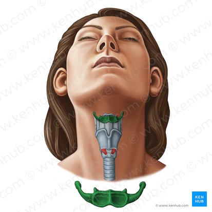
anterior triangle of neck
Name the space outlined in purple

inferior border of mandible
superior border of anterior triangle of the neck

anterior border of sternocleidomastoid
lateral border of anterior triangle of the neck

median line of neck
medial border of anterior triangle of the neck

investing layer of deep cervical fascia
roof of anterior triangle of the neck
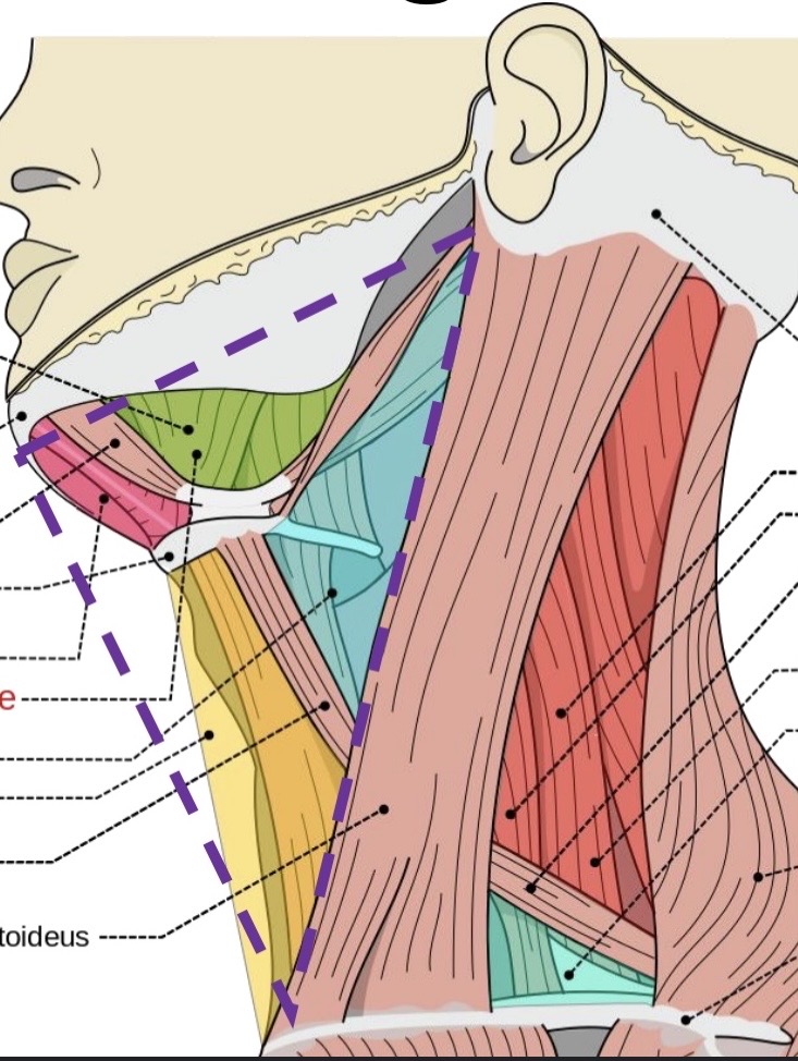
pretracheal layer of deep cervical fascia
floor of anterior triangle of the neck

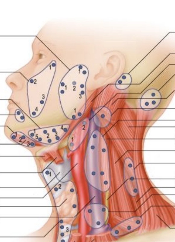
lymph nodes of head and neck
peripheral lymphoid organs connected to vascular system by afferent and efferent lymphatic vessels
ovoid or bean-shaped
2-20 mm in longitudinal diameter, usually occur in groups
~300 in head and neck
superficial located in superficial fascia/subcutaneous connective tissue
deep lie deep to fascia and muscles
function: play an important role in the defense mechanism of the body by filtering out micro-organisms and foreign substances
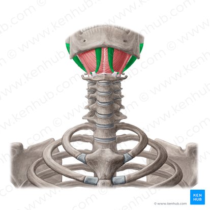
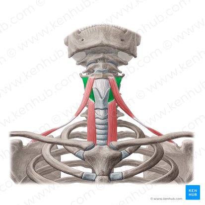
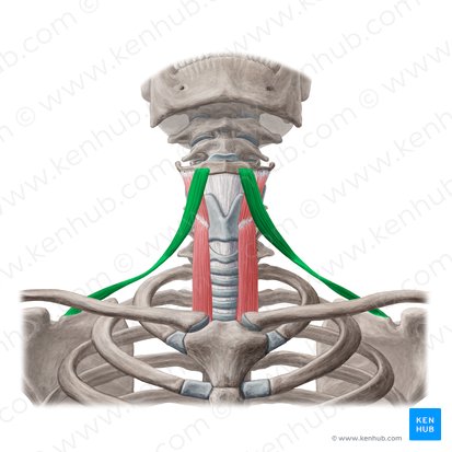
digastric
mylohyoid
stylohyoid
geniohyoid
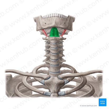
Name the 4 suprahyoid muscles
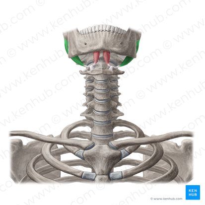
digastric
anterior belly of digastric
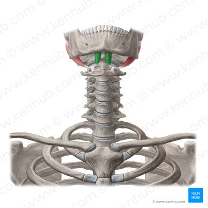
posterior belly of digastric
mylohyoid
forms the floor of the mouth
floor of submental triangle and submandibular triangle
oral diaphragm =
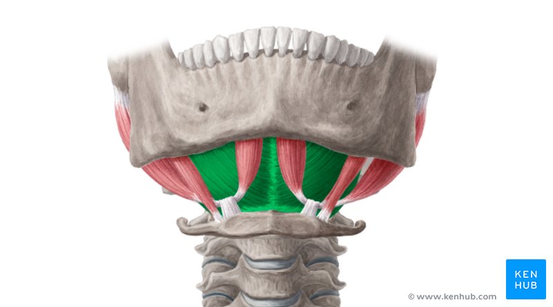
stylohyoid
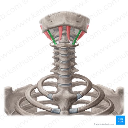
geniohyoid
sternohyoid
sternothyroid
thyrohyoid
omohyoid
Name the 4 infrahyoid muscles
strap muscles
What is another name for the infrahyoid muscles?
sternohyoid
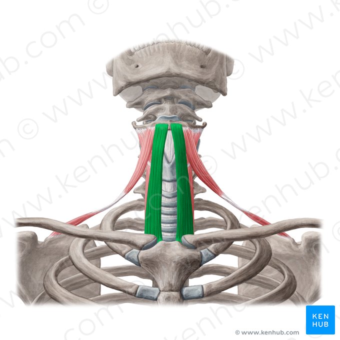
sternothyroid
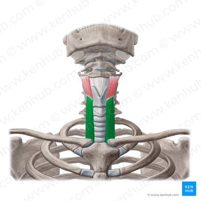
thyrohyoid
omohyoid
mastication
a vital function that ensures that ingested food is broken down into pieces and prepared for digestion
chewing
What is another name for mastication?
hyoid
suspended inferior to the mandible
situated at the root of the tongue
not directly articulated to any other bone
attached to thyroid cartilage by thyrohyoid membrane
attached to styloid process of temporal bone by stylohyoid ligament
functions: supports the tongue, which is located superior to it; holds up the larynx which is inferior to it; transmits the force of muscles that assist in opening the mouth (depressing the mandible)
submental triangle
Name the space outlined in blue

anterior bellies of digastric
lateral borders of submental triangle

hyoid bone
inferior border of submental triangle

submental lymph nodes
small veins
contents of submental triangle

carotid triangle
Name the space outlined in green

posterior belly of digastric
superior border of carotid triangle

medial border of sternocleidomastoid
lateral border of carotid triangle

superior belly of omohyoid
anterior/inferior border of carotid triangle

larynx
pharynx
thyroid gland
common carotid artery
external carotid artery
internal jugular vein
cervical plexus
ansa cervicalis
CN X
CN XI
CN XII
deep cervical lymph nodes
Name the 12 contents of carotid triangle

common carotid artery
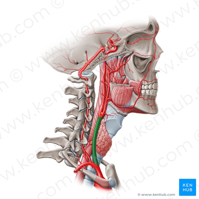
external carotid artery
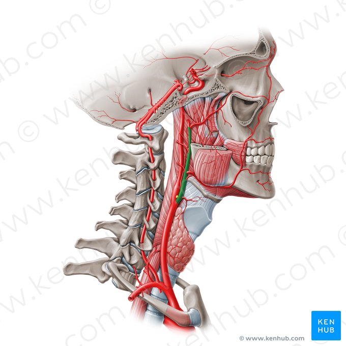
internal carotid artery
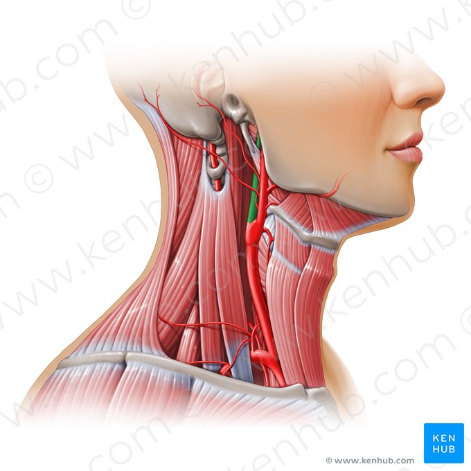
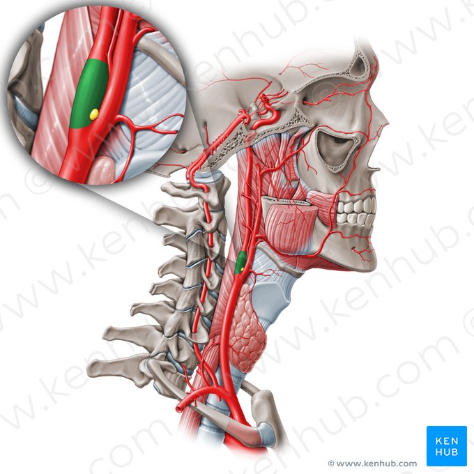
carotid body
small cluster of chemoreceptor cells at bifurcation of the common carotid artery
function: detects decreases in partial pressure of O2 and CO2; thereby modulates cardiovascular and respiratory function
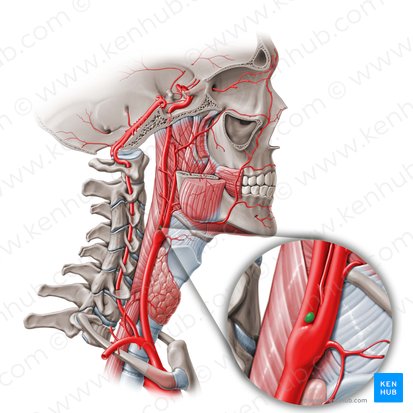
carotid sinus
a neurovascular structure at the dilation at the beginning of the internal carotid artery
crucial role in the control of blood pressure and heart rate
function: parasympathetic baroreceptor that is sensitive to pressure changes in the arterial blood pressure
muscular triangle
Name the space in yellow

hyoid bone
superior border of muscular triangle

midline of neck
medial border of muscular triangle

superior belly of omohyoid
superolateral border of muscular triangle

inferior portion of sternocleidomastoid
inferolateral border of muscular triangle

infrahyoid muscles
thyroid gland
parathyroid glands
contents of muscular triangle

submandibular triangle
Name the space outlined in orange

body of mandible
superior border of submandibular triangle

anterior belly of digastric
anterior border of submandibular triangle

posterior belly of digastric
posterior border of submandibular triangle

submandibular gland
lymph nodes
CN XII
mylohyoid nerve
facial artery and vein
contents of submandibular triangle

submandibular gland
in the submandibular triangle
lies along the body of the mandible
partly superior and partly inferior to posterior half of mandible
partly superficial and partly deep to the mylohyoid